Here, we describe a magnetic separation-assisted high-speed homogenization method for large-scale production of endosome-derived nanovesicles as a new type of exosome mimics (EMs) that share the same biological origin and similar structure, morphology, and protein composition of native extracellular vesicles (EVs).
Extracellular vesicles (EVs) have attracted significant attention in physiological and pathological research, disease diagnosis, and treatment; however, their clinical translation has been limited by the lack of scale-up manufacturing approaches. Therefore, this protocol provides a magnetic separation-assisted high-speed homogenization method for the large-scale production of endosome-derived nanovesicles as a new type of exosome mimics (EMs) derived from the endosomes, which have about 100-time higher yield than conventional ultracentrifugation method. In this method, magnetic nanoparticles (MNPs) were internalized by parental cells via endocytosis and were subsequently accumulated within their endosomes. Then, MNPs-loaded endosomes were collected and purified by hypotonic treatment and magnetic separation. A high-speed homogenizer was utilized to break MNP-loaded endosomes into monodisperse nanovesicles. The resulting endosome-derived vesicles feature the same biological origin and structure, characterized by nanoparticle tracking analysis, transmission electron microscope, and western blotting. Their morphology and protein composition are similar to native EVs, indicating that EMs may potentially serve as a low-cost and high-yield surrogate of native EVs for clinical translations.
Extracellular vesicles (EVs) are small vesicles secreted by almost all cells with a size range of 30-150 nm, containing abundant bioactive substances. Depending on the cell of origin, EVs show high heterogeneity, possessing multiple components specific to parent cells1. EVs are released into body fluids and transported to distant sites where they are taken up by target cells for action2, which can be utilized to deliver a wide range of bioactive molecules and drugs for tissue repairing, tumor diagnosis and treatment, and immune modulation3,4. However, other biological nanoparticles (e.g., lipoproteins) and nanovesicles (e.g., EVs derived from non-endosomal pathways) with similar biophysical properties in body fluids inevitably affect EV isolation and purification. To date, ultracentrifugation remains the gold standard for EV isolation, and other isolation methods, including sucrose density gradient centrifugation, ultrafiltration, polyethylene glycol precipitation, chromatography, and immunomagnetic bead isolation, have been developed5. The current bottleneck limiting clinical translation and commercialization of EV therapeutics is the severe lack of isolation techniques that allow for highly scalable and reproducible isolation of EVs6,7,8. Traditional EV isolation techniques (e.g., ultracentrifugation and size exclusion chromatography) suffer from low yield (1 x 10⁷-1 x 10⁸/1 x 106 cells), long production cycle (24-48 h), poor reproducibility of product quality, and require expensive and energy-intensive production equipment that cannot meet the current clinical demand for EVs6.
Exosome mimics (EMs), synthetic surrogates of native EVs, have attracted important attention due to their highly similar structure, function, and scalability in production. The main source of EMs is from the direct extrusion of whole parental cells with continuous sectioning9,10, demonstrating potent biological functions as native EVs11,12. For instance, EMs derived from human umbilical cord mesenchymal stem cells (hUCMSCs) exert similar wound-healing effects as native EVs and are richer in protein composition13. Though EMs derived from whole cells have the biological complexity of EVs, their main drawback is the heterogeneity of products because they are inevitably contaminated by various cellular organelles and cell debris. Protein localization analysis further revealed that EMs derived from whole-cell extrusion contain many non-EVs-specific proteins from mitochondria and the endoplasmic reticulum13. Moreover, most methods for manufacturing EMs still require ultracentrifugation, a highly time and energy-consuming process14. Considering the fact that exosomes are exclusively derived from cellular endosomes, we hypothesized that bioengineered endosome-derived nanovesicles may better recapitulate the biological homology between exosomes and EMs in comparison with the well-established cell membrane-derived EMs produced by whole cell extrusion method14. Nevertheless, the manufacture of endosome-derived nanovesicles is difficult due to the lack of viable approaches.
Clinical studies have been carried out by utilizing EVs as a surrogate of cell-free therapy and a nanoscale drug delivery system for the treatment of various diseases. For instance, EVs derived from bone marrow mesenchymal stem cells have been used to treat severe pneumonia caused by COVID-19 and have achieved promising results. Recently, genetically engineered EVs carrying CD24 proteins have also demonstrated potent therapeutic benefits for treating COVID-19 patients15,16. However, the clinical requirement of EV therapy still cannot be met with traditional isolation methods because of the low yield and cost. This study reports the large-scale production of endosome-derived nanovesicles via a magnetic separation-assisted high-speed homogenization approach. It takes advantage of the endocytosis pathway of MNPs to isolate MNP-loaded endosomes via magnetic separation, followed by high-speed homogenization to formulate endosomes into monodisperse nanovesicles. Since the types of endosomes collected by this protocol are diverse, further in-depth research is still required to establish good manufacturing practices (GMP) in the industry. This novel EM preparation approach is more time efficient (5 min of high-speed homogenization) to obtain nanovesicles homologous to native EVs. It produces exponentially more vesicles from the same amounts of cells than ultracentrifugation, which can be generally applied to various cell types.
NOTE: A schematic of the method is shown in Figure 1.
1. EM preparation and isolation
2. EM characterization (Figure 2 and Figure 3)
3. In vitro EM function detection
The workflow of EM preparation by magnetic separation-assisted high-speed homogenization is shown in Figure 1. Cells internalize 10 nm polylysine-modified IONPs, which are specifically accumulated in endosomes via endocytosis (Figure 3A). After being treated with hypotonic buffer and homogenized, the IONP-loaded endosomes are released from the cells and subsequently collected by magnetic separation. The isolated endosomes are further reconstituted into monodisperse nanovesicles, also known as EMs, by high-speed homogenization. We explored multiple key parameters (e.g., homogenization speed and time) to identify optimized EM preparation conditions (Figure 2). Finally, a homogenization speed of 140 x g for 5 min was chosen as the optimized condition by considering the particle size and yield of produced EMs. Free IONPs and IONP-loaded EMs are eventually removed from the final product solution by a second round of magnetic separation to obtain IONP-free EMs. The method produces highly uniform and monodispersed nanovesicles from parental cell endosomes, sharing the same biological origin as native EVs.
To compare EVs obtained by ultracentrifugation with EMs generated by this method, BMSC and 293T were prepared for EVs and EMs. The diameter and morphology of EMs were analyzed by NTA and TEM. The morphology of BMSC-EMs has the feature of a typical bowl-shaped vesicle-like structure and is delimited by a lipid bilayer (Figure 3A). As analyzed by NTA, both BMSC-EMs and 293T-EMs have a similar hydrodynamic diameter to native EVs (BMSC-EVs and 293T-EVs) (Figure 3B). The BMSC-EMs yields of high-speed homogenization were 8.16 × 10⁸-1.42 × 10⁹/1 × 106cells, and the yield of native EVs prepared by ultracentrifugation was only 7.2 × 10⁷-1.12 × 10⁸/1 × 106cells. Similarly, the 293T-EMs yields of high-speed homogenization were 3.71 × 10⁸-7.58 × 10⁸/1 × 106cells, which reaches up to approximately 100-fold higher than those of native 293T-EVs prepared by the conventional ultracentrifuge method (≈5.5 × 10⁶/1 × 106 cells) (Figure 4A).
Moreover, western blotting results showed that BMSC-EMs contain the same protein biomarkers as EVs (CD63 and Annexin). Both EMs and EVs are negative for Calnexin expression, suggesting that EMs produced by this method had almost no plasma membrane contamination (Figure 3C). There is no significant difference in protein concentration between EM and EV, BMSC-EMs and BMSC-EVs exhibited similar total protein concentrations, 11.15 µg/1 × 109 particles and 14.71 µg/1 × 109 particles via the BCA protein assay kit. Moreover, 293T-EMs and 293T-EVs exhibited total protein concentrations of approximately 31.8 µg/1 × 109 particles and 9.95 µg/1 × 109 particles, respectively (Figure 4B). These results indicate that EMs have a similar protein composition as native EVs. To detect whether EMs can be endocytosed, PKH26-labeled EMs and EVs were co-incubated with BMSC and ID8 at a concentration of 1 × 10⁹ particles/mL for 8 h, and the cells were observed under confocal fluorescence microscopy to confirm that EMs could be readily taken up by the cells for action as well as EVs (Figure 5).
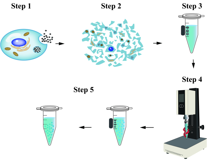
Figure 1: Schematic diagram of the magnetic-assisted high-speed homogenization method. Step 1: Cells internalize IONPs into endosomes through endocytosis. Step 2: Collect organelles, including IONP-loaded endosomes, after hypotonic treatment and homogenization. Step 3: Purify IONP-loaded endosomes by magnetic separation. Step 4: The endosomes are homogenized and reconstituted into monodisperse nanovesicles. Step 5: Remove free IONPs and IONP-loaded EMs by magnetic separation to collect IONP-free EMs. Please click here to view a larger version of this figure.
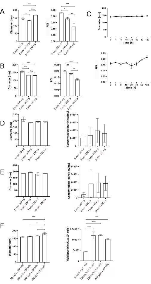
Figure 2: The optimization of EM preparation conditions. (A) The diameter and PDI of BMSC-EMs in response to homogenization speed changes when time is set at 5 min were analyzed by DLS. (B) The diameter and PDI of BMSC-EMs in response to homogenization time changes when the homogenization speed is set at 140 x g were analyzed by DLS. (C) The diameter and PDI of BMSC-EMs at different storage time points were analyzed by DLS. (D) The diameter and concentration of BMSC-EMs in response to homogenization speed changes when time is set at 5 min were analyzed by NTA. (E) The diameter and concentration of BMSC-EMs in response to the homogenization time change when the homogenization speed is set at 140 x g were analyzed by NTA. (F) The diameter and yield of BMSC-EMs in response to BMSC cells co-incubation with IONPs of different concentrations were analyzed by NTA. ****p < 0.0001. Please click here to view a larger version of this figure.
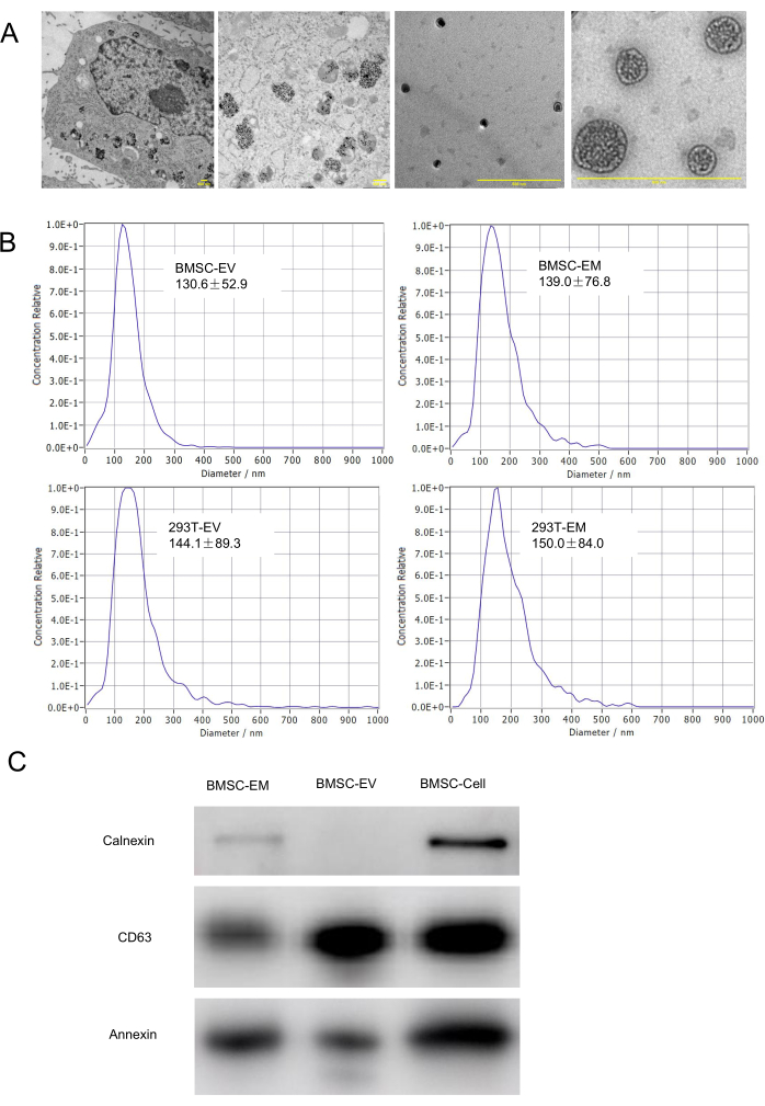
Figure 3: Characterization of EMs. (A) TEM characterization of the morphology of parental cells with endocytosed IONPs, endosomes, and EMs. The scale bars represent 500 nm. (B) Hydrodynamic diameters of BMSC-EM, BMSC-EV, 293T-EM, and 293T-EV are characterized by NTA. (C) Western blot analysis results of BMSC-EMs, BMSC-EVs, and BMSC cell lysates. Please click here to view a larger version of this figure.
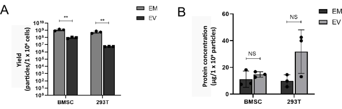
Figure 4: The yield of EMs is higher than EVs. (A) The yield of EMs and EVs prepared from BMSC and 293T was analyzed by NTA. (B) Protein yield of EMs and EVs prepared from BMSC and 293T. **p < 0.01. Please click here to view a larger version of this figure.
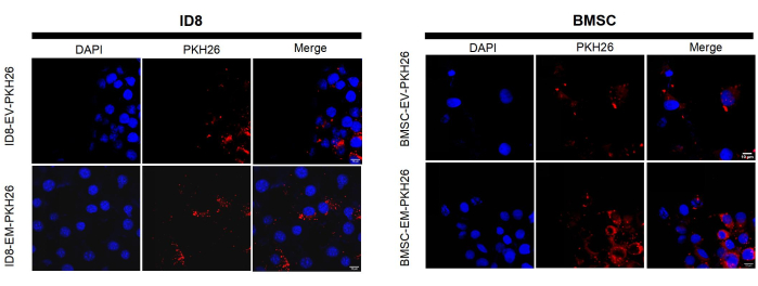
Figure 5: Representative fluorescent microscope images of PKH26-labeled EVs and EMs internalized by BMSC and ID8. The scale bars represent 10 µm. Please click here to view a larger version of this figure.
As a surrogate of cell-free therapy and a nanoscale drug delivery system, EVs have yet to meet their clinical expectations, and a main obstacle is the lack of scalable and reproducible production and purification methods6. Therefore, various types of EMs have been developed as EV analogs with similar biological complexity14. To date, the most commonly used EM example is cell plasma membrane-derived nanovesicles. The preparation of such nanovesicles is relatively easy and straightforward by directly extruding whole parent cells17. However, cell plasma membrane-derived nanovesicles cannot recapitulate native EVs owing to two drawbacks: First, the biological origin of these nanovesicles is from cell plasma membrane, which contains different lipid and protein compositions in comparison with native EVs; Second, the contaminations, including proteins, nucleic acids and lipids from non-EV organelles and cell debris, causing inevitable EM heterogeneity. In this method, we incubated cells with IONPs and collected IONP-loaded endosomes through magnetic separation, then generated monodisperse nanovesicles via high-speed homogenization, eventually obtaining IONP-free EMs by a second round of magnetic separation. Moreover, we optimized the EM preparation conditions by exploring the impact of different parameters, such as homogenization speed and time, on EM size and yield. This protocol takes advantage of an interesting biological phenomenon: MNPs (e.g., 10 nm IONPs) can be efficiently internalized by almost all cells via endocytosis and exclusively accumulated in endosomes, not other organelles. This unique biological process enabled the isolation of MNP-loaded endosomes without non-EV contaminations via magnetic separation. In return, nanovesicles prepared by these purified cell endosomes could more faithfully recapitulate the biological complexity of native EVs.
Moreover, the golden standard of the EV isolation method is ultracentrifugation, which costs massive cell culture media and a long processing time of over 5 h and only produces 1 × 10⁷-1 × 10⁸ particles from 1 x 10⁶ cells6. Traditional whole parental cell extrusion methods include multiple steps involving ultra-high pressure membrane extrusion and ultracentrifugation, which have a relatively higher yield but require expensive equipment17. However, the EM production yield, efficiency, cost, and manpower are substantially improved by utilizing our protocol. The high-speed homogenizer is a common and low-cost equipment for a short processing time of 5 min in this protocol, and this method can produce up to 100 times more EMs from 1 × 10⁶ cells in comparison with native EVs. However, this method has some limitations, such as loss of endosomal proteins during high-speed homogenization, and it has been shown that MNPs endocytosed by cells can be transferred to lysosomes, which would be a potential contamination18.
In perspective, EMs can be engineered to exert important biological functions as their native counterparts, which may serve as a novel and promising type of biological therapeutics. Bioactive substances (e.g., proteins and nucleic acids) can be integrated into EMs via selecting specific parental cells (e.g., stem cells19) or genetic modification (e.g., fusion proteins20). For example, cell-derived nanovesicles by extruding living embryonic stem cells have a positive effect on the recovery or wound-healing process19. Genetic modification is an alternative approach to generate EMs with specific biological functions. For example, when cells were transfected with vectors of anti-epidermal growth factor receptor (EGFR) nanobodies fused to glycosylphosphatidylinositol (GPI) anchor signal peptides, the EGFR nanobodies can be anchored on the surface of EVs for targeting EGFR-expressing tumor cells20. EMs have performed well as a surrogate of cell-free therapy and a nanoscale drug delivery system in multiple preclinical studies14, but there is a lack of a comprehensive mechanistic understanding of EM or EV-based therapy with concerns about the heterogeneity and reproducibility of EVs and EMs in clinic investigations. Taken together, the biological functions of EMs and their clinical implications warrant further investigations.
The authors have nothing to disclose.
The authors acknowledge the use of instruments at the Shared Instrumentation Core Facility at the Institute of Basic Medicine and Cancer (IBMC), Chinese Academy of Sciences. This study was supported by the National Natural Science Foundation of China (NSFC; 82172598), the Natural Science Foundation of Zhejiang Province, China (LZ22H310001), the 551 Health Talent Training Project of Health Commission of Zhejiang Province, China, the Agricultural and Social Development Research Project of Hangzhou Municipal Science and Technology Bureau (2022ZDSJ0474) and Qiantang Interdisciplinary Research Grant.
Annexin antibody | ABclonal | A11235 | Western blotting |
BCA assay kit | Beyotime | P0012 | Protein concentration assay |
Calnexin | GeneTex | HL1598 | Western blotting |
CD63 antibody | ABclonal | A19023 | Western blotting |
Cell lysis buffer for Western and IP | Beyotime | P0013 | Western blotting |
Centrifuge | Beckman | Allegra X-30R | Cell centrifuge |
CO2 incubator | Thermo | Cell culture | |
Confocal laser scanning fluorescence microscopy | NIKON | A1 HD25 | Photo the fluorescence picture |
DMEM basic (1x) | GIBCO | C11995500BT | Cell culture |
Dynamic light scattering (DLS) | Malvern | Zetasizer Nano ZS ZEN3600 | Diameter analysis |
Electric glass homogenizer | SCIENTZ(Ningbo, China) | DY89-II | Low-speed homogenization |
Exosome-depleted FBS | system Bioscience | EXO-FBS-50A-1 | Cell culture |
High-speed homogenizer | SCIENTZ(Ningbo, China) | XHF-DY | High-speed homogenization |
Magnetic grate | Tuohe Electromechanical Technology (Shanghai, China) | NA | Magnetic separation |
PKH26 Red Fluorescent Cell Linker Kit for General Cell Membrane Labeling | Sigma-Aldrich | PKH26GL-1KT | The kit contains PKH26 cell linker in ethanol and Diluent C |
Polylysine-modified iron oxide nanoparticles (IONPs) | Zhongke Leiming Technology (Beijing, China) | Mag1100-10 | Cell culture |
Potassium chloride | Aladdin | 7447-40-7 | Cell hypotonic treatment |
Protease inhibitor cocktail | Beyotime | P1030 | Proteinase inhibitor |
Sodium citrate | Aladdin | 7447-40-7 | Cell hypotonic treatment |
Transmission electron microscopy (TEM) | JEOL | JEM-2100plus | Morphology image |
Ultracentrifuge | Beckman | Optima XPN-100 | Exosome centrifuge |
ZetaView nanoparticle tracking analyzers | Particle Metrix | PMX120 | Nanoparticle tracking analysis |