Source: Honkanen, R. A. et al, A Rabbit Model of Aqueous-Deficient Dry Eye Disease Induced by Concanavalin A Injection into the Lacrimal Glands: Application to Drug Efficacy Studies. J. Vis. Exp. (2020).
This video describes the procedure to induce dry eye disease in a rabbit model through an injection of Concanavalin A into the orbital lobe of the superior lacrimal gland. The developed model is useful to study the effectiveness of various drugs for treating dry eye disease.
All procedures involving animal models have been reviewed by the local institutional animal care committee and the JoVE veterinary review board.
1. Animal preparation
NOTE: The portions of the orbital lacrimal gland system are injected.
2. Injection of the orbital superior lacrimal gland (OSLG)
NOTE: OSLG follows in rapid succession.
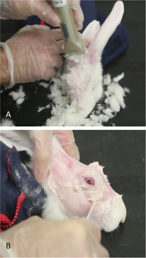
Figure 1: Preparation of rabbit for concanavalin A injections. (A) Small shears are used to remove fur, allowing easier visualization of landmarks to identify the orbital superior lacrimal gland. (B) Nair is used to remove hair that remains after shearing.
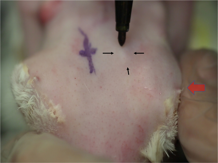
Figure 2: Localization of the orbital superior lacrimal gland. Changes in skin contours indicate the location of the OSLG as it protrudes through the posterior incisure. Alternating medial pressure on the globe (large arrow) causes the superior orbital gland to prolapse, which is seen as a small elevation in the skin. This elevation will increase in size each time the pressure is applied (small arrows). The location of this gland is usually in line with the posterior orbital rim.
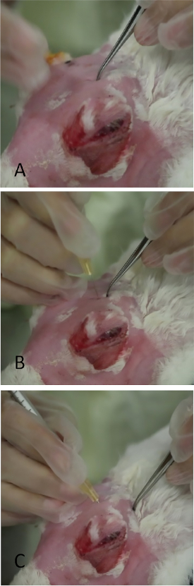
Figure 3: Injection of the orbital superior lacrimal gland. (A) Application of gentle pressure to the skull with fine-toothed forceps in the area which prolapsed as in Figure 2. A thin slit-like opening in the skull can be palpated. Leaving a small indentation mark with the forceps greatly aids placement of the needle during injection. (B) The needle is being inserted perpendicularly through the incisure. If placed incorrectly, its passage is stopped by the bony skull. (C) The needle is in final position angled towards the lateral canthus.
The authors have nothing to disclose.
100 mm macro lens | Canon EF 100mm f/2.8L IS USM | 3554B002 | |
26 gauge needles (5/8) | Becton Dickinson and Company, Franklin Lakes, NJ |
305115 | Needles for injecting ConA into the lacrimal glands |
27 gauge needles (5/8) | Becton Dickinson and Company, Franklin Lakes, NJ |
305921 | Needles for injecting ConA into the lacrimal glands |
Aceproinj (acepromazine) | Henry Schein Animal Health, Dublin, OH |
NDC11695-0079-8 | 0.1ml/kg subcutaneously injection for rabbit sedation |
Anesthesia vaporizer | VetEquip, Pleasanton, CA | Item #911103 | |
Bishop Harmon Forceps | Bausch and Lomb (Storz), Bridgewater, NJ |
E1500-C | Tissue forceps |
Caliper | Bausch and Lomb (Storz), Bridgewater, NJ |
E-2404 | Caliper used to measure length of needle during ConA injection |
Concanavalin A | Sigma, St. Louis, MO | C2010 | Make 5mg/ml in PBS for injection into rabbit lacrimal glands |
Rabbit, New Zealand White or Dutch Belted (as described in text) |
Charles River Labs, Waltham, MA | 2-3 kg | Research animals |
Surgical Loupes +1.50 | Designs for Vision, Bohemia, NY | Specialty item | Provide magnificantion of ocular surface while observing tear break up and performing Concanavalin A injections. |