Summary

Micro-dissection of Rat Brain for RNA or Protein Extraction from Specific Brain Region

Published: August 30, 2007
doi:

Summary

Micro-dissection of rat brain into various regions is extremely important for the study of different neurodegenerative diseases. This video demonstrates micro-dissection of four major brain regions include olfactory bulb, frontal cortex, striatum and hippocampus in fresh rat brain tissue. Useful tips for quick removal of respective regions to avoid RNA and protein degradation of the tissue are given.

Abstract

Micro-dissection of rat brain into various regions is extremely important for the study of different neurodegenerative diseases. This video demonstrates micro-dissection of four major brain regions include olfactory bulb, frontal cortex, striatum and hippocampus in fresh rat brain tissue. Useful tips for quick removal of respective regions to avoid RNA and protein degradation of the tissue are given.

Protocol

Step 1

Sacrifice the rat according to established protocols. Remove the brain from the skull and rinse it in ice cold DEPC treated Milli Q water to remove any surface blood.

Step 2

Place on cold metal plate. Cut the brain bi-half into right and left hemisphere.

269_Figure-01

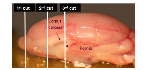
Figure 1.  The cutting positions from the medial view of rat hemisphere
 

Step 3

Collect the olfactory bulb right after the first cut. Flash freeze the specimen in liquid nitrogen and store at -80°C.

269_Figure-02

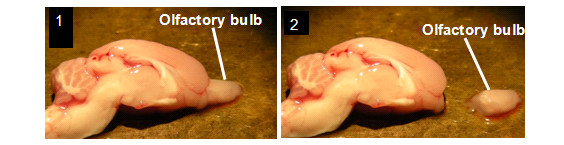
Figure 2. Olfactory bulb dissection

Step 4

Collect the frontal cortex right after the second cut, using a blade and a Dumont No. 5 forceps. Flash freeze the specimen in liquid nitrogen and store at -80°C.

269_Figure-03

Figure 3. Frontal cortex dissection

Step 5

Collect the striatum (caudate nucleus) right after the third cut with the help of two Dumont No.5 forceps. Flash freeze the specimen in liquid nitrogen and store at -80°C.

269_Figure-04

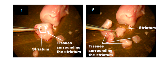
Figure 4. Striatum (caudate nucleus) dissection

Step 6

To collect the hippocampus, put the ventral side of the brain up and then remove midbrain to expose the hippocampus. Dissect the hippocampus from the cortex using two Dumont No.5 forceps. Flash freeze the specimen in liquid nitrogen and store at -80°C.

269_Figure-05

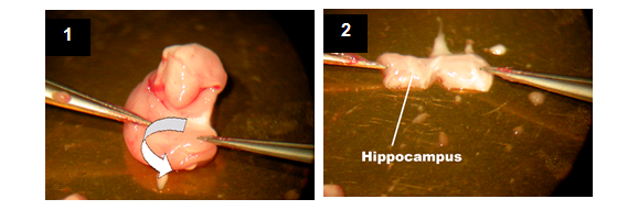
Figure 5. Hippocampus dissection

Discussion

Different brain regions associate with different neurodegenerative diseases. For example, frontal cortex and striatum relate with Parkinson s disease while hippocampus relates with Alzheimer’s disease. Precise micro-dissection of the brain allows us to detect the mRNA and protein changes in diseased region more accurately.

Disclosures

The authors have nothing to disclose.

Materials

Material Name Type Company Catalogue Number Comment
Microscope   Olympus    
Dissection tools       Metal plate, forceps, blade, etc. Individually wrapped with aluminum foil and heated at 180oC for eight hours to eliminate RNase. Made ice cold before use.

References

  1. Palkovits, M. . Maps and guide to micro-dissection of the rat brain. , (c.1988).
  2. Swanson, L. W. . Brain maps: structure of the rat brain. 3rd revised edition. , (2004).
check_url/269?article_type=t

Play Video

Cite This Article
Chiu, K., Lau, W. M., Lau, H. T., So, K., Chang, R. C. Micro-dissection of Rat Brain for RNA or Protein Extraction from Specific Brain Region. J. Vis. Exp. (7), e269, doi:10.3791/269 (2007).

View Video