A biochemical approach is described to identify in vivo protein-protein interactions (PPI) of membrane proteins. The method combines protein cross-linking, affinity purification and mass spectrometry, and is adaptable to almost any cell type or organism. With this approach, even the identification of transient PPIs becomes possible.
Membrane proteins are essential for cell viability and are therefore important therapeutic targets1-3. Since they function in complexes4, methods to identify and characterize their interactions are necessary5. To this end, we developed the Membrane Strep-protein interaction experiment, called Membrane-SPINE6. This technique combines in vivo cross-linking using the reversible cross-linker formaldehyde with affinity purification of a Strep-tagged membrane bait protein. During the procedure, cross-linked prey proteins are co-purified with the membrane bait protein and subsequently separated by boiling. Hence, two major tasks can be executed when analyzing protein-protein interactions (PPIs) of membrane proteins using Membrane-SPINE: first, the confirmation of a proposed interaction partner by immunoblotting, and second, the identification of new interaction partners by mass spectrometry analysis. Moreover, even low affinity, transient PPIs are detectable by this technique. Finally, Membrane-SPINE is adaptable to almost any cell type, making it applicable as a powerful screening tool to identify PPIs of membrane proteins.
To understand the function of a protein it is essential to know its interaction partners. Several classical techniques are available for the identification of interaction partners of soluble proteins. However, these techniques are not easily transferable to membrane proteins due to their hydrophobic nature4. To overcome this limitation, we have developed the Membrane Strep-protein interaction experiment (Membrane-SPINE)6. It is based on the SPINE method, which was only suitable for soluble proteins7.
Membrane-SPINE benefits from two advantages of the cross-linking agent formaldehyde: first, formaldehyde can easily penetrate membranes and therefore generates a precise snapshot of the interactome of a living cell8. Second, formaldehyde cross-links can be reversed by boiling9. Here, these two advantages are used to identify not only permanent but also transient PPIs of membrane proteins6.
In brief, a Strep-tag is fused to the C-terminus of the integral membrane bait protein. Cells expressing the membrane bait protein are incubated with formaldehyde which cross-links prey proteins to the membrane bait protein (Figure 1). Modifications of prey proteins are not needed. Next, the membrane fraction is prepared. Therefore, membrane proteins are solubilized by detergent treatment and bait proteins are co-purified with its prey proteins using affinity purification. Subsequently, the cross-link is reversed by boiling, and the bait and its co-eluted prey proteins are separated by SDS-PAGE. Finally, prey proteins can be identified by immunoblot analysis or mass spectrometry.
Note: Detailed information regarding buffers indicated in the protocol is available in Table 1.
1. Fixation of Protein-protein Interactions by Formaldehyde Cross-linking in Living Cells
2. Purification of Strep-tag Membrane Protein (Bait)
3. Purification of Strep-tag Membrane Bait Protein and SDS-PAGE
4. Immunoblot Analysis to Confirm Interaction Partners
5. NanoLC-ESI-MS/MS High Resolution Experiments to Identify Interaction Partners
Membrane SPINE analysis allows the co-purification of membrane proteins and transiently interacting protein partners. The co-purification is achieved by using the cross-linking agent formaldehyde. Two parameters are critical to prevent unspecific cross-links: the formaldehyde concentration and the cross-linking time. The sufficient, but not excessive use of formaldehyde can easily be controlled by immunoblotting. Formaldehyde cross-linked protein complexes can be separated by boiling but not by SDS treatment. Hence, they can be visualized after blotting of the Strep-tagged membrane bait protein as smear in the upper section of the immune-blot of an unboiled sample.
A representative result of a Membrane SPINE assay, including all required controls, is presented in Figure 2. As bait protein the integral membrane protein CpxA of Escherichia coli was used6,11. CpxA is a sensor kinase and consists of an N-terminal sensor domain with two transmembrane domains (TMD) integrating a large extracytoplasmic sensor domain and a C-terminal highly conserved cytoplasmic catalytic domain12. After stimulation, CpxA activates its cognate response regulator CpxR. Activated CpxR diffuses off to mediate the response. For Membrane SPINE, the Strep-tag was fused to the C-terminus of CpxA (CpxA-Strep). CpxA-Strep cross-linked to other proteins or protein complexes is detected as a smear in the unboiled formaldehyde-treated sample (Figure 2, line 1 versus line 3) indicating sufficient cross-linking. Moreover, a direct protein-protein interaction of CpxA-Strep with its cognate response regulator protein CpxR as the prey protein is only detectable in the presence of formaldehyde (Figure 2, line 2 versus line 4) supporting the specificity of formaldehyde cross-linking.
A representative result of a Membrane SPINE analysis is presented in Figure 3. Samples corresponding to those of Figure 2 were silver stained. The arrow marks a band that is specific for the boiled sample. Due to the background, MS analysis also identified other proteins besides CpxR6. Therefore, the non-formaldehyde treated sample should always be analyzed, to assign background noise and specific interaction partner.
Table 1: Buffers and reagents required for membrane-SPINE.
Buffer / Reagent / Medium | Working concentration | comment | |
---|---|---|---|
LB | 10 g Tryptone 5 g yeast extract 10 g NaCl to 1 L, adjust pH to 7.0 |
Luria Broth medium | |
IPTG | 1 M Isopropyl-β-D-thiogalactopyranoside | 0.5 mM | |
Tris-buffer | 20 mM Tris-HCl, pH 8 | Adjust pH to 8.0 using NaOH | |
0,1 M EDTA, pH 8 | 0.1 M EDTA | Adjust pH to 8.0 using NaOH | |
PMSF | 1 M Phenylmethylsulfonylfluoride in 100% Isopropanol | 10 mM | PMSF is stable in 100% isopropanol but not in water! Stock solution can be stored at -20 °C; PMSF has to be adapted to room temperature before diluting in buffer; PMSF containing buffers should be used in within 10 min of preparation. |
P1 | 20 mM Tris-HCl, pH 8.0 0.5 M Sucrose |
||
P2 | 2 mg/ml lysozyme in 0.1 M EDTA, pH 7.5 | ||
P3 | 20 mM Tris-HCl, pH 8.0 10 mM PMSF |
Use immediately after preparation | |
Detergent | 20% Triton X-100 | 2% for solubilization | |
Buffer W | Fill up 10 ml 5x concentrate to 50 ml add 150 µl 20% Triton X-100 |
100 mM Tris-HCl, pH 8.0 150 mM NaCl 1 mM EDTA 0.06% Triton X-100 |
5x concentrate is part of Strep-tag protein purification buffer set (IBA) |
Buffer E | Fill up 1 ml 5x concentrate to 10 ml add 30 µl 20% Triton X-100 |
100 mM Tris-HCl, pH 8.0 150 mM NaCl 1 mM EDTA 2.5 mM Dethiobiotin 0.06% Triton X-100 |
5x concentrate is part of Strep-tag protein purification buffer set (IBA) |
5x SDS-PAGE loading dye | 0.3125 M Tris-HCl, pH 6.8 10% SDS 0.5 M DTT 50% glycerol 0.05% bromophenol blue |
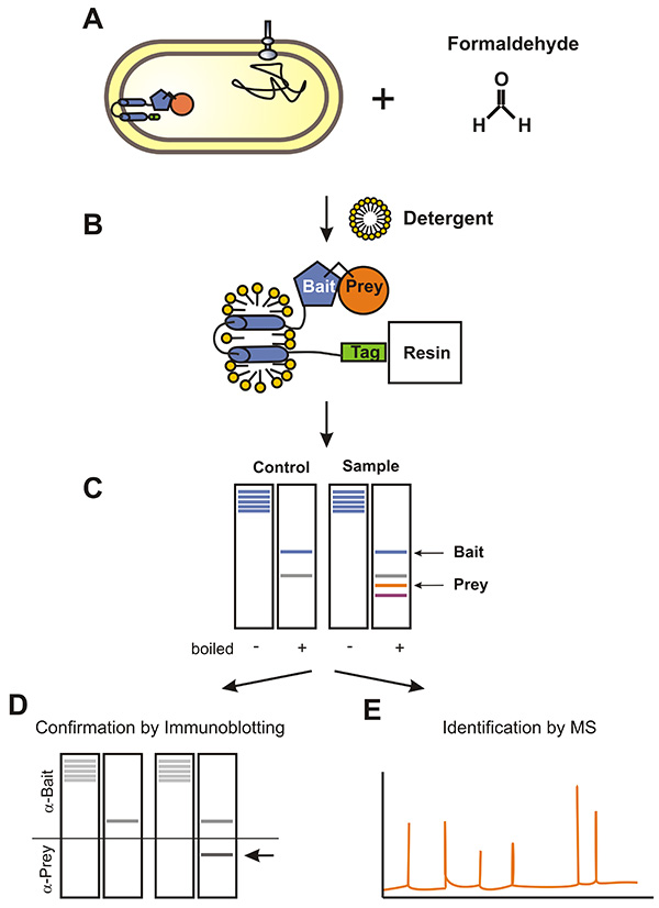
Figure 1. Flow chart of the Membrane-SPINE procedure using an Escherichia coli membrane protein as bait protein. A) Bacteria expressing the Strep-tagged membrane bait protein are treated with formaldehyde. Formaldehyde penetrates membranes and cross-links prey proteins to the membrane bait protein. B) The membrane fraction is prepared and membrane proteins are solubilized by detergent treatment. Subsequently, prey proteins are co-purified with the bait protein. C) Formaldehyde cross-links are reversed by boiling and proteins are separated by SDS-PAGE. Finally, prey proteins are either monitored by immunoblotting (D) or identified by MS-analysis (E). Click here to view larger image.
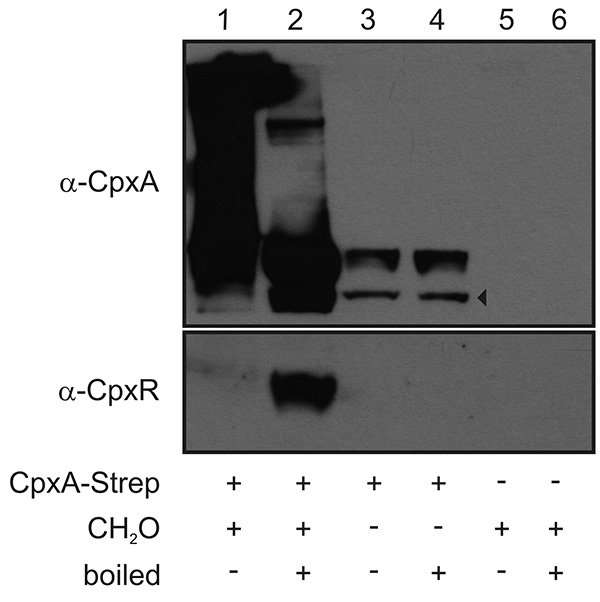
Figure 2. Representative immunoblot used to monitor a PPI of a membrane protein. Bacteria producing CpxA-Strep from a plasmid as membrane bait protein, were grown in LB medium to an OD600 of 1 and exposed to formaldehyde (CH2O) for 20 min. The inner membrane fraction was prepared, membrane proteins were solubilized by detergent treatment and CpxA-Strep was purified (lanes 1 and 2). Bacteria producing either CpxA-Strep without formaldehyde treatment (lanes 3 and 4) or carrying the empty vector with formaldehyde treatment (lanes 5 and 5) served as controls. Aliquots of each sample were boiled at 95 °C for 20 min (lanes 2, 4, and 6) to separate cross-linked proteins from CpxA-Strep. Proteins were separated in a 12.5% SDS-PAGE. Immunoblotting was performed and the blot was separated according to the size of CpxA-Strep (51 kDa) and the respective prey proteins CpxR (26 kDa). The two parts of the immunoblot were incubated with polyclonal antibodies raised against CpxA and CpxR, respectively. Subsequently, the blots were further treated with an anti-rabbit horse (HRP) antibody and developed using the SuperSignal West Pico Chemiluminescent substrate. The arrowhead marks degradation products of CpxA. Click here to view larger image.
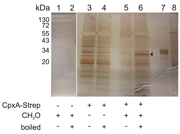
Figure 3. Representative silver-stained SDS-PAGE used for the identification of an interaction partner of a membrane protein by MS analysis. Samples corresponding to those of Figure 2 were silver stained. The arrow marks a band which was analyzed by MS analysis and which confirmed CpxR as the interaction partner of CpxA5. Lane 7 shows purified CpxR-His6 and lane 8 shows purified CpxA-His6 protein. Click here to view larger image.
Membrane SPINE analysis is a biochemical approach that enables one to confirm and to identify to this point unknown interaction partners of membrane proteins. Membrane SPINE combines in vivo cross-linking by formaldehyde with purification of a Strep-tagged membrane bait protein. The combination with immunoblotting facilitates the confirmation of predicted interaction partners (Figure 2). Additionally, the combination with MS analysis permits the identification of unknown interaction partners (Figure 3). For both applications, there is no requirement for modifying your prey protein. Moreover, Membrane SPINE is sufficiently sensitive to allow the detection of endogenous prey proteins6.
Here, we present a protocol optimized for membrane proteins of gram-negative bacteria. The envelope of gram-negative bacteria separates the cytoplasm from the environment and posses, in addition to the cytoplasmic membrane, an outer membrane and a murein sacculus. Hence, our protocol includes the removal of the outer membrane and the murein sacculus, resulting in so-called spheroplasts. Because such protocols are available for most cell types, our protocol should be adaptable for most membrane proteins. In addition, the following points should be considered.
First of all, the copy number of the vector used to overproduce the membrane bait protein has to be balanced between a high level to allow sufficient membrane protein purification and a low level to prevent unspecific interactions
Second, for some membrane proteins an N-terminal fusion might be more optimal than a C-terminal fusion. In both cases, the functionality of the fusion should be confirmed by trans-complementation.
Third, other affinity purification protocols are also compatible with subsequent MS analysis, such as FLAG purification and tandem affinity purification (TAP). We prefer the Strep-tag II because of its small size with only 8 amino acids (WSHPQFEK).
Fourth, the concentration of formaldehyde used and the cross-linking time should be optimized to prevent unspecific cross-linking. A sufficient but not excessive use of formaldehyde can be monitored by immunoblotting of unboiled samples as the ratio between cross-linked and nonlinked membrane bait protein (Figure 2). Cross-linked proteins migrate as a smear in the upper part of the immunoblot. Un-linked proteins migrate as untreated protein. The ratio between cross-linked and unlinked protein should be about 3:1.
Fifth, there is no "general detergent" for all membrane proteins for solubilization. Hence, in some cases the appropriate detergent for solubilization of the membrane bait protein has to be determined. Thereby, the chosen detergent must be compatible with the Strep-tag column.
Finally, the silver staining procedure has to be compatible with subsequent MS analysis. MS compatible silver staining kits are available from different suppliers. We used different ones with comparable good results.
The authors have nothing to disclose.
This research was supported by grants of the Deutsche Forschungsgemeinschaft GraKo1121, Hu1011/2-1 and SFB940 to S.H.
37% Formaldehyde | Roth | 4979.1 | Should not be older than one year |
DNaseI | Sigma | DN25 | |
BCA protein assay kit | Pierce | 23225 | |
Micromagnetic rod | Roth | 0955.2 | 5 mm in length, 2 mm in diameter |
Triton X-100 | Roth | 6683.1 | standard detergent for solubilization |
n-Dodecyl-β-maltoside | Glycon | D97002-C | the best detergent to solubilze CpxA |
1 ml Strep-Tactin superflow gravity flow column | IBA | 2-1207-050 | |
Strep-tag protein purification buffer set | IBA | 2-1002-001 | Contains buffer W and buffer E |
Amicon Ultra-4 centrifugal filter | Millipore | UFC803024 | |
SuperSignal West Pico Chemiluminescent substrate | Pierce | 34080 | |
SilverQuest Silverstaining kit | Invitrogen | LC6070 | |
FireSilver Staining Kit | Proteome Factory | P-S-2001 | |
Ultracentrifuge |