1. Silanization and Biomolecule Functionalization of Slide or Polycarbonate Membrane
Note: Many of the chemicals and solutions within this protocol have high evaporation rates (ethanol (EtOH), acetone, etc.). Other steps entail long incubation times for low evaporation rates. Paraffin film is recommended to seal containers. Caution: Many of the chemicals (including: sulfuric acid, acetone, (3-aminopropyl)triethoxysilane, glutaraldehyde, EtOH) are considered hazardous, or volatile. Consult material safety data sheet (MSDS) of each material for proper storage, handling, and disposal before use.
- Place new glass slide measuring 75 mm by 50 mm by 1 mm, in clean, glass slide staining dish without regard to orientation, ensuring full immersion. Use container for steps 1.1-1.12.
Note: For co-culture, include polycarbonate (PC) membrane with 0.4 micron pore size, cut to size of 75 mm by 50 mm, for all subsequent steps for PC functionalization and EC seeding. - Immerse slide in sulfuric acid (20%), O/N.
Caution: Consult MSDS for sulfuric acid before use. - Wash slide by immersing in deionized water (DI) for 5 min, three times, changing DI each time.
- Immerse slide in acetone for 30 min.
Caution: Consult MSDS for acetone before use. - Immerse slide in 6% (3-aminopropyl)triethoxysilane / acetone solution O/N.
Caution: Consult MSDS for (3-aminopropyl)triethoxysilane and acetone before use. - Immerse slide in acetone for 5 min, three times, changing acetone each time.
Caution: Consult MSDS for acetone before use. - Wash slide by immersing in DI for 5 min, three times, changing DI each time.
- Immerse slide in 1.5% glutaraldehyde / DI solution for 60 min.
Caution: Consult MSDS for glutaraldehyde before use. - Wash slide by immersing in DI for 5 min, two times, changing DI each time.
- Place slides into 70% ethanol (EtOH) for 30 min in sterile, laminar flow hood.
- Remove EtOH and allow remaining residue to evaporate. Immerse slides in sterile DI and drain in order to rehydrate the slides.
Note: Slides or PC membrane may be sealed in glass slide holder and stored at 4 °C up to one week. Upon use for cell culture, repeat steps 1.10 and 1.11. - Remove the slide from the slide holder and place it into a 100 mm by 100 mm square petri dish in the laminar flow hood.
Note: This container will be used for all remaining steps.- Prepare slide or PC for endothelial cell seeding for monoculture experiment.
- Coat the functionalized slide or PC (step 1.11) with 1 ml fibronectin solution (25 μg/ml), on one side, covering the approximate area exposed to cell culture chamber.
- Incubate slides or PC in sterile incubator set at 37 °C for 1-2 hr.
- After incubation, aspirate the remaining solution using a glass aspirator pipette connected to a vacuum.
Note: Slides may now be stored O/N at 4 °C for future use.
- Follow 1.12.2 steps only for preparing EC/SMC co-culture. Prepare and seed smooth muscle cell (SMC) culture for co-culture experiment.
- Place silicone gasket on top of the slide (step 1.11), with dimensions of 75 mm by 50 mm outer diameter, inner diameter of 50 mm by 30 mm, thickness of 0.5 mm, and perforations which align with flow chamber vacuum.
- Prepare 1 ml SMC in 10% FBS/DMEM suspension. Neutralize 1 ml solution of 2 mg/ml collagen type-I with 7% NaHCO3 and 0.1 M NaOH solution to pH of 7.4, and mix with SMC suspension with final cell density of 2 x 10⁶ cells/ml.
- Spread SMC solution within gasket and incubate slides at 37 °C and 5% CO2 for 30 min.
- After incubation, cover the slide with 25 ml 0.1% fetal bovine serum (FBS) mixed in Dulbecco’s Modified Eagle Medium (DMEM) for 72 hr.
- Prepare slide or PC for endothelial cell seeding for monoculture experiment.
- Culture endothelial cells (ECs) on glass slide/polycarbonate membrane.
- Seed 1 ml ECs in 10% FBS/DMEM, with initial concentration of 6.0 x 10⁵ cells/ml, on fibronectin surfaces of glass slide or polycarbonate membrane.
- Incubate cells in an incubator at 37 °C, 5% CO2, and 10% FBS for 120 min.
- Cover slides containing cells with additional 10% FBS/DMEM, and place in incubator in 37 °C, 5% CO2 O/N, until 70%-80% confluent.
2. Determination of Fluid Viscosity and Volumetric Flow Rate
Note: Rotating viscometers are sensitive equipment, and the viscometer user manual should be consulted before calibrating, zeroing, or performing measurements.
- Determine desired fluid shear (τ) from literature of the experiment’s targeted vasculature.
- Measure 1% FBS/DMEM viscosity (μ), using a rotational viscometer.
- Determine required volumetric flow rate (Q) from Poiseuille equation:
 ,
,
where τ = fluid shear, μ = viscosity, Q = vol. flow rate, w = chamber width, and h = chamber height). - Set pump to flow rate derived in step 2.3, and use for all subsequent steps.
3. Determination of Pulsatility Index
Note: All connection ports within the system should be connected with appropriately sized lock-ring-to-barb, or female luer-to-barb connections. Connecting PVC tubing may then be connected to barb fittings, and circuit completed.
- Connect flow circuit as in Figure 2, with damping chamber (Figure 3) and ultrasonic flow meter in correct flow direction.
Note: Ultrasonic flow meter is a sensitive piece of equipment, and the user manual should be consulted before use. - Fill flow circuit and reservoir with DI water, by ensuring reservoir outlet tube (to pump) is submerged within reservoir volume. Visualize flow waveform using a flow meter.
- Open air release valve on the damping chamber to change fluid/air volume ratio. Close air release valve at different fluid level intervals and calculate pulsatility index (PI = (VMax – VMin) / VMean) by using flow waveform peak (VMax), trough (VMin), and mean (VMean). Record PI values in notebook.
- Mark resulting fluid level and PI on the damping chamber, using a felt tipped, permanent ink pen for future use. Determine desired PI levels from pathology literature11.
- Silicon Tube Formation- Optional, more advanced method of controlling pulsatility
- Mix the silicone elastomer base and curing agent at different ratios (e.g., a base-to-crosslinker ratio from 10:1 to 36:1) for varied targeted elastic moduli, as illustrated previously.2
- Fabricate silicone tubes by repeated dip-cure process, briefly dipping steel cannula (14 G) into silicone prepolymer mixture with a predetermined base-to-crosslinker ratio.
- Place the prepolymer-coated cannula into an oven set at 60 °C for 4 hr to cure the polymer coating on the cannula, switching the direction in the middle of curing process.
- Repeat the dipping process with the same silicone elastomer mixture and place back in the oven for additional 4 hr, which results in ~0.3 mm thick tubes.
- Remove the ultrathin silicone tube from the stainless steel cannula.
- Connect tubes to flow circuit instead of damping chamber, and test PI for each as in 3.3.
4. Pump Sterilization
- Immerse the flow chamber (Figure 2), PVC tubing, and gaskets in 10% hydrogen peroxide (H2O2). Wash with sterile DI before step 4.
- Place the blood pump on a spare incubator shelf.
Note: Incubator shelves should be stainless steel, and be able to support the weight of the entire flow circuit. - Fill the flow circuit and reservoir with 10% H2O2 by ensuring reservoir outlet tube (to pump) is submerged within reservoir volume. Circulate H2O2 by pumping through circuit for 20 min in the laminar flow hood with UV light on.
- Aspirate all H2O2 from tubing, and wash with sterile PBS by pumping through flow circuit.
5. Flow Chamber Assembly
Note: The flow chamber consists of an acrylic, custom made plate, with vacuum ports and inlet and outlet flow ports (see Figure 2). Chamber assembly consists of placing the flow chamber and gaskets on top of culture slides, properly aligned, and is described below.
- Warm up 1% FBS/DMEM in a 37 °C water bath and prepare flow chamber.
- Take EC slide out of media (from step 1.13) and place gasket on top with cell side up.
- (Optional) Take PC membrane out of media (from step 1.13) and place gasket on top with cell side up. Place co-culture slide with seeded SMC (step 1.12.2) underneath PC membrane.
- Align vacuum channel of the flow chamber with the holes in the gasket (Figure 2).
- Attach vacuum tube to vacuum ports (Figure 2) ensuring no media leaks to vacuum.
- Place polycarbonate sheet underneath glass slide and clamp flow chamber assembly with spring clamps, one on each side of flow chamber (Figures 2 and 5).
- Place flow system in incubator at 37 °C and 5% CO2, and fill circuit with media by pumping from filled reservoir at low pulsatility. Maintain this flow for 4 hr for preconditioning cells.
- Adjust fluid volume in damping chamber to marked PI level, and culture for desired time.
Maintenance of flow conditions is reliant on correct assembly of the flow circuit (Figure 1). Tubing diameter is an important selection in assembly, with larger diameters reducing flow resistance and subsequent pressure drop before and after the culture chamber. To ensure intended pressure and flow velocity, assemble the system with flow meter before experiment with intended tubing. Alignment of culture chamber vacuum channel (Figure 2), with perforations on silicon gasket (not pictured), maintains seal within the flow circuit. To ensure seal, spring clamps may be used to apply additional pressure on the gasket. Glass slides should be protected by polycarbonate sheet, cut to fit chamber area (Figure 6). Because flow velocity may additionally contribute to complications in maintaining seals, media may be altered through the addition of dextran to increase media viscosity. Through increased viscosity, fluid shear can be maintained at lower velocity. System response and integrity may be checked in the middle of the experiment simply by observing fluid levels and fluid color within the damping chamber and reservoir (Figure 3). The fluid level should maintain a constant value. Initial fluid level should be marked on both reservoir and damping chamber for checkup comparison. Adjustments can be made by the addition of media at the experiment initiation, or allowing entrance of air to damping chamber. Figure 4 illustrates a representative recording of flow velocity. Phenol red coloration should decrease over the course of the experiment. Upon completion of the experiment, media should be allowed to drain from the circuit, and stored at -80 °C freezer for future measure or use. Media may be analyzed for metabolite content or released cytokines, or used to monitor paracrine signaling to cellular responses by using it for cell culture. Cells may be imaged under phase contrast microscopy for morphology and confluency (Figure 5). Spindle like morphology is observed in ECs after HPF over 24 hr (Figure 7).
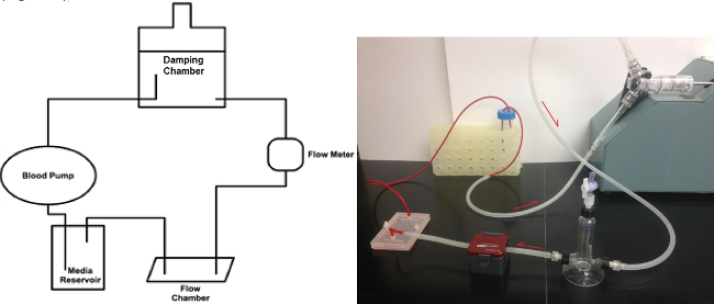
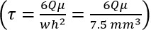
Figure 1. Flow circuit schematic. The flow meter labeled within the scheme is used to measure pulsatility index levels set by the damping chamber. Flow waveform should be measured with intended connection tube diameters to ensure system pressure remains physiologic. Please click here to view a larger version of this figure.
Figure 2. Flow chamber design and measurements. Outer channel is connected to vacuum. Center picture demonstrates clamping of the chamber. Picture at right illustrates gasket design. Please click here to view a larger version of this figure.
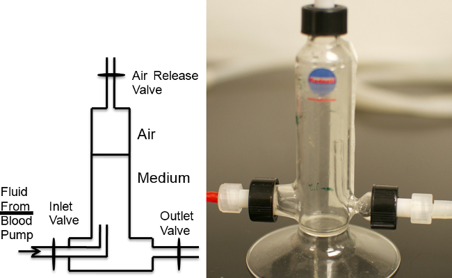
Figure 3. Flow Damping Chamber. Flow damping chamber schematic indicates intended flow direction and connection. Air release valve is opened, outlet valve is closed, and fluid fills the chamber to desired PI level. Upon opening of outlet valve and closing of air release valve, flow resumes and fluid levels within the chamber maintains levels. Please click here to view a larger version of this figure.
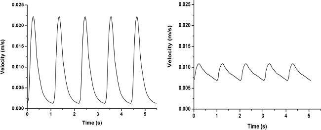
Figure 4. Sample flow waves with various pulsatility indices. High Pulsatility Flow (Left) and Low Pulsatility Flow (Right). Please click here to view a larger version of this figure.
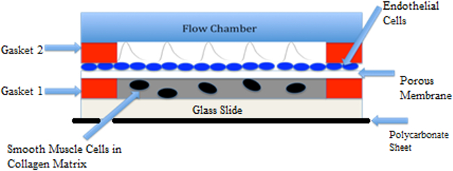
Figure 5. Co-culture Flow Schematic. Co-culture shows pulsatile flow over endothelial cells, with smooth muscle cells in collagen gel. Cytokine release from cells may cross through the porous polycarbonate membrane. Please click here to view a larger version of this figure.
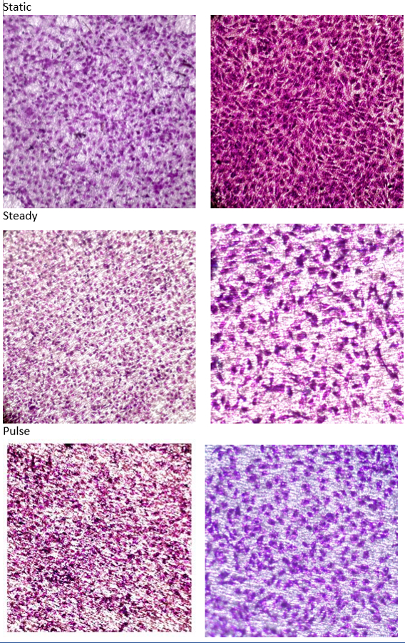
Figure 6. Endothelial Cell Morphology Changes with Flow Conditions. Confluent endothelial cell culture on functionalized polycarbonate membrane. Shown are before flow (left) and after flow (right). Flow is shown under differing conditions, static (no velocity), steady (constant velocity), high pulse (high pulsatility). Cells are stained with 2% crystal violet. Please click here to view a larger version of this figure.
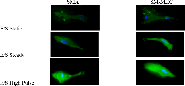
Figure 7. Smooth Muscle Cell Protein Expression Changes with Flow Conditions. Smooth muscle cells expression of α-smooth muscle actin (SMA) and myosin heavy chain (SM-MHC) under differing conditions, static (no velocity), steady flow (constant velocity), and high pulse. Please click here to view a larger version of this figure.
