1. Designing and Ordering Oligonucleotides with the Appropriate Sticky Ends
- Identify potential rho-independent terminators through genomic analysis using programs that are freely available online.7
- When working with double stranded DNA, determine the orientation of the terminator to be tested.7 The pGR-Blue plasmid only verifies terminators ligated in the 5' to 3' direction on the top (forward strand).
- Convert a bottom (reverse) strand terminator to its reversed complement to reorient the sequence for testing in pGR-Blue using free online software, e.g., ApE.
- After the orientation is determined to be correct, add the sticky end " 5'-CGAC-3' " to the 5' end of the top strand. Add the sticky end " 5'-CCGC-3' " to the 5' end of the bottom strand. 8 This will ensure that ligated insert will be inserted in the appropriate orientation using GGA.
- Once sequences are determined, order as single stranded oligonucleotides commercially.
2. Annealing Oligonucleotides (Applies Only to Freeze-dried DNA)
- Re-suspend individual oligonucleotides in nuclease free water to a concentration of 100 µM.
- Make 10x annealing buffer: 1 M NaCl and 100 mM Tris-HCl pH 7.4. Annealing buffer can be stored at 4 °C for several weeks. Once annealing buffer is made and oligonucleotides are re-suspended, begin the annealing8.
- For each terminator to be tested, add 16 µl of ultra-pure water to a 1.5 ml microcentrifuge tube.
- Add 2 µl of 10x annealing buffer to each microcentrifuge tube.
- Add 1 µl of the top strand oligonucleotide (100 µM) from the desired terminator.
- Add 1 µl of the bottom strand oligonucleotide (100 µM) from the desired terminator.
- Create a boiling water bath using a 1,000 ml beaker containing approximately 400 ml of tap water and place on a hot plate.
- Place the microcentrifuge tubes from step 2.6 in a float and boil for 4 min. After 4 min, turn off the heat plate but leave the tube and float in the water bath to slowly cool O/N (18 hr) 8. Store the annealed terminators at -20 °C.
3. Golden Gate Assembly (Standard Mix — Cost Efficient)
- Make a 40 nM dilution of the annealed terminators. Add 124 µl of nuclease free water to 1 µl of annealed terminators mix (100 µM).
- Centrifuge samples for 30 sec at 10,000 x g and store on ice.
- Into a new tube appropriate for the thermocycler being used, add 6 µl of nuclease free water.
- Add 1 µl of commercial DNA Ligase Buffer [300 mM Tris-HCl (pH 7.7-7.8), 100 mM MgCl2, 100 mM DTT, and 10 mM ATP]. Perform all subsequent steps on ice.
- Add 1 µl of pGR-Blue (35-50 ng/µl) destination plasmid to the microcentrifuge tube.
- Add 1 µl of the 40 nM annealed terminators to the microcentrifuge tube.
- Add 0.5 µl commercial high-fidelity BsaI restriction endonuclease to the microcentrifuge tube.
- Add 0.5 µl of commercial DNA ligase to the microcentrifuge tube.
- Centrifuge the reaction mixture at 10,000 x g for 120 sec. Then place the tube directly into the thermocycler.
- Run the thermocycler program: 20 cycles of 1 min at 37 °C followed by 1 min at 16 °C, followed by 1 cycle of 15 min at 37 °C. Use samples immediately or freeze and store at -20 °C.8
4. Golden Gate Assembly (Commercial Master Mix — Time Efficient)
- Add 124 µl of nuclease free water to 1 µl of the 100 µM annealed terminators created in step 2. This dilutes the terminator DNA concentration to 40 nM. Perform this step in a new, labeled 1.5 ml microcentrifuge tube.
- Into a new tube appropriate for the thermocycler, add 14 µl of nuclease free water. Make sure the new tube is labeled accordingly.
- Add 2 µl of Commercial Master Mix Buffer to the microcentrifuge tube.
- Add 1 µl of pGR-Blue plasmid (35-50 ng/µl) to the microcentrifuge tube.
- Add 2 µl of the 40 nM annealed terminators to the microcentrifuge tube.
- Add 1 µl of Commercial Master Mix to the microcentrifuge tube.
- Centrifuge the 40 nM tube at 10,000 x g for 120 sec. Then place the tube directly into the thermocycler.
- Run the thermocycler program: 1 cycle of 60 min at 37 °C followed by 1 cycle of 15 min at 55 °C. Use samples immediately or freeze and store at -20 °C.
5. Transformation and Colony Selection
- Prepare Luria Base (LB) agar Petri plates containing 1 mM concentration of ampicillin (Amp) and a 10 mM concentration of arabinose (arb). Prepare at least 10 ml of 1 M L-arabinose stock solution because this stock solution will be used in later steps.
- Use any high-efficient (108 – 109 transformants/µg ) chemically competent E. coli (JM109) cells for transformation.9 For best results, add 3-5 µl of the ligation reaction to 50 µl of competent cells. When using chemical competent cells, follow the transformation protocol specific to the cells being used.
- Spread half (25 µl) of the transformed cells onto pre-warmed LB (Amp/arb) agar plates using a sterile L-shaped "hockey stick" and incubate O/N at 37 °C. Due to ampicillin's rapid rate of decomposition, do not incubate at 37 °C over 24 hr.
- Perform colony selection based on color by visual inspection. A successful ligation should be white/yellow in visible light and fluoresce green under blue (450 nm) or UV light. An unsuccessful ligation will produce a colony that is blue in color under visible light after 18 – 20 hr.
6. Verification and Quantification of Terminator
- Prepare 5 ml sterile LB-broth tubes with 1 mM ampicillin and 10 mM arabinose. The number of tubes needed is dependent on the number of terminators being tested plus controls (cells with ampicillin resistance that do not fluoresce and cells containing the uncut pGR-Blue plasmid).
- Under visible light pick 2-3 white/yellow colonies (step 5.4) using a sterile loop. Allow the samples to incubate in broths on a shaker at 37 °C and 160 rpm for 16-18 hr. Do not incubate these samples for more than 24 hr.
- Optionally, use a spectrophotometer to determine optical cell density at 600 nm (OD600) to ensure adequate cell growth. Generally, an OD600 of ~0.8-1.0 is observed after 16-18 hr of growth.
- Use a sterile 96-well plate for the remaining steps. Conduct these steps in an aseptic environment and in triplicate. Add 199 µl of sterile LB+ Amp/arabinose broth (1 mM ampicillin/10 mM arabinose) to each well. Include room on the plate for three separate controls.
- Use the following three controls: A well (1) containing LB + Amp/arabinose broth only and a well (2) for untransformed competent cells (blank) to serve as a blank. For quantification (3) of the terminator strength, cells containing uncut original pGR or containing pGR-Blue ligated with a non-terminating sequence should be included (reference control for quantification). See Figure 1 for plate reader layout.
- Pipet 1 µl of the cell solution from the O/N broths into individual wells of the 96-well plate.
- Seal plates with a breathable cover and shake for 18-20 hr at 37 °C and 160 rpm.
- Using a plate reader, determine OD600 by reading absorbance at 600 nm. Measure GFP fluorescence as follows: excitation 395 and emission 509. Measure RFP fluorescence as follows: excitation 575 and emission 605. After 18-20 hr, the OD600 should be approximately 0.5.
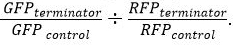
- To normalize for variation in growth, use the equation:
 for both GFP and RFP.
 - After normalization, determine relative terminator strength using the equation
This protocol will produce cells containing pGR-Blue with a terminator ligated between GFP and RFP using Golden Gate Assembly (Figure 2). Positive colonies containing ligated insert can be selected based on color. In visible light positive colonies will be white/yellow and false positives will produce blue colonies after 18-20 hr of incubation at 37 °C (Figure 3).
After colony selection, a plate reader can be used to determine terminator strength. As proof of concept, six putative terminators (T1-6) were bioinformatically identified in the mycobacteriophage Bernal13. The results in Figure 4 show relative terminator strength with cells expressing uncut/unmodified pGR as a control. If a predicted terminator has a GFP/RFP ratio similar to pGR (~0.0) or below 0.1, then the sequence was determined to not be a terminator (T5 and T6). However, numerically each tenth increase represents a fold increase in terminator strength relative to the control. For example, T3 had a high ratio of GFP expression relative to RFP and showed a 5-fold increase (0.5 on graph) in terminator strength as compared to the control and was considered a strong terminator (Figure 4). Data was collected in triplicate and averaged for visual representation.
To determine the best reference control for quantitating terminator strength, we compared raw data using the original pGR plasmid containing no terminator and pGR-Blue cloned with a sequence known not be a terminator. Both controls showed similar levels of GFP and RFP expression (Figure 5) and, more importantly, produced similar GFP/RFP ratios.
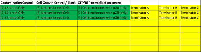
Figure 1. Schematic Detailing Plate Reader Layout. Room should be included on the plate reader for three separate controls (Green). A well (1) containing LB + Amp/arabinose broth only and a well (2) for untransformed competent cells (blank) to serve as a blank. For quantification (3) of the terminator strength, cells containing uncut original pGR or containing pGR-Blue ligated with a non-terminating sequence should be included (reference control for quantification). The remainder of the plate (yellow) can be used to test putative terminators. It is suggested all cells be grown in triplicate and averaged before analysis. Please click here to view a larger version of this figure.
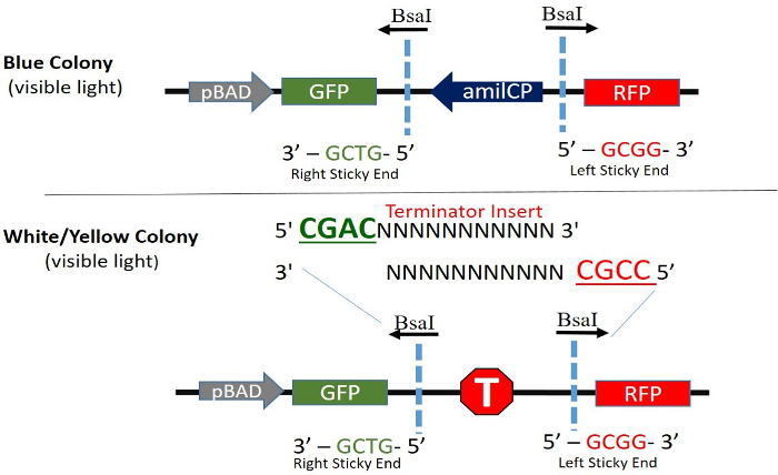
Figure 2. Cloning Site Orientation and Expression of pGR-Blue. Constitutive expression of AmilCP (Blue Chromo Protein) is driven in the opposite (bottom strand) direction of GFP and RFP. When digested with BsaI, AmilCP and the BsaI recognition sites are removed and the terminator to be tested is permanently ligated into the plasmid. The top strand and bottom strand of a terminator with the appropriate sticky ends are shown. The AraC regulator and ampicillin resistance genes are in the plasmid but not shown. Please click here to view a larger version of this figure.
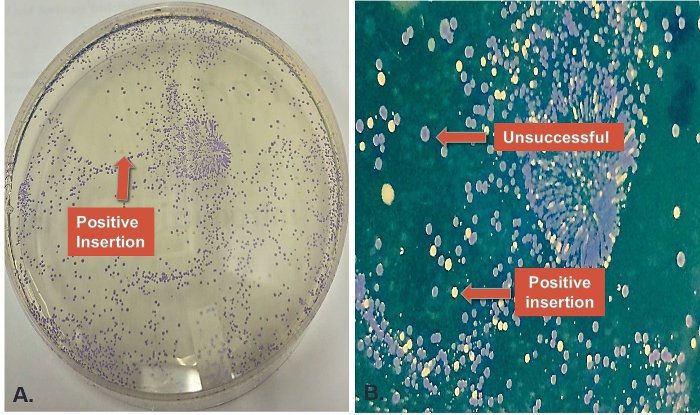
Figure 3. Colony Selection after Cloning. (A) Representative plate after pGR-Blue / terminator ligation (standard mix) and cell transformation. Blue colonies represent unsuccessful ligations and white/yellow colonies (indicated by arrow) are successful ligations. (B) A zoomed in image of the same plate showing representative colonies after transformation. The background color was digitally altered to further highlight colony color differences. Transformed cells (E.coli-JM109) were plated on LB agar plats containing ampicillin and arabinose. Please click here to view a larger version of this figure.
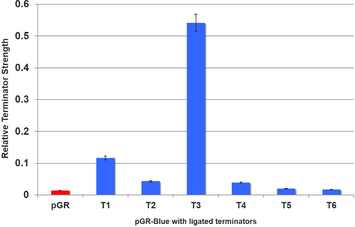
Figure 4. Quantification of Six Different Terminators in pGR-Blue. Six putative terminators (T1-6) were bioinformatically identified in the mycobacteriophage Bernal13. Results show relative fluorescence. All results were normalized using cells containing uncut/unmodified pGR as a control. T3 was found to be a strong terminator. All results represent individual colonies grown in triplicate and averaged for visual representation. Error bars represent deviations between averaged data points. Please click here to view a larger version of this figure.
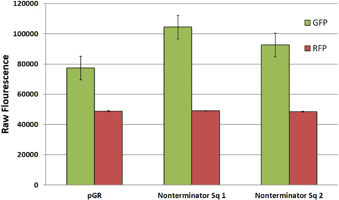
Figure 5. GFP and RFP Expression in Controls. Expression of GFP and RFP in uncut/unmodified pGR and two different non-terminator sequences inserted into pGR-Blue. All results represent individual colonies grown in triplicate and averaged for visual representation. Error bars represent deviations between averaged data points. Please click here to view a larger version of this figure.
