Accurate identification and location of epithelial cells along the intestinal mucosal lining are essential to define different cell lineages. Proper imaging of intestinal tissues is crucial for identification of protein expression patterns with maximum resolution. This study aims to delineate the optimal methods and conditions for processing mouse intestinal tissues.
Understanding the role of factors that regulate intestinal epithelial homeostasis and response to injury and regeneration is important. The current literature describes several different methodological approaches to obtain images of intestinal tissues for data validation. In this paper, we delineate a common protocol relating to the derivation and processing of mouse intestinal tissues. Proper fixation of intestinal tissues and Swiss-roll techniques that enhance intestinal epithelial morphology are discussed. Postresection processing and reorientation of embedded intestinal tissues are critical in obtaining paraffin-embedded blocks that display intact intestinal structural features after sectioning. The Swiss-rolling technique helps in histological assessment of the complete intestinal or colonic sections examined. An ability to differentiate intestinal structural features can be vital in quantitative measurements of intestinal inflammation and tumorigenesis along the entire length. Finally, paraffin-embedded sections are ideal for robust processing using both immunohistochemical and immunofluorescent detection methods. Nonfluorescent immunohistochemical sections provide a vibrant image of the tissue detailing different cellular structural features but do not provide flexibility for intracellular co-localization experiments. Multiple fluorescent channels can be appropriately utilized with immunofluorescent detection for co-localization experiments, lending support to mechanistic studies.
The mammalian intestinal epithelium comprises a single layer of columnar cells. In the small intestine, the proliferative cells are confined to the crypts while differentiated cells occupy the villus region. However, because there are no villi in the large bowel, the proliferative cells are localized to the bottom of the crypts and differentiated cells occupy the upper region of the crypts. The intestinal epithelium undergoes rapid replenishment (about 3 – 5 days) that is driven by continuous division of the proliferative cells within the crypts. The proliferative cells of the crypts are not a homogeneous population and are further subdivided into stem cells and transit-amplifying (TA) cells1. The stem cells reside at the bottom of the crypt, within the first 4 – 5 cells from the very bottom2. The current model supports the existence of two types of stem cells: crypt base columnar (CBC) stem cells and reserve quiescent stem cells. The CBC stem cells are actively proliferating and are marked by Leucine-rich repeat-containing G-protein coupled receptor 5 (Lgr5)3, Olfactomedin 4 (Olfm4)4 and Achaete scute-like 2 (Ascl2)5. On the other hand, reserve quiescent stem cells are labeled by B cell-specific Moloney murine leukemia virus integration site 1 (Bmi1)6, mouse telomerase reverse transcriptase (mTert)7, HOP Homeobox (Hopx)8, Doublecortin-Like And CAM Kinase-Like 1 (Dclk1)9, and Leucine-Rich Repeats And Immunoglobulin-Like Domains 1 (Lrig1)10. The actively proliferating stem cells give rise to TA cells then undergo further differentiation into absorptive cells (enterocytes) and secretory cells (enteroendocrine, goblet, Paneth, and Tuft cells). Continuous cell division in the proliferative zone results in upward movement of epithelial cells along the crypt-villus axis until they reach top of the villi, where they undergo apoptosis and are sloughed off from the surface of the epithelium. The different types of intestinal epithelial cells are marked by the expression of distinct proteins (e.g., intestinal goblet cells can be recognized by staining with antibody against Muc2 and Paneth cells with antibody against lysozyme). We study the role of Krüppel-like factors (KLFs) in the homeostasis and pathobiology of the intestinal epithelium11-13. The results presented here supporting the feasibility of a modified Swiss-rolling technique are based on previous studies of the role of Krüppel-like factor 5 (KLF5) in the maintenance of the actively proliferating intestinal epithelial stem cells14. KLF5 is a zinc-finger transcription factor that is highly expressed in the active intestinal stem and TA cells12. Previous studies demonstrated that KLF5 is co-expressed with Ki-67, a known proliferative marker in the intestinal crypts.
The gastrointestinal tract is not a structurally or functionally homogeneous tissue. The small intestine is divided into duodenum, jejunum, and ileum and the large intestine into cecum and colon, with the latter further divided into proximal, middle, and distal portions. Each of these sections has unique histological features and plays distinct roles15. As such, the effects of insults and the degree of the response of the intestinal epithelium may depend on the region of studied tissue16. Additionally, various mice strains demonstrate diversity of the response at the histological level based on the type of insult used in the studies16. Thus, befitting tissue preparation is necessary to permit appropriate histological and molecular analysis of the intestinal tissues. As such, the Swiss-roll technique grants analysis of the complete length of the intestinal epithelium at one time and thus ascertains well-informed conclusions based on comprehensive information.
The Swiss-roll technique was first mentioned by Magnus17, and described in detail by Moolenbeck and Ruitenberg and Park et al. as a method for preparing tissues and performing histological analyses of the rodent intestine18,19, respectively. The protocol delineated in this publication presents an improved version of the original method that permits for timely and reliable tissue preparation for diagnostic purposes. This modified technique allows for efficient collections and preparation of the intestinal epithelium for universally used techniques, such as immunohistochemistry, immunofluorescence, as well as in situ hybridization (fluorescent and chromogenic20). Furthermore, the modified tissue specimen preparation method utilizes readily available and relatively inexpensive reagents while offering a method of rapid tissue fixation and allows for recovery of protein, DNA, and RNA for additional evaluation. Taken together, this technique is excellent for comprehensive assessment of histopathological, pathological, and molecular features of the intestinal epithelium.
1. Mice
2. Tissue Preparation and Swiss Rolling
3. Histological Analysis and Immunofluorescence Staining
Perform histological analysis and immunofluorescence staining as previously described23,24, with modifications. For enhanced green fluorescent protein (EGFP) immunofluorescence:
The Swiss rolling technique in combination with immunohistochemical staining allows for comprehensive analysis of small or large intestinal tissue. The example of H&E staining of a large bowel of a C57BL/6 mouse (Figure 1) is an illustration of the feasibility and the effectiveness of this technique. As shown in Figure 1, the image is able to capture all portions of the colon: proximal, middle, and distal. Thus, it allows for comprehensive histological assessment. The Swiss rolling and the ability to capture the entire length of the small or large intestinal tissue is extremely helpful for heterogeneous gene expression and marker staining or a variable response of the intestinal tissue to the insult.
An example is the EGFP staining pattern in Lgr5-EGFP/CreERT2 mice. In this mouse model, expression of EGFP is driven by the Lgr5 promoter, which is active only in the actively proliferating intestinal crypts. Additionally, this mouse model is characterized by low (approximately 5 – 10%) penetrance of transgene expression. As shown in Figure 2, the EGFP expression pattern in the intestinal tissue is variable. Consequently, the capability to capture a large view of the tissue helps to identify the region of interest.
The technique described here is powerful especially with application of multifluorophore staining. Here, we show an example of trifluorophore staining of the intestinal tissue that was prepared using the Swiss-roll technique. The main aim of this study was to investigate the role of Klf5 in the maintenance of intestinal stem cells expressing Lgr5 marker. Therefore, we deleted Klf5 from the Lgr5-positive intestinal stem cells in Lgr5-EGFP/CreERT2 mice and collected intestinal tissues on day 14 after first tamoxifen injection. For immunohistological analysis, the tissues were prepared according to the protocol presented in this manuscript, and staining for EGFP (Lgr5 marker), Klf5, and Ki-67 was performed. In the control mice, denoted as Lgr5-EGFP/CreERT2, both EGFP-positive (marked with blue arrows) and EGFP-negative crypts (marked with white arrows) exhibited co-staining for Klf5 and Ki-67 at two examined time points, as shown in Figure 3D. In contrast, in Lgr5-EGFP/CreERT2/Klf5fl/fl small intestinal tissue, Klf5/Ki-67 co-staining was missing from the EGFP-positive CBC stem cells (marked by blue arrows) but present in the EGFP-negative crypts adjoining the green crypts (marked by white arrows), as shown in Figure 3H. This is an excellent example that the tissue preparation techniques presented here does not negatively influence staining quality.
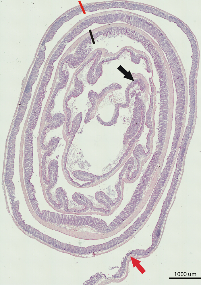
Figure 1. H&E Staining of a Large Bowel from a C57BL/6 Mouse. Shown is the composite image of the whole length of the large bowel. The portion between the black arrow and black line marks the proximal part, between the black and orange lines marks the middle, and between the orange line and orange arrow marks the distal part of the large bowel. Scale bar = 1,000 μm. Please click here to view a larger version of this figure.
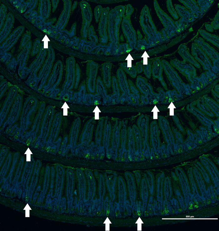
Figure 2. Immunofluorescence Staining of a Small Bowel of an Lgr5-EGFP/CreERT2 Mouse. Composite image of EGFP (labeling Lgr5-positive epithelial cells) staining of small intestine sections of an Lgr5-EGFP/CreERT2 mouse. Example of heterogeneous immunofluorescence staining of protein marker (EGFP) that labels Lgr5-postive epithelial cells in the crypts of the Lgr5-EGFP/CreERT2 mouse. Nuclei are visualized with Hoechst. White arrows mark crypts positive for EGFP expression. Scale bar = 500 μm. Please click here to view a larger version of this figure.
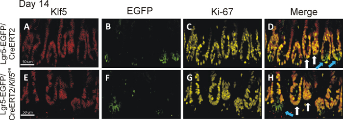
Figure 3. Klf5 Deletion in Lgr5-EGFP/CreERT2/Klf5fl/fl Mice Persists Long Term in Lgr5-EGFP-positive Crypts. The top and bottom set of images are representative small intestinal tissue from the Lgr5-EGFP/CreERT2 control and Lgr5-EGFP/CreERT2/Klf5fl/fl mice on day 14 day after first tamoxifen injection, respectively. Panels A-D are representative of staining from tissue collected from Lgr5-EGFP/CreERT2 control mice, while panels E-H show staining representative of Lgr5-EGFP/CreERT2/Klf5fl/fl mice. Panels A and E display Klf5 immunofluorescent staining in red; panels B and F show EGFP staining in green; panels C and G show Ki-67 staining in yellow; and panels D and H show merged images of Klf5, EGFP and Ki-67 stains. Blue arrows point to green crypts; white arrows point to nongreen crypts in the merged images. Lgr5-EGFP/CreERT2/Klf5fl/fl mice showed long-term loss of Klf5 only in the EGFP-labeled CBC cells14. Scale bar = 50 μm. Please click here to view a larger version of this figure.
The Swiss rolling technique is a powerful method for preparing intestinal tissue for histological and morphological assessment on a large scale. In contrast to the previously described Swiss-rolling technique, which was originally developed for preparation of frozen sections18,19, the procedure presented here allows prompt intestinal tissue preparation and fixation for formalin fixation and paraffin embedding (FFPE). Compared to frozen tissue, FFPE tissue has much longer shelf life and is the preferred type of tissue for histological analysis because of better tissue integrity. Critical parts of the Swiss-rolling protocol involve tissue flexibility for rolling and maintaining the Swiss-roll integrity and tissue quality for staining postfixation. The standard Swiss-roll technique for FFPE of intestinal tissue is typically laborious and time consuming (2 days)18,19. Additionally, this standard method usually yields intestinal tissue that is relatively stiff and not easy to form in a Swiss roll. This is because overnight incubation of the dissected intestinal tissue in 10% buffered formalin is required before tissue rolling. Other modifications of the technique for FFPE purposes have been also developed, but they have a major problem of the tendency of intestinal tissue to unroll and/or for the center of the roll to be distorted. This is because the tissue used in these techniques is fresh unfixed tissue, which is slippery from the mucus produced by the intestine. The new modified technique shown here overcomes these problems by using a modified form of Bouin's fixative. Original Bouin's fixative consists of a mixture of acetic acid, ethanol, picric acid and paraformaldehyde (or formalin). The two most hazardous components, picric acid and paraformaldehyde (or formalin), have been eliminated to allow much safer use of the fixative with an acetic acid/ethanol mix. Picric acid presents an explosion hazard, and paraformaldehyde (or formalin) is a cancer hazard. The significance of using the modified Bouin's fixative is that it (1) is less hazardous compared to its original formula, (2) is easy to prepare with relatively nonexpensive reagents that are readily available in most labs, and (3) allows for quick, almost instant, fixation of the tissue when used for flushing. To knowledge, there are no known limitations to using this modified technique. Additionally, the quick fixation with this mixture immensely reduces the slipperiness of the intestinal tissue, allowing much faster and easier tissue rolling. This fixative also allows for excellent preservation of tissue and cell integrity for histological and immunostaining analysis. Thus, in comparison to other methods of intestinal tissue preparation, this improved technique overcomes several technical issues and grants greatly enhanced speed and ease of use.
Also, this modified fixative is useful to use with other thick tissues that require quick fixation. Because of the nature of acetic acid and of ethanol, that have the capacity of quick penetration of thick tissue while at the same time undergoing fixation. We have used it successfully to quickly fix thick tissue such as liver, spleen and kidneys. For example, fixation of an adult mouse whole liver using the modified Bouin's fixative is usually achieved within 15 – 20 min. This can be easily judged by cutting a cross section into the liver and observing whether the deep regions of the liver had changed color from blood-red to gray. The change in color is indicative of fixation. This fixation is then followed by crosslinking using buffered formalin for 24 – 48 hr with no fear of altered cellular/tissue composition, as the tissue is already fixed at this point.
By comparison, the use traditional buffered-formalin fixation method alone on an adult mouse whole liver would require 24 – 48 hr to achieve deep-tissue fixation, with the risk of altered cellular composition of deep tissue. The quick fixation methods of thick tissues has the superior advantage of reducing to a minimum the alteration of cellular components in response to stress conditions, such as hypoxia, following removal of the organ/tissue from the body. We anticipate that the modified fixative can be used in animal perfusion as an even faster way of fixing thick tissue/organs is desired. Thus, in conclusion, this method and modified fixative provide multiple advantages over traditional methods used for intestinal tissue harvesting and fixation and the modified fixative can be used to quickly fix other thick tissues/organs.
The authors have nothing to disclose.
We would like to thank Ainara Ruiz de Sabando for providing H&E images. This work was supported by grants from the National Institutes of Health (DK052230, DK093680 and CA172113) awarded to Dr. Vincent W. Yang.
Stainless Steel Dissecting Kits | VWR | 25640-002 | |
Decloaking Chamber | Biocare Medical | DC2012 | |
Syringe 10ml | VWR | 89215-218 | |
Swingsette Tissue embedding/processing cassette with lid | Simport | M515 | |
Superfrost Plus Slides [size: 25x75x1mm] | VWR | 48311-703 | |
Manual Slide Staining Set | Tissue-Tek/Sakura | 4451 | |
Staining Dish Green | Tissue-Tek/Sakura | 4456 | |
Staining Dish White | Tissue-Tek/Sakura | 4457 | |
24-Slide Slide Holder with Detachable Handle | Tissue-Tek/Sakura | 4465 | |
Oven | Thermo Scientific | 6243 | for baking slides at 65 degree |
Dissection microscope | Zeiss | Stemi 2000C | |
Fluorescence Microscope | Nikon | Eclipse 90i | Bright and fluoerescent light, with objectives: 10x, 20x |
PAP Pen Super-Liquid Blocker Mini | Fisher Scientific | DAI-PAP-S-M | |
Ethanol 200 proof | AAPR | 111000200 | |
Methanol | VWR | BDH1135-4LP | |
Glacial acetic acid | AAPR | 281000ACS | |
Xylene | Fisher Scientific | X5P-1GAL | |
Hydrogen peroxide 25% solution in water | ACROS | 202465000 | |
10% bufered formalin | Fisher Scientific | 22-026-213 | |
Bovine serum fraction V, heat shock | Roche | 3116956001 | |
Tween 20 | Sigma Aldrich | P7949 | |
Sodium citrate | Fisher Scientific | S279 | |
Gavage needle | VWR | 20068-624 | |
Rabbit anti Klf5 antibody | Santa Cruz Biotechnology | sc-22797 | Dilution 1: 150 |
Chicken anti EGFP antibody | Millipore | AB16901 | Dilution 1: 500 |
Rabbit anti Ki67 antibody | Biocare Medical | CRM325B | Dilution 1: 500 |
Mach3 rabbit AP polymer detection kit | Biocare Medical | M3R533L | |
Warp red chromogen kit | Biocare Medical | WR806 H | |
Lgr5-EGFP/CreERT2 mice | Jackson labs | 008875 | |
Automated processor | Leica | Leica TP1020 |