We describe an in vitro protocol to co-culture gut microbiome and intestinal villi for an extended period using a human gut-on-a-chip microphysiological system.
Here, we describe a protocol to perform long-term co-culture of multi-species human gut microbiome with microengineered intestinal villi in a human gut-on-a-chip microphysiological device. We recapitulate the intestinal lumen-capillary tissue interface in a microfluidic device, where physiological mechanical deformations and fluid shear flow are constantly applied to mimic peristalsis. In the lumen microchannel, human intestinal epithelial Caco-2 cells are cultured to form a 'germ-free' villus epithelium and regenerate small intestinal villi. Pre-cultured microbial cells are inoculated into the lumen side to establish a host-microbe ecosystem. After microbial cells adhere to the apical surface of the villi, fluid flow and mechanical deformations are resumed to produce a steady-state microenvironment in which fresh culture medium is constantly supplied and unbound bacteria (as well as bacterial wastes) are continuously removed. After extended co-culture from days to weeks, multiple microcolonies are found to be randomly located between the villi, and both microbial and epithelial cells remain viable and functional for at least one week in culture. Our co-culture protocol can be adapted to provide a versatile platform for other host-microbiome ecosystems that can be found in various human organs, which may facilitate in vitro study of the role of human microbiome in orchestrating health and disease.
The human intestine harbors a stunningly diverse array of microbial species (<1,000 species) and a tremendous number of microbial cells (10 times more than the human host cells) and genes (100 times more than the human genome)1. These human microbiomes play a key role in metabolizing nutrients and xenobiotics, regulating immune responses, and maintaining intestinal homeostasis2. Not surprisingly, given these diverse functions, the commensal gut microbiome extensively modulates health and disease3. Thus, understanding the role of gut microbiome and host-microbe interactions are of great importance to promote gastrointestinal (GI) health and explore new therapeutics for intestinal disorders4. However, existing in vitro intestine models (e.g., static cultures) restrict host-microbiome co-culture to a short period of time (<1 day) because microbial cells overgrow and compromise intestinal barrier function5. Surrogate animal models (e.g., germ-free6 or genetically engineered mice7) are also not commonly used to study host-gut microbiome crosstalk because the colonization and stable maintenance of human gut microbiome are difficult.
To overcome these challenges, we recently developed a biomimetic human "Gut-on-a-Chip" microphysiological system (Figure 1A, left) to emulate the host-gut microbiome interactions that occur in the living human intestine5,8. The gut-on-a-chip microdevice contains two parallel microfluidic channels separated by a flexible, porous, extracellular matrix (ECM)-coated membrane lined by human intestinal epithelial Caco-2 cells, mimicking the intestinal lumen-capillary tissue interface (Figure 1A, right)9. Vacuum-driven cyclic rhythmical deformations induce physiological mechanical deformations that mimic changes normally induced by peristalsis (Figure 1A, right). Interestingly, when Caco-2 cells are grown in the gut-on-a-chip for more than 100 hr, they spontaneously form three-dimensional (3D) intestinal villi with tight junctions, apical brush borders, proliferative cells limited to basal crypts, mucus production, increased drug metabolizing activity (e.g., cytochrome P450 3A4, CYP3A4), and enhanced glucose reuptake8. In this 'germ-free' microenvironment, it was possible to co-culture the probiotic Lactobacillus rhamnosus GG or a therapeutic formation of a probiotic bacterial mixture with host epithelial cells for up to two weeks5,10.
In this study, we describe the detailed protocol to perform host-gut microbiome co-culture in the gut-on-a-chip device for an extended period. In addition, we discuss critical issues and potential challenges to be considered for a broad application of this host-microbiome co-culture protocol.
1. Microfabrication of a Gut-on-a-chip Device
Note: The gut-on-a-chip is a microfluidic device made by transparent, gas-permeable silicone polymer (polydimethylsiloxane, PDMS), containing two parallel microchannels (1 mm width x 150 µm height x 1 cm length) separated by a flexible porous (10 µm in pore diameter, 25 µm in spacing pore to pore) PDMS membrane5,9. Fabricate the gut-on-a-chip (Figure 1A, left) following the steps provided.
2. Growth of Microengineered Intestinal Villi in the Gut-on-a-chip Device
3. Host-gut Microbiome Co-culture in a Gut-on-a-chip Microdevice
To emulate the human intestinal host-microbiome ecosystem in vitro, it is necessary to develop an experimental protocol to reconstitute the stable long-term co-culture of gut bacteria and human intestinal epithelial cells under physiological conditions such as peristalsis-like mechanics and fluid flow. Here, we utilize a biomimetic gut-on-a-chip microdevice (Figure 1A) to co-culture living microbial cells in direct contact with living human villi for periods of a week or more in vitro. The intestinal epithelial cells spontaneously form well differentiated villi when cultured on one side of a porous ECM-coated membrane in one channel of a 2-channel microfluidic device in the presence of physiologically relevant fluid flow and cyclic mechanical deformations5. The microengineered villi replicate structures and functions of human small intestinal villi, including formation of columnar epithelial cells lined by an apical brush border, basal proliferative crypts with migration and differentiation of daughter cells progressing from the crypt to villus tip, high levels of mucus production, enhanced drug metabolizing activity, and increased glucose reuptake8. Living endothelial cells can be cultured on the opposite side of the same membrane to recreate the tissue-tissue interface of the intestinal wall, and immune cells can be cultured in the system as well10. To validate the protective function of intestinal epithelial cells, tight junction barrier of intestinal villi formed in a gut-on-a-chip is intermittently quantitated by measuring the transepithelial electrical resistance (TEER)5,10,12. Routine monitoring of TEER value is required to estimate the integrity of a cell monolayer or intestinal villi at every juncture (e.g., prior to adding bacteria into the lumen).
To reestablish the host-microbe ecosystem, living microbial cells resuspended in the antibiotic-free cell culture medium (cell density, ~1.0 x 10⁷ CFU/ml) are inoculated into the lumen of the epithelial microchannel (Figure 1B, "Inoculation"). After microbial cells adhere on the apical surface of villi in the absence of flow for ~1.5 hr (Figure 1B, "Attachment"), physiological flow (40 µl/hr) is resumed through both channels with cyclic mechanical deformations (10% strain at 0.15 Hz) to remove unbound gut bacteria and supply nutrients to both bacterial and villus epithelial cells (Figure 1B, "Co-culture"). This co-culture protocol allows the stable colonization of multiple species of probiotic bacteria in the intervillus space, with viable bacterial microcolonies being maintained for up to two weeks in co-culture (Figures 2A, 2B). In this study, a commercial probiotic formulation that contains a mixed population of 8 different facultative or obligate anaerobic, probiotic strains of Bifidobacterium breve, B. longum, B. infantis, Lactobacillus acidophilus, L. plantarum, L. paracasei, L. bulgaricus, and Streptococcus thermophiles is used.
This co-culture method can be broadly applied to emulate the human intestinal host-microbe ecosystem. For instance, non-pathogenic GFP E. coli can be co-cultured on the surface of intestinal villi grown in a gut-on-a-chip device. Based on the same protocol described above, adhered GFP E. coli cells on the villi grow from single cells at 1 hr to multiple microcolonies with 3 days (Figure 3A, 1 hr vs. 72 hr) that locate in the intervillus spaces (Figure 3B, 1 hr vs. 72 hr) when cultured under trickling flow (40 µl/hr) in the presence of peristalsis-like deformations (10%, 0.15 Hz).
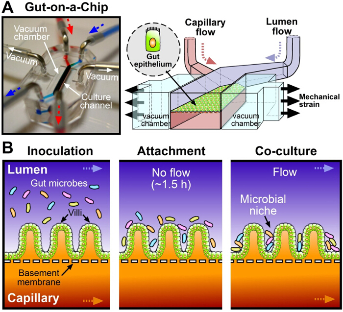
Figure 1. The human Gut-on-a-Chip microphysiological system for host-gut microbiome co-culture. (A) A photograph (left) and a schematic (right) of a 2-channel, gut-on-a-chip microfluidic device. Arrows indicate the direction of culture medium flow (blue, top microchannel; red, bottom microchannel). (B) Schematic diagrams of the procedure of host-gut microbiome co-culture in the gut-on-a-chip. After intestinal epithelial cells form villi (~100 hr), microbial cells resuspended in the cell culture medium are introduced to the lumen microchannel (Inoculation), then the flow of culture medium is suspended for ~1.5 hr (Attachment). After microbial cells adhere to the apical surface of the intestinal villi, flow is resumed (Co-culture). Please click here to view a larger version of this figure.
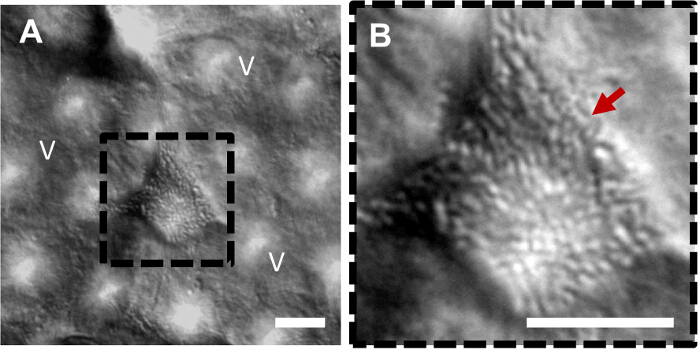
Figure 2. Co-culture of multiple probiotic bacteria with intestinal villi in the gut-on-a-chip. (A) A differential interference contrast micrograph shows a microcolony of probiotic bacterial cells growing between the villi in the gut chip. (B) A higher power magnification view of A (a black dotted square) showing the microcolony of probiotic bacterial cells (a red arrow). V, villi; Scale bar = 20 µm. Please click here to view a larger version of this figure.
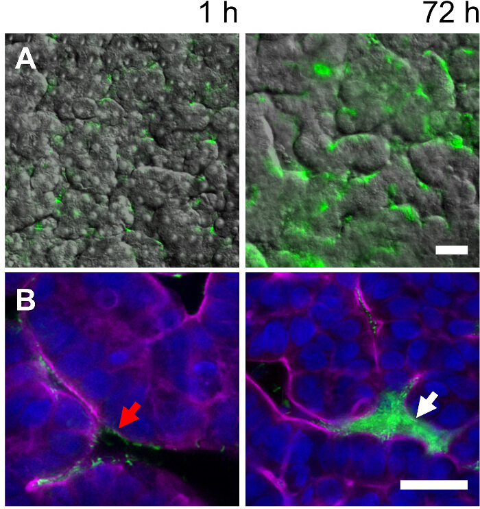
Figure 3. Co-culture of non-pathogenic, GFP-labeled E. coli with intestinal villi in the gut-on-a-chip. (A) Overlaid fluorescence and DIC microscopic images taken at 1 and 72 hr, displaying the colonization of GFP E. coli on the intestinal villi grown in the gut-on-a-chip. (B) High power magnification views of overlaid fluorescence confocal micrographs taken at 1 and 72 hr showing the growth of GFP-labeled E. coli from a single cell (a red arrow) to a microcolony (a white arrow). Blue, nuclei; magenta, F-actin; Scale bar = 30 µm. Please click here to view a larger version of this figure.
Understanding host-microbiome interactions is critical for advancing medicine; however, traditional cell culture models performed in a plastic dish or a static well plate do not support the stable co-culture of human intestinal cells with living gut microbes for more than 1-2 days because microbial cells mostly overgrow the mammalian cells in vitro. The overgrowing microbial population rapidly consumes oxygen and nutrients, subsequently producing excessive amount of metabolic wastes (e.g., organic acids), which seriously compromise intestinal barrier functions and cause intestinal epithelial cell death. Hence, preventing the microbial overgrowth that causes the depletion of nutrients and the accumulation of microbial wastes is crucial for sustaining viable host-microbiome coexistence for an extended period (from days to weeks) in the co-culture microenvironment.
To overcome these challenges, a microphysiological gut-on-a-chip device5,8 has been developed to form fully differentiated human intestinal villi and to maintain the steady-state microenvironment of the intestinal lumen in vitro. The design and functional units (e.g., microfluidic flow cell chambers, vacuum-driven cyclic deformations) of a gut-on-a-chip microfluidic device are modified to precisely emulate the physiological, physical and chemical microenvironment for the microbial co-culture5,10. The shear stress applied in the gut-on-a-chip microfluidic culture was determined by the physiological range of luminal flow in the human intestine5,13. For the host-microbe co-culture, maintaining the "steady-state" of microbial population in the luminal microchannel of the gut-on-a-chip is critical in supporting chemically and nutritionally feasible conditions. In the gut-on-a-chip microsystem, it is possible to estimate the intra-luminal steady-state in the gut-on-a-chip by measuring the pH of the culture medium and the microbial cell density. Because fresh culture medium continuously flows through the microchannels, both microbial and epithelial cells do not undergo nutrient depletion when the initial seeding density of microbial cells and the volumetric flow rate of the culture medium are optimized. The conditioned medium passing through the microchannel flows out at a defined volumetric flow rate (here, 40 µl/hr), so metabolic wastes (e.g., organic acids) as well as cellular secretomes (e.g., cytokines) are continuously removed from the co-culture microenvironment. Thus, the gut-on-a-chip device can be considered as a "continuous bioreactor" with a steady-state microenvironment necessary and sufficient to effectively wash out unbound or overgrown microbial cells from the lumen of the intestinal microchannel, which facilitates generation of a stable microbial niche within 2-3 days.
There are crucial factors that must be considered as troubleshooting for the successful co-culture of microbial cells on the intestinal villi. First, it is necessary to determine the optimized incubation time required for the microbial cells to attach to the surface of villi because microbial adhesion can vary between microbial species. For instance, probiotic bacteria, such as Lactobacillus rhamnosus GG5, generally require ~1.0-1.5 hr to attach, but some other microbial species may require shorter (e.g., pathogenic microbes) or longer times, depending on their adhesion kinetics14. Second, the initial seeding density of the microbial cells should be identified because excessive microbial cell numbers may lead to the outgrowth of bacteria in the early stage of co-culture, which can interfere with the achievement of a stable steady-state. To optimize this seeding density, it is recommended to characterize the growth profile of the target microbial strain in the antibiotic-free cell culture medium at various seeding densities. Finally, adjustment of the volumetric flow rate may be required depending on the microbial species. For example, increased flow rates will be required if the microbial cells proliferate very rapidly. It was experimentally confirmed that flow rates up to 300 µl/hr (0.2 dyne/cm2) do not compromise the barrier function and cell morphology of the microengineered villi (data not shown). Although Lactobacillus rhamnosus GG5, over-the-counter probiotic mixture, VSL#3, non-pathogenic lab strain E. coli as well as the pathogenic enteroinvasive E. coli strain10 were successfully applied for the long-term co-culture in the gut-on-a-chip, it is still required to test a variety of microbial species from commensal gut microbiota to pathogenic infectious microorganisms. The cultivability of microbial cells in vitro should be contemplated in the presence or the absence of host cells in the context of symbiosis and evolution, where the test of cultivating the unculturable gut microbiome is an intriguing challenge. Finally, a robust protocol for the co-culture of both aerobic and anaerobic microbes in the gut-on-a-chip is a critical unmet need to be discovered in the future.
There are steps that can be taken to further improve the model, such as the use of primary intestinal epithelial or undifferentiated stem cells from individual human subjects who have specific gastrointestinal diseases such as Crohn's disease or colorectal cancer; or induced pluripotent stem cells (iPSCs). However, with the burgeoning interest in the role of human microbiome in orchestrating human health and disease, our host-microbiome co-culture method has an enormous significance and potential to be applied to mimic other host-microbiome ecosystems found in the human body (e.g., oral cavity15, skin16, or urogenital tract17). Finally, this co-culture method may innovate the conventional drug development process by improving the predictability in terms of how the gut microbiome influences on the bioavailability, efficacy, and toxicity of new drug compounds. Taken together, the gut-on-a-chip microphysiological system can provide a robust platform to establish a stable steady-state intestinal microenvironment that can be used to reconstitute host-microbiome ecosystem.
The authors have nothing to disclose.
We thank Sri Kosuri (Wyss Institute at Harvard University) for providing the GFP-labeled E. coli strain. This work was supported by the Defense Advanced Research Projects Agency under Cooperative Agreement Number W911NF-12-2-0036, Food and Drug Administration under contract #HHSF223201310079C, and the Wyss Institute for Biologically Inspired Engineering at Harvard University. The views and conclusions contained in this document are those of the authors and should not be interpreted as representing the official policies, either expressed or implied, of the Army Research Office, Army Research Laboratory, Food and Drug Administration, or the U.S. Government. The U.S. Government is authorized to reproduce and distribute reprints for Government purposes notwithstanding any copyright notation hereon.
Dulbecco's Modified Eagle Medium (DMEM) containing 25 mM glucose and 25 mM HEPES | Gibco | 10564-011 | Warm it up at 37°C in a water bath. |
Difco Lactobacilli MRS broth | BD | 288120 | Run autoclave at 121°C for 15 min. |
Poly(dimethylsiloxane) | Dow Corning | 3097358-1004 | 15:1 (w/w), PDMS : cureing agent |
Caco-2BBE human colorectal carcinoma line | Harvard Digestive Disease Center | Human colorectal adenocarcinoma | |
Heat-inactivated FBS | Gibco | 10082-147 | 20% (v/v) in DMEM |
Trypsin/EDTA solution (0.05%) | Gibco | 25300-054 | Warm it up at 37℃ in a water bath. |
Penicillin-streptomycin-glutamine | Gibco | 10378-016 | 1/100 dilution in DMEM |
4′,6-Diamidino-2-phenylindole dihydrochloride | Molecular Probes | D1306 | Nuclei staining |
Phalloidin-CF647 conjugate (25 units/mL) | Biotium | 00041 | F-actin staining |
Flexcell FX-5000 tension system | Flexcell International Corporation | FX5K | Peristalsis-like stretcing motion (10% cell strain, 0.15 Hz frequency) |
Inverted epifluorescence microscope | Zeiss | Axio Observer Z1 | Imaging, DIC |
Scanning confocal microscope | Leica | DMI6000 | Imaging, Fluorescence |
UVO Cleaner | Jelight Company Inc | 342 | Surface activation of the gut-chip |
Type I collagen | Gibco | A10483-01 | Extracellular matrix component for cell culture into the chip |
Matrigel | BD | 354234 | Extracellular matrix component for cell culture into the chip |
1 mL disposable syringe | BD | 309628 | Cell and media injection stuff |
25G5/8 needle | BD | 329651 | Cell and media injection stuff |
Syringe pump | Braintree Scientific Inc. | BS-8000 | Injection equipment into the chip |
VSL#3 | Sigma-Tau Pharmaceuticals | 7-45749-01782-6 | A formulation of 8 different commensal gut microbes |
Reinforced Clostridial Medium | BD | 218081 | Anaerobic bacteria culture medium |
GasPak EZ Anaerobe Container System with Indicator | BD | 260001 | Anaerobic gas generating sachet |
4% paraformaldehyde | Electron Microscopy Science | 157-4-100 | Fixing the cells for staining |
Triton X-100 | Sigma-Aldrich | T8787 | Permeabilizing the cells |
Bovine serum albumin | Sigma-Aldrich | A7030 | Blocking agent for staining of the cells |
Corona treater | Electro-Technic Products | BD-20AC | Plasma generator for fabrication of the chip |
Steriflip | Millipore | SE1M003M00 | Degasing the complete culture medium |
Disposable hemocytometer | iNCYTO | DHC-N01 | For manual cell counting |