NOTE: A schematic of the overall protocol described here is given in Figure 1. All samples used for the development of this method are surplus clinical diagnostic samples and have ethical approval from the London Bloomsbury Ethics committee.
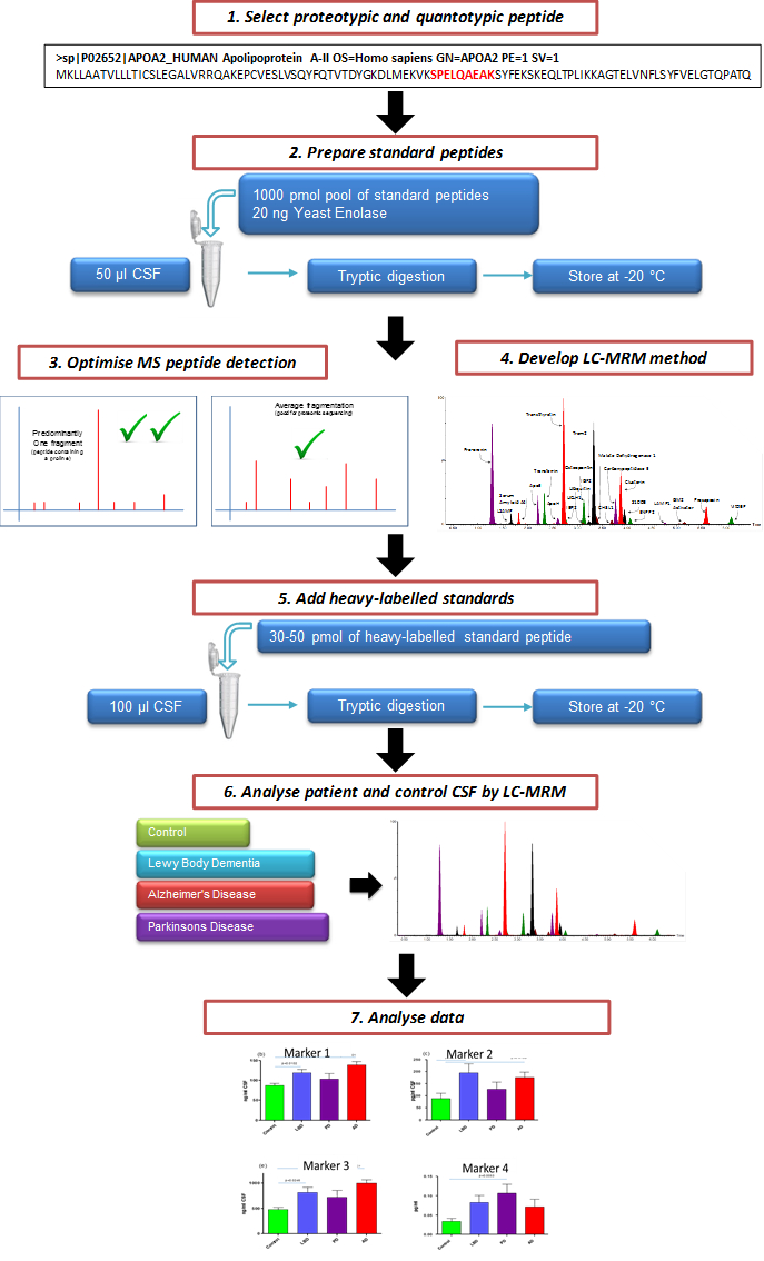
Figure 1. Schematic Illustrating the Overall Process of Creating a Targeted CSF MRM LC-MS/MS Assay. Candidate marker peptides for evaluation are selected from protein targets. Through the use of custom synthesized peptides, a targeted LC-MS/MS multiplex method is created. After evaluation, the assay can be used to assess the efficacy of potential markers of neurodegeneration.
1. Peptide Selection and Design
NOTE: Criteria for a marker peptide is that it must be unique (proteotypic) and representing the quantitative abundance of the protein (quantotypic). To determine if a peptide is unique or not, the 'blast' search tool on the Uniprot website (http://www.uniprot.org/blast/) can be used.
- Define a list of target proteins (markers), i.e., ApoE (see Table 2).
NOTE: If the marker has been identified from previous proteomic profiling experiments8, then select the peptide that gives the best response from that data set. - If this information is unavailable, use open source websites, such as in silico trypsin digestion of target proteins, using a software tool such as MS-Digest.
- Choose the peptide which is tryptic and not susceptible to post translational modifications.
NOTE: This information can be checked on the Uniprot website www.uniprot.org. Avoid peptides prone to chemical modification during LC-MS sample preparation. - Order the custom synthesis of the chosen peptides.
NOTE: Marker peptides can be custom synthesized by various commercial companies. For ApoE the quantotypic peptide used to measure total ApoE levels was determined to be AATVGSLAGQPLQER. The proteotypic peptide sequences used to determine the E2 and E4 variants are the tryptic peptides for position 158 (RLAVYQAGAR and CLAVYQAGAR) and position 112 (LGADMEDVCGR and LGADMEDVR), respectively.
2. Preparation of Standard Peptides
NOTE: In order to select the best quantitative transitions, the detection of the matrix (CSF) needs to be optimized. The most efficient way of optimizing multiplexed peptides is to create pools of the peptides at known concentrations. These pools can then be used for method development and standard curves.
- Resuspend synthetic peptides (peptide details are given in Table 2) to 1 mg/ml stock concentration according to manufactures instructions. By default, if instructions are not available, resuspend peptides in 50:50 (v/v) acetonitrile (ACN)/H2O.
- Prepare the 1:10 dilutions of the peptide from the stock concentration and pool 1,000 pmol of each peptide into a low binding microcentrifuge tube. Dry down in a speed-vac concentrator the final pool and store at -20 °C. Prepare several pools for future use.
- Aliquot 100 µl of CSF into low binding tubes. Freeze-dry the CSF.
- Resuspend an aliquot of pooled 1,000 pmol peptides in digestion buffer (100 mM Tris HCl, pH 7.8, 6 M Urea, 2 M Thiourea, 2% ASB14) to obtain concentrations of 10 and 1 pmol/µl.
- Spike the pooled peptides into the freeze-dried 100 µl aliquots of CSF at 0, 1, 2, 5, 10 and 15 pM concentrations. Add 20 ng of intact unrelated protein such as yeast enolase to act as an internal standard and control for digestion efficiency of trypsin.
- Top up the CSF aliquots with digest buffer to a final amount of 20 µl. Vortex.
- Add 1.5 µl of dithiothreitol (30 mg in 1 ml of 100 mM Tris HCl, pH 7.8) and shake at room temperature for 1 hr.
- Add 3 µl of iodoacetamide (35 mg in 1 ml of 100 mM Tris HCl, pH 7.8) and shake at room temperature for 45 min in the dark.
- Add 165 µl of ddH2O.
- Add 10 µl of 0.1 µg/µl sequencing grade modified trypsin solution resuspended in 50 mM ammonium bicarbonate buffer, pH 7.8. Incubate in a water bath at 37 °C O/N and stop the digestion by freezing samples. Store digests at -20 °C until ready to analyze.
3. Optimization of MS Peptide Detection
- Copy and paste the sequence of the peptide to be optimized, into an appropriate software, e.g., Skyline9. Click on the peptide sequence to obtain the information of expected precursor ion mass and product ions.
- Dilute the peptides from stock concentration (see section 2.1) further to 1 ng/ml concentration for MS method development.
- Directly infuse the peptides in the mass spectrometer at optimized flow rate (usually 0.1 - 0.8 ml/min) and with 0 – 5% collision energy.
- Acquire MS spectrum in order to identify the experimental multiply charged precursor ions8. Choose the precursor ion (m/z) that gives the most intensity (doubly- or triply-charged precursor ions are advised).
- Reinfuse the peptide and apply collision energy to fragment the peptide by collision-induced-dissociation (CID). Optimize the cone and collision energies to obtain the best fragmentation pattern (fragment ions singly or doubly charged, and mass of fragment ion preferably bigger than parent ion, are advised).
- Verify that the experimentally obtained transitions match transitions generated in silico (as described in step 3.1). Save the transition list in the MRM method file using at least 2 most intense transitions per precursor ion. Include 2 transitions: 1 for quantitation and 1 for confirmation in the final assay.
NOTE: Some MS manufacturers have a function (i.e., Waters Intellistart) for automated MRM or SRM analysis optimization. If possible infuse peptides with combined mobile phases at 50 – 70% ACN with 0.1% FA.
4. LC-MRM Method Development
NOTE: Analyze the mixture of synthetic peptides by an Ultra Performance Liquid Chromatography (UPLC) system coupled to triple quadrupole mass spectrometer. Ensure the source is clean. Solvent A is ddH20 with 0.1% FA; Solvent B is ACN with 0.1% FA.
- Use the LC-MS system equipped with a UPLC column packed with C-18 phase (1.6 µm diameter, 90 Å pores, 2.1 mm x 50 mm length) and attached to a pre-column of the same phase.
- Defrost CSF digests on ice, centrifuge at 16,000 g for 10 min and transfer 60 µl into 300 µl glass insert vials and store the rest back at -20oC.
- Inject the highest concentration from standard curve point using 10 min 1 – 40% ACN linear gradient (see Table 1 for gradient settings)
- Open the resulting chromatogram and note retention time and top two most intense (quantitative) transitions per peptide with all transitions created in step 3.
- Based on this information, update a 10 min MRM method (created in step 3.6) with timed channels to measure peptides (see Figure 2 for an example). To maintain sensitivity, keep each channel with points per peak greater than 8 and dwell time greater than 0.01 sec for at least one transition for each peptide.
- Include 'solvent delays' in the MRM method: one at the beginning until 10 sec before the first peak elution and another at the end of the method, 20 sec after the last peak elution. Do this by selecting "solvent delays" in method events in MS method file.
- Run the standard curve through the timed MRM method and ensure there are no interfering nonspecific peaks with transitions (generated in step 3.6) by checking for linearity.
- Determine if the peptides are detectable by running non-spiked pooled control and disease CSF through the method.
- Remove the peptides that are below the limit of detection from the assay.
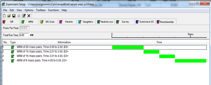
Figure 2. Example of a Dynamic MRM MS Method. Timed channels of peptide transitions can be grouped according to established retention times. Enabling MRMs to be incorporated into a timed fashion as the selected marker peptides elute from the chromatography column, minimizes the number of transitions over a time period and increases the sensitivity of the assay. Please click here to view a larger version of this figure.
5. Addition of Internal Standards
NOTE: As described previously10, stable isotope-labeled internal standards can be included in the assay. Due to the expense of these standards, it is advised to first assess the peptides in the matrix.
- Determine the ideal peptides optimized for detection in CSF: choose the peptides which are the most recurrent, with the highest intensity and without interference peaks.
- Design corresponding peptides to include heavy 13C 15N amino acid labels that increase the mass of the peptide by at least 6 Da relative to endogenous peptide. Also, add a tag of 4 – 6 additional amino acids to either the N- or C-terminus of the heavy peptide to control for tryptic digestion.
- Dilute stable isotope-labeled internal standards in digestion buffer (see step 2.4). Determine the ideal amount of stable isotope-labeled internal standard which will be spiked in CSF by spiking in various levels depending on the abundancies previously observed during development. Aim to achieve approximately 1:1 ratio of stable isotope-labeled standard to endogenous peptide.
NOTE: Stable isotope-labeled internal standards can be synthesized by various commercial companies.
6. LC-MRM Assay of CSF Patient Samples
- Spike optimized amount of stable isotope-labeled standards into 100 µl of CSF and freeze-dry the mixture.
- Resuspend the CSF in 20 µl of digest buffer and perform the digestion as described in points 2.7 – 2.10.
- Analyze the samples using LC-MRM method developed in step 4.
- For quantitative analysis, run the standard peptides in concentrations of 0 -15 pmol in a standard curve. See step 2.5 for the preparation of standard curve.
7. Data Analysis
NOTE: Quantitative data is based on the intensity ratio of baseline peak internal standards (heavy labeled peptides). Detailed information regarding SRM/MRM data analysis was previously described10. Ratio data can then be used in a standard curve to determine absolute levels or calculate them from the added concentration of heavy-labeled peptide.
- Analyze LC-MRM data using the mass spectrometers manufacturers' standard software10. Alternatively, use Skyline software to analyze quantitative MRM data10.
- Check the sensitivity of the run by checking the response of an internal standard such as the spiked yeast enolase or a stable isotope-labeled standard in each run. Ensure that the coefficient of variation (CV) is not larger than 25%.
- Manually review the data annotation to ensure accuracy. Analyze each peptide and ratio to an appropriate stable isotope-labeled internal standard, i.e., use the stable isotope-labeled version of the peptide if available.
- To obtain absolute values of pmol per 100 µl of CSF, run the ratio data through appropriate standard curves that were run at the same time.
- Calculate the coefficient of variation (CV). CV for each peptide should be below 25% and below 15% for high abundant peptides.
NOTE: Absolute value pmol per 100 µl CSF values can used in subsequent downstream statistical analysis.
8. Apolipoprotein E Isoform Status
NOTE: To determine ApoE isoform status, the presence of the corresponding peptides can be performed by determining the presence of each isoform.
- Consider the ApoE in 100 µl of CSF threshold of > 1,000 signal-to-noise as positive for that peptide. See Figure 5 for the peptides required/absent to determine a patient's isoform status. Determine the allelic expression by calculating the % of each isoform to total ApoE expression.
Using the method described above, a high throughput 10 min multiplex assay consisting of 74 peptides from 54 proteins was developed, as an assay for markers of the neurodegenerative disorders Alzheimer's Disease and Lewy Body Dementia (LBD)8. Figure 3 shows a multiplex chromatogram published previously8 of the significant peptide markers from the assay. The peptides included in the assay and their quantitative transitions are given in Table 2. The data generated by this method gives standardized ratios to the relevant stable isotope-labeled internal standard. These values can then be put through a standard curve to determine absolute pmol/100 µl CSF concentrations. These values can then be statistically analyzed for changes in clinical samples.
As described previously8, quantitation of the 74 peptides included in this CSF assay revealed that 25 of these markers were altered significantly in the CSF of dementia patients. To illustrate the effectiveness of this assay, results from previously described dementia markers Pro-Orexin and YKL chitinase 3-like protein (YKL-40)11,12 are given in Figure 4. The integrated ApoE assay identifies the ApoE isoform/allele status of the patient as well. Apo E4 is a known risk factor for Alzheimer's Disease, therefore integrating this into the assay will provide valuable information. The detection of the ApoE isoforms is explained in Figure 5 and is based on the detection of the corresponding peptides for the amino acid changes for isoforms E2 (R158C) and E4 (C112R). Figure 5A shows the peak pattern expected for each isoform combination and Figure 5B shows the result of a CSF tested with the assay on patient samples.
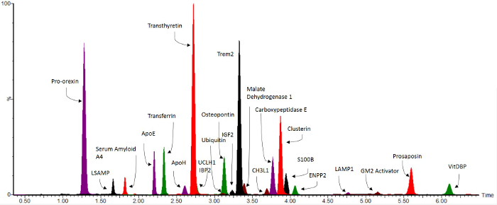
Figure 3. Representative Overlaid MRM Chromatograms. Reprint from Heywood et al.8 Biomarkers significant for the neurodegenerative disorders Lewy Body Dementia and Alzheimer's Disease8 are shown over a 10 min LC gradient. Please click here to view a larger version of this figure.
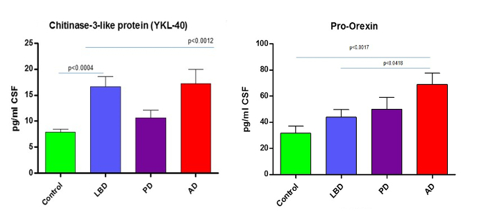
Figure 4. Example Data. Reprint from Heywood et al.8 Graphs show how the described MRM LC-MS/MS method can reliably quantitate and discriminate from controls the known neurodegenerative markers such as YKL chitinase 3-like protein (YKL-40)11,12 and the AD marker Pro-Orexin 13. AD = Alzheimer's Disease, LBD = Lewy Body Dementia and PD = Parkinson's Disease. Data previously published. Please click here to view a larger version of this figure.
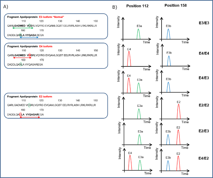
Figure 5. Illustration of How the Apo E Isoform Status of a Patient Can Be Determined. A. Indicates the peptides covering the 112 amino acid sequence LGADMEDVCGR for neutral (E3a) or LGADMEDVR for presence of E4 and for position 158 to detect RLAVYQAGAR the neutral (E3b) or CLAVYQAGAR for the E2 isoform. B. Peptides from the ApoE sequence are shown in the left hand panel. The different combinations of the peptides detected in the CSF can indicate the ApoE isoform status. Please click here to view a larger version of this figure.
| Time | Flow (ml/min) | % A | % B | Curve |
| Initial | 0.8 | 97 | 3 | initial |
| 0.2 | 0.8 | 97 | 3 | 6 |
| 7 | 0.8 | 60 | 40 | 6 |
| 7.01 | 0.8 | 0.1 | 99.9 | 6 |
| 8 | 0.8 | 0.1 | 99.9 | 6 |
| 8.01 | 0.8 | 97 | 3 | 1 |
| 10 | 0.8 | 97 | 3 | 1 |
Table 1. UPLC Gradient Settings for a 10 min Method. A = ddH20 0.1% FA, B= ACN, 0.1% FA
Table 2. Peptides Included in the CSF MRM LC MS/MS Assay. Shown are all the included peptides used in the method, which are described and published 8. Indication of whether the marker was reliably detected in 100 µl of CSF is indicated. Transitions labeled in bold are the ones used for quantitative data. Please click here to download this table.
