We describe a microsurgical approach for the generation of an arteriovenous (AV) loop as a model for analyzing vascularization in vivo in an isolated and well-characterized environment. This model is not only useful for the investigation of angiogenesis, but is also optimally suited for engineering axially vascularized and transplantable tissues.
A functional blood vessel network is a prerequisite for the survival and growth of almost all tissues and organs in the human body. Moreover, in pathological situations such as cancer, vascularization plays a leading role in disease progression. Consequently, there is a strong need for a standardized and well-characterized in vivo model in order to elucidate the mechanisms of neovascularization and develop different vascularization approaches for tissue engineering and regenerative medicine.
We describe a microsurgical approach for a small animal model for induction of a vascular axis consisting of a vein and artery that are anastomosed to an arteriovenous (AV) loop. The AV loop is transferred to an enclosed implantation chamber to create an isolated microenvironment in vivo, which is connected to the living organism only by means of the vascular axis. Using 3D imaging (MRI, micro-CT) and immunohistology, the growing vasculature can be visualized over time. By implanting different cells, growth factors and matrices, their function in blood vessel network formation can be analyzed without any disturbing influences from the surroundings in a well controllable environment.
In addition to angiogenesis and antiangiogenesis studies, the AV loop model is also perfectly suited for engineering vascularized tissues. After a certain prevascularization time, the generated tissues can be transplanted into the defect site and microsurgically connected to the local vessels, thereby ensuring immediate blood supply and integration of the engineered tissue. By varying the matrices, cells, growth factors and chamber architecture, it is possible to generate various tissues, which can then be tailored to the individual patient’s needs.
Most tissues and organs in the human body are dependent on a functional blood vessel network that supplies nutrients, exchanges gases and removes waste products. Malfunction of this system caused by local or systemic vascular problems can lead to a multitude of severe diseases. Moreover, in research areas such as tissue engineering or regenerative medicine, a functional blood vessel network within artificially generated tissues or transplanted organs is indispensable for successful clinical application.
For decades researchers have been investigating the exact mechanisms involved in the growing vasculature to gain deeper insight into pathological situations in order to find novel therapeutic interventions and provide better prevention of vascular disorders. In the first step, basic processes such as cell-cell interactions or the effect of molecules on cells of the vascular system are usually investigated by in vitro 2D or 3D experiments. Traditional 2D models are easy to perform, are well established and have contributed greatly to a better understanding of these processes. For the first time in 1980, Folkman et al. reported in vitro angiogenesis seeding of capillary endothelial cells on gelatin coated plates1. This immediately gave way to publication of a multitude of further 2D angiogenesis experiments on endothelial cell tube formation assay2, migration assay3 and the co-culturing of different cell types4, as well as others. These assays are still used today and accepted as standard in vitro methods.
However, this experimental setup is not always appropriate for the study of in vivo cell behavior since most cell types require a 3D environment to form relevant physiological tissue structures5. It could be shown that the architecture of the 3D matrix is decisive for capillary morphogenesis6 and that cell-extra cellular matrix (ECM) interactions and 3D culture conditions regulate important factors involved in tumor angiogenesis7. The 3D matrix provides complex mechanical inputs, can bind effector proteins and establish tissue-scale solute concentration gradients. Moreover, it is considered necessary in order to imitate in vivo morphogenetic and remodeling steps in complex tissues5. In these systems, both angiogenesis and vasculogenesis can be studied. While angiogenesis describes the sprouting of capillaries from preexisting blood vessels8, vasculogenesis refers to the de novo formation of blood vessels through endothelial cells or their progenitors9,10. Maturation of vessels is described in a process called 'arteriogenesis' via recruitment of smooth muscle cells11. A typical angiogenic in vitro model is the sprouting of endothelial cells from existing monolayers seeded as a monolayer on gel surfaces, on surface of microspheres embedded within a gel or by building endothelial cell spheroids12. In vasculogenic models single endothelial cells are entrapped in a 3D gel. They interact with adjacent endothelial cells to form vascular structures and networks de novo, typically in combination with supportive cells12.
However, even complex 3D in vitro models cannot mimic in vivo settings completely given the multitude of cell-cell and cell-ECM interactions13. Substances with high in vitro activity do not automatically show the same effects in vivo and vice versa14. For a comprehensive analysis of vascularization processes there is an urgent need to develop in vivo models that better simulate the situation in the body. A large range of in vivo angiogenesis assays are described in the literature, including the chick chorioallantoic membrane assay (CAM), the zebrafish model, the corneal angiogenesis assay, the dorsal air sac model, the dorsal skinfold chamber, the subcutaneous tumor models14. However, these assays are often associated with limitations, such as rapid morphological changes, problems in distinguishing new capillaries from already existing ones in the CAM assay, or the limited space in the corneal angiogenesis assay15. Furthermore, non-mammalian systems are used (e.g., the zebrafish model16), which leads to problems in xenotransplantation17. In the subcutaneous tumor model, angiogenesis originating only from the tumor itself cannot be analyzed since the adjacent tissue greatly contributes to the vascularization process. Moreover, the surrounding tissue can have a decisive role in shaping the tumor microenvironment18.
Not only for studying angiogenesis or vasculogenesis is there a strong need for a standardized and well-characterized in vivo model but also for studying different vascularization strategies in tissue engineering and regenerative medicine. Today, the generation of complex artificial organs or tissues is possible both in vitro and in vivo. 3D bioprinting provides an on-demand fabrication technique for generating complex 3D functional living tissues19. Furthermore, bioreactors can be used for generating tissues20 or even the own body can be used as bioreactor21. However, the main barrier to successful application of artificially generated tissues is the lack of vascularization within the engineered constructs. Immediate connection to the host's vasculature after transplantation is a major prerequisite for survival, especially in the case of large-scale artificial tissues or organs.
Different in vitro or in vivo prevascularization strategies were developed to establish a functional microvasculature in constructs prior to implantation22. The implantation of a scaffold with in vitro preformed engineered capillaries onto the dorsal skin of mice led to rapid anastomosis of the mice vasculature within a day23. In contrast, a spheroid co-culture consisting of human mesenchymal stem cells and human umbilical vein endothelial cells assembled into a three-dimensional prevascular network developed further after in vivo implantation. However, anastomosis with the host vasculature was limited24. Above all, in poorly vascularized defects, such as necrotic or irradiated areas, this so-called extrinsic vascularization — the ingrowth of vessels from the surrounding area into the scaffold — often fails. Intrinsic vascularization, on the other hand, is based on a vascular axis as a source of new capillaries sprouting into the scaffold25. Using the axial vascularization approach, the engineered tissue can be transplanted with its vascular axis and connected to local vessels at the recipient site. Immediately after transplantation, the tissue is adequately supported by oxygen and nutrients, which creates the right conditions for optimal integration.
Due to the limited availability of models for investigating in vivo angiogenesis and in recognition of the growing importance of generating axially vascularized tissue, we developed the microsurgical approach of Erol and Spira further to generate an arteriovenous (AV) loop in the animal model26. The use of a completely closed implantation chamber makes this method very well suited to study blood vessel formation under "controlled", well characterized in vivo conditions (Figure 1). This model is not only useful for the investigation of angiogenesis but is also optimally suited for the axial vascularization of scaffolds for tissue engineering purposes.
The Animal Care Committee of the Friedrich-Alexander University of Erlangen-Nürnberg (FAU) and the Government of Middle Franconia, Germany, approved all the experiments. For the experiments, male Lewis rats with a body weight of 300 – 350 g were used.
1. The Arteriovenous Loop Model in the Rat
Tissue Engineering
For bone tissue engineering purposes, a number of different bone substitutes were implanted in the small animal rat AV loop model27,28,33,34. Vascularization could perfectly be demonstrated by 3D micro-computed tomography (micro-CT) (Figure 3A). Vascularization of a processed bovine cancellous bone (PBCB) matrix was significantly higher in the loop group compared to the group without vascularization. A constantly growing and maturating blood vessel network developed within the implantation chamber over 8 weeks. Between 4 and 8 weeks, continuous growth of the vascularized tissue towards the center of the constructs was observed, whereas no increase was detected in the non-vascularized group33. Prevascularization of the PBCB matrix for 6 weeks in the AV loop model led to superior survival of the injected osteoblasts compared to the control osteoblasts. In contrast to the control groups, expression of bone-specific genes was detected in the AV loop group with implanted osteoblasts28. As a further matrix, sintered bioactive glass together with fibrin gel was implanted in the AV loops of the rats. After 3 weeks, a dense network of newly formed vessels has developed demonstrated by micro-CT and histology27.
The implantation chamber was modified in order to accelerate scaffold vascularization. By using a perforated titanium chamber, intrinsic vascularization was supported by extrinsic vessels from the surrounding tissue. At just 2 weeks after implantation of a β-tricalciumphosphate hydroxyapatite (β-TCP/HA)/fibrin matrix, 83% of the vessels were connected to the AV loop with continuous increase over time and reached 97% connection after 8 weeks34. With the implantation of 5 x 10⁶ bone marrow derived mesenchymal stem cells (MSC) and bone morphogenetic protein 2 (BMP-2), a significant increase in bone formation compared to the BMP-2 or MSC alone groups could be induced. At 6 and 12 weeks, the fibrin matrix was completely degraded and replaced by highly vascularized connective tissue in all groups (Figure 3B 6 week implantation). There was a significant decrease in vessel number in the BMP-2/MSC group between 6 and 12 weeks and after 12 weeks in the other groups. This was probably due to maturation of the vascular network or the compact arrangement of bone structures leading to a limited vascular network formation32.
Besides bone, other tissues such as muscle or liver can also be engineered in the AV loop model.
For engineering axially vascularized muscle tissue, experiments with primary myoblasts in an AV loop fibrin matrix were carried out. After a prevascularization time of 2 weeks for 2, 4, and 8 weeks, 1 x 10⁶ myoblasts were transplanted into the AV loop chamber. Transplanted myoblasts could be redetected even after 8 weeks using carboxyfluorescein diacetate succinimidyl ester (CFDA) labeling. The cells kept their myogenic phenotype within the fibrin matrix and expression of the muscle-specific markers MEF-2 and desmin was positive after 4 weeks. However, myogenic marker gene expression was negative after 8 weeks, which was probably due to the absence of myogenic stimuli and rapid absorption of the fibrin matrix35. To increase myogenic stimulation, a new modification of the rat AV loop was developed using the epigastric vein instead of the saphenous vein in order to achieve a more proximal positioning of the isolation chamber. Hence, additional incorporation of the obturator motor nerve was geometrically facilitated. By using this AV loop modification, which is referred to as the EPI loop, we could show myogenic differentiation of co-implanted myoblasts and MSC36.
For hepatic tissue engineering 4 x 10⁶ pkh-26 labeled fetal liver cells were transplanted within a fibrin matrix in the rat AV loop model for 2 weeks. In the control group, matrices without an AV loop and cell-free matrices were implanted. Functional capillaries arose from the AV loop vessels and highly vascularized neo-tissue was observed within the chamber after 14 days of implantation, as shown by CD31 staining and India ink labeling. There was no difference between the cell-free and hepatocyte AV loop group. The AV loop vascularized the fibrin matrix densely and viable fetal cells could be detected after explantation by positive pkh-26 staining and liver cell-specific cytokeratin 18 (CK-18) immunohistology mainly in the proximity of the major vascular axis. mRNA levels of CK-18 were elevated in the AV loop cell group. In contrast, no CK-18 expression could be detected in constructs without a loop or cells37.
Angiogenesis Studies
The AV loop consists of three segments: the vein, the arterial graft and the interpositional venous graft (IVG) segment (Figure 1). Three-dimensional evaluation of the vascular system demonstrated that newly formed vessels originated both from the venous and arterial portion as well as from the venous interponate. A great number of newly formed vessels were observed from the IVG33. With in vivo MRA, scanning electron microscopy of corrosion casts and immune histology, the onset of angiogenesis in a fibrin matrix was observed between day 10 and 14. Above all, the venous and IVG segments gave rise to many capillaries and larger vessels. A gradual reduction in luminal caliber as a sign of arterialization of the IVG due to the increase in endovascular pressure and shear stress was detected from day 7 on38. In further studies, it could be confirmed that vascular sprouting mainly takes place at the non-arterial graft39.
The exact analysis of angiogenesis processes and the stimulation and inhibition of blood vessel formation could be visualized in the AV loop implantation chamber. The growth factors vascular endothelial growth factor A (VEGFA) and basic fibroblast growth factor (bFGF) induced a higher absolute and relative vascular density and faster resorption of the fibrin matrix compared to the growth factor-free control group31. Further, remodeling phenomena and maturation of the vascular network within the isolation chamber were visualized over an implantation period of 8 weeks. In AV loop chambers processes of intercapillary interconnection and intussusceptive angiogenesis as well as possible lymphatic growth were identified immunohistologically as parameters of neovascular maturation39. By applying the PHD (prolyl hydroxylase domain) inhibitor DMOG (dimethyloxallyl glycine) systemically in rats, it could be shown that the concentration of the hypoxia-inducible factor alpha (HIF-α) correlates with the growing vascularization in the AV loop and is a stimulus for vessel outgrowth40.
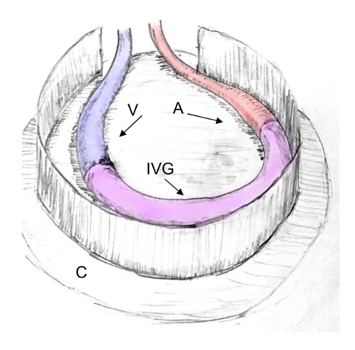
Figure 1: Scheme of an AV Loop in the Rat Model. The AV loop consists of three segments: the vein (V), the arterial (A) graft and the interpositional venous graft (IVG) segment. The AV loop can be embedded into a closed implantation chamber (C) for induction of intrinsic vascularization. Please click here to view a larger version of this figure.
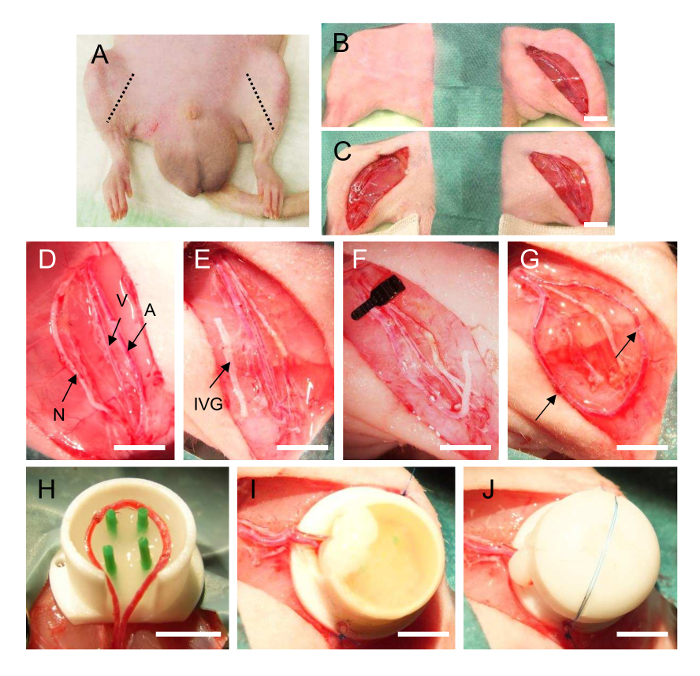
Figure 2: AV Loop Operation in the Rat. (A): Localization of the femoral bundle on the inner side of the hind limbs of the rat. (B/C): Preparation of the femoral vascular bundle in the left and right groin of the rat. The vessels are separated (D), the interpositional vein graft is harvested from the right side (E) and anastomosed with the femoral vein (F) and femoral artery of the left side into an AV loop (G, arrow indicates the anastomoses). The loop vessels are transferred into the implantation chamber prefilled with a matrix (H) and after complete filling (I) the lid is closed (J). A = femoral artery, V = femoral vein, N = femoral nerve, IVG = interpositional venous graft. Scale bar 5 mm (D-J). Please click here to view a larger version of this figure.
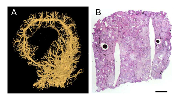
Figure 3: Visualization of Vascularization in the Rat AV Loop Model. (A): Micro-CT after perfusion with a contrast agent (yellow perfused vessels). (B): Hematoxylin-Eosin staining of a β-TCP/HA bone substitute with MSC implanted in the AV loop model rat for 6 weeks. The AV loop vessels are perfused with India ink (black color). Scale bar 1 mm. Please click here to view a larger version of this figure.
For over a decade, we have successfully used the arteriovenous (AV) loop for tissue engineering purposes and studying angiogenesis in vivo in the small animal model. We could demonstrate that this microsurgical model is very well suited for engineering different tissues and that it can also be used for angiogenesis or antiangiogenesis studies.
Significance of the Technique with Respect to Existing/Alternative Methods
Engineered tissues or organs require a functional blood vessel network to supply the nutrients and oxygen they need for their survival and successful integration after transplantation into the defect site41. A number of different prevascularization strategies have been developed over the past decades, which can be differentiated according to in vitro vs. in vivo and extrinsic vs. intrinsic approaches.
Scaffolds can be fabricated with tubular-shaped structures and seeded with vascular cells such as endothelial cells or progenitor cells in vitro42. On the other hand, for in vivo prevascularization, scaffolds are implanted in a highly vascularized area such as subcutaneous or muscle tissue21. Afterwards, these extrinsically vascularized constructs can be transplanted into the defect site. However, the drawback of these approaches is the lack of microsurgical connection with the recipient's vessels after transplantation. Particularly in the case of large-scale constructs, immediate connection to the host vasculature is essential for immediate supply of the engineered tissue43. The obvious and most promising solution to this problem lies in the generation of an intrinsically vascularized tissue or organ by a vascular axis, such as the AV loop model.
Besides using the AV loop method as described above, axial vascularization can also be induced by using AV bundles instead44 or only one vessel such as the epigastric artery45. However, in several publications the AV loop model proved to be superior with regard to degree of vascularization and the amount of de novo tissue formation. Tanaka et al. compared both methodological approaches and observed significantly higher tissue formation and a greater degree of developing capillaries in the loop compared to the bundle group46. Dong et al. also conducted a study using the AV loop or AV bundle approaches for bone tissue engineering in a rabbit model, which likewise showed significantly higher vascular density in the loop compared to the bundle group47. We were able to confirm these results as well as show in a previous study that the AV loop model has a higher capacity for angiogenesis48.
To the best of our knowledge, there is no comparable model for analyzing vascularization in vivo in an isolated and well-characterized environment. Therefore, the AV loop model represents a powerful tool for evaluating how different cell types or growth factors contribute to vessel network formation or vascularization processes in different tissues without disturbances from surrounding structures, such as invading cells or growth factors.
Limitations of the Technique
However, one significant challenge of the proposed model is the high complexity of the surgery. For one, the treatment of defects using the AV loop model requires a two-step procedure – prevascularization of the scaffold and transplantation into the defect site. This means that the patient has to undergo two surgeries. In addition, microsurgical skills are a necessary prerequisite for successful anastomosing submillimeter vessels49. Therefore, the AV bundle is sometimes considered more useful for clinical application since it also offers promising, although less, potential for angiogenesis and tissue generation compared to the AV loop46. However, this operation can be learned step-by-step even by non-surgeons, using small caliber silicon tubes for the training in the beginning and afterwards the vessels of dead animals (e.g., chicken legs) before doing the AV loop operation in a live animal. In contrast, most practiced micro-surgeons can perform this operation with only a short time of training.
Critical Steps within the Protocol
In general, due to the small caliber of the vessels there is the risk of thrombus formation and closure of the loop vessels. However, in the rat model 80%-100% of the loops on average were patent using only short-time heparin anticoagulation post-surgery28,30,31,34,38,39.
Furthermore, due to the high complexity of the surgery it will take a couple of hours (depending on the expertise of the surgeon). It is essential to check proper anesthesia of animals during the whole operation and to adequately supply infusion for maintaining an adequate blood pressure. During the postoperative period it is of high importance to check the health of the animal several times, to administer analgesics/antibiotics and to check the operation wound. Since in most cases implantation of an isolated chamber is performed, it is possible that infection in the inner of the chamber occurs without noticing. Therefore, it is very important to maintain sterility during the whole operation and administration of antibiotics should carefully be done over a period of 3 – 5 days.
Modifications of the Protocol
The chamber can be individually adjusted to the size and shape of the defect. Furthermore, also membranes can be used for enclosing the AV loop as performed by Manasseri et al.50. In addition, the scaffold, supplemented cells and growth factors can be chosen according to the different tissue types. Recently, Miomas et al. combined gene therapeutic approaches successfully with the AV loop model and could induce enhancement of vessel growth by transduction with VEGF165 51. Recently, we adjusted the rat AV loop model for muscle tissue engineering purposes. Instead of the femoral vessels, the epigastric vein and saphenous artery were used, which enabled implantation of the obturator nerve in the axially vascularized scaffold for motoric innervation ("EPI loop model")36. Besides implantation of a motoric nerve, the neurotization of bone tissue engineered constructs with sensory nerves is reported to be beneficial for enhanced osteogenesis and better repair of bone defects52. The AV loop induces minimal donor site morbidity and can be created at various sites of the body52. It would be possible to use superficial vessels at other sites of the body for generation of the AV loop or even to use other animals such as the rabbit or the mouse model.
Future Applications or Directions after Mastering this Technique
Recently, our working group implanted a well-characterized murine embryonal endothelial progenitor cell (EPC) line (T17b) expressing the guanylate binding protein-1 (GBP-1) – a marker and intracellular inhibitor of endothelial cell functions such as proliferation, migration and invasion – in the rat AV loop model. The antiangiogenic capacity of differentiated GBP-1-EPC could be demonstrated by a significant reduction of blood vessel density in the AV loop constructs. With regard to clinical application, the proinflammatory antiangiogenic GTPase GBP-1 could open up new avenues of antiangiogenic therapies, e.g., for cancer or other diseases53. Based on this study, it is conceivable that the AV loop model can be used for establishing a pathological vascularization network for further analysis and possible modulation. For example, this model provides an optimal opportunity to gain a better understanding of tumor angiogenesis, its influencing factors and the exact role of the different cells involved in tumor vessel network formation such as EPCs, tumor cells and stem cells54. In vivo cancer models are often carried out in genetically modified mice to simulate the processes and growth characteristics of different human cancer types and have proven to be excellent for drug development and preclinical trials55. Furthermore, there are xenograft models for transplanting tumors into experimental animals such as immunocompromised mice56. A more clinically related approach involves transplantation from the patient's tumor, known as "personalized mouse models" or "patient-derived tumor xenografts models"57. However, these models are not practical for studying the influence of one single cell source or growth factor without effects from the surrounding tissue.
The AV loop model makes it possible to use tissue engineering methods to study tumor biology. This is defined as "tumor engineering" by Ghajar et al. and involves "the construction of complex culture models that recapitulate aspects of the in vivo tumor microenvironment to study the dynamics of tumor development, progression, and therapy on multiple scales"58. A tumor environment can be constructed within the isolated implantation chamber, which allows a precise analysis of cell-cell interactions, angiogenesis, modulation, enhancement and inhibition. Furthermore, the AV loop model may prove beneficial for developing or validating therapies concerning the interruption of neoangiogenesis or the inhibition of tumor growth.
Using this approach for inducing vascularization, it is possible to engineer tissues in a clinically relevant size. In further studies, we were able to generate axially vascularized bone tissue for transplantation with a significant volume of about 15 cm³ in a relatively short time of 12 weeks59,60. In order to translate these findings to clinical practice, a proof of principle study using the tibia defect model will be performed in the near future prior for application in humans. As a first step, we could successfully demonstrate in situ bone tissue engineering in a large volume defect in a clinical scenario with long-term stability61. Applying the described AV loop model makes it is possible to provide a therapy tailored to the individual patient's requirements. Based on our results the idea of the human body itself serving as a living bioreactor still holds great promise for the future.
The authors have nothing to disclose.
We would like to thank the following institutions for supporting our AV loop research: Staedtler Stiftung, Dr. Fritz Erler Fonds, Else Kröner Fesenius Stiftung, Baxter Healthcare GmbH, DFG, IZKF/ELAN/EFI/Office for Gender and Diversity, the Forschungsstiftung Medizin, Friedrich-Alexander University of Erlangen-Nürnberg (FAU), AO Foundation, Manfred Roth Stiftung, Xue Hong, Hans Georg Geis Foundation, Deutscher Akademischer Austauschdienst (DAAD), Germany, and the Ministry of Higher Education and Scientific Research, Iraq. We would like to thank Stefan Fleischer, Marina Milde, Katrin Köhn and Ilse Arnold-Herberth for their excellent technical support.
0.9% sodium chloride | Berlin-Chemie AG | 34592508 | |
11-0 Ethilon / polyamide 6/6 | Ethicon | EH7438G | |
4-0 Vicryl / polygalactin 910 | Ethicon | V392H | |
6-0 Prolene / polypropylene | Ethicon | 8695H | |
aluminium spray | Pharma Partner Vertriebs-GmbH | 1020 | |
antiseptics | BODE Chemie GmbH | ||
Catheter | B Braun Meslungen AG | 4251612-02 | |
contrast agent | Flowtech | MV-122 | |
embutramide, mebezonium iodide, tetracaine hydrochloride injectable solution | Intervet International GmbH | ||
encre de chine intense indian ink | Lefranc & Bourgeois | ||
Enrofloxacin | Bayer AG | ||
eye ointment | Bayer AG | ||
Formalin 4 % | Carl Roth GmbH & Co. KG | P087.4 | |
Heparin | Ratiopharm GmbH | ||
isoflurane | Abbott Laboratories | 6055482 | |
Lewis rat, male | Charles River Laboratories | ||
Metamizol-Natrium | Ratiopharm GmbH | ||
papaverine / Paveron N | Linden Arzneimittel-Vertrieb-GmbH | ||
tramadol / Tramal | Grünenthal GmbH |