Many vision-threatening ocular diseases are associated with dysfunctional retinal microvessels. Therefore, the measurement of retinal arteriole responses is important to investigate the underlying pathophysiological mechanisms. This article describes a detailed protocol for mouse retinal arteriole isolation and preparation to assess the effects of vasoactive substances on vascular diameter.
Vascular insufficiency and alterations in normal retinal perfusion are among the major factors for the pathogenesis of various sight-threatening ocular diseases, such as diabetic retinopathy, hypertensive retinopathy, and possibly glaucoma. Therefore, retinal microvascular preparations are pivotal tools for physiological and pharmacological studies to delineate the underlying pathophysiological mechanisms and to design therapies for the diseases. Despite the wide use of mouse models in ophthalmic research, studies on retinal vascular reactivity are scarce in this species. A major reason for this discrepancy is the challenging isolation procedures owing to the small size of these retinal blood vessels, which is ~ ≤ 30 µm in luminal diameter. To circumvent the problem of direct isolation of these retinal microvessels for functional studies, we established an isolation and preparation technique that enables ex vivo studies of mouse retinal vasoactivity under near-physiological conditions. Although the present experimental preparations will specifically refer to the mouse retinal arterioles, this methodology can readily be employed to microvessels from rats.
Disturbances in retinal perfusion have been implicated in the pathogenesis of various ocular diseases, such as diabetic retinopathy, hypertensive retinopathy, and glaucoma1,2,3. Thus, studies aimed at measuring vascular reactivity in the retina are important to understand the pathophysiology of these diseases and to develop new treatment approaches.
Due to the possibility of gene manipulation in the murine genome, the mouse has become a widely used animal model for studies of the cardiovascular system4. However, because of the small size of retinal blood vessels (≤ 30 µm), measurement of vascular reactivity in the mouse retina is challenging. For example, stereomicroscopic techniques for in vivo measurement are limited in their optical resolution and therefore only allow to exactly detect changes in diameter or blood flow in small blood of less than ≤ 30 µm diameter when equipped with additional sophisticated devices, such as a confocal microscope using fluorescent dyes or the Adaptive Optics Scanning Light Ophthalmoscope5,6. Moreover, the interpretation of in vivo measurements aimed at identifying local signaling mechanisms in retinal blood vessels can be confounded by anaesthetics, changes in systemic blood pressure and the influence of retrobulbar blood vessels.
Therefore, we developed a method to measure responses of mouse retinal blood vessels with high optic resolution ex vivo. The technique presented herein allows visualization of retinal arterioles via transmitted light microscopy. This method, which can also be used in rats, provides access to the advantages of gene targeting technology in ocular vascular research.
The experimental procedures of this study were approved by the Animal Care Committee of Rhineland-Palatinate, Germany. Animal care conformed to the institutional guidelines and The Association for Research in Vision and Ophthalmology (ARVO) statement for the use of animals in ophthalmic and vision research. Animals were treated according to the EU Directive 2010/63/EU for animal experiments. Male C57BL/6J mice (The Jackson Laboratory, Bar Harbour, ME, USA) aged 3-4 months were used for the experiments. Animals were housed under standard conditions (temperature 23 ± 2 °C, humidity range 55 ± 10% and 12 h light/dark cycles), and had access to standard mouse chow and water ad libitum.
1. Isolation of Mouse Retina
2. Mounting the Retina in the Perfusion Chamber
NOTE: The perfusion chamber used for retinal arteriole experiments is home-made. It consists of a transparent reservoir with an in- and outflow tube (Figure 5).One end of a silicone tube is glued to the bottom of the chamber with histoacryl adhesive and the other end attached to a three-way stopcock. Connect a syringe containing fresh Krebs buffer to the three-way stopcock and fill the tube with buffer.
3. Preparation of Retinal Arterioles for the Experiment
4. Performing the Experiment
U-46619 produced concentration-dependent vasoconstrictor responses in retinal arterioles from wild-type mice of the C57Bl/6J background. At a concentration of 10-6 M, reduction in luminal diameter was ≈50% from resting diameter. Figure 9A shows a representative concentration-response curve of one retinal arteriole. In arterioles preconstricted with U46619, cumulative administration of acetylcholine evoked concentration-dependent increases in luminal diameter to ≈25% from the preconstricted diameter at 10-5 M, indicative of an intact vascular endothelium (Figure 9B).
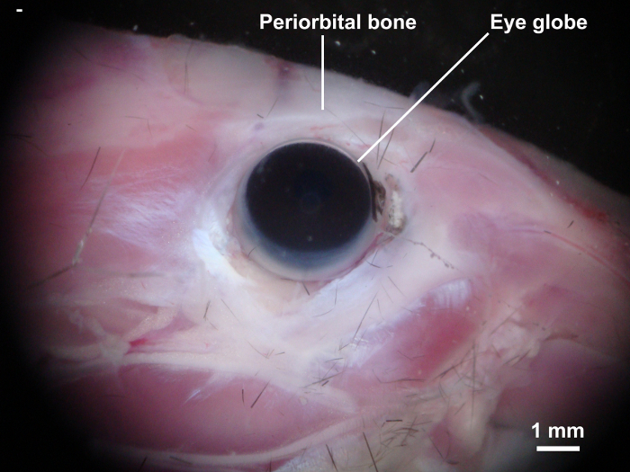
Figure 1: Dissection of the mouse skull. Skin on the decapitated mouse head is removed to expose the eyes and orbital cavity. Please click here to view a larger version of this figure.
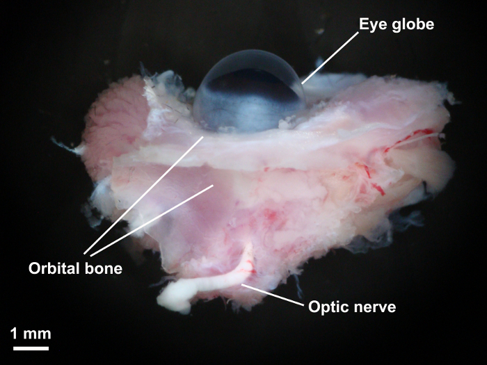
Figure 2: Isolation of eye globe and orbital tissue. The orbital bone was cut with eye scissors and the eye globe together with the retrobulbar tissues and optic nerve were carefully isolated. Please click here to view a larger version of this figure.
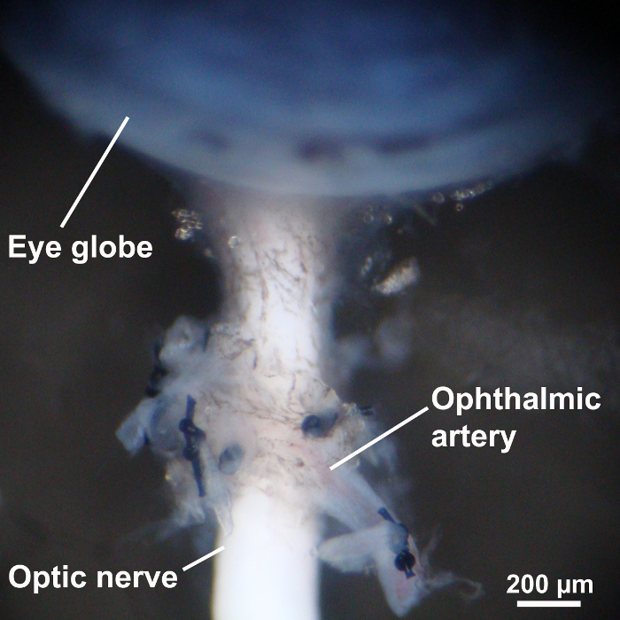
Figure 3: The eye globe and preparation of the ophthalmic artery. Once the surrounding orbital tissues were dissected with fine microscissors, the ophthalmic artery was carefully exposed and their small branches ligated with 10-0 nylon monofilament sutures. Please click here to view a larger version of this figure.
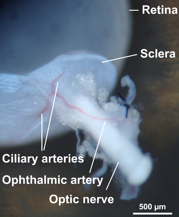
Figure 4: Dissection of ocular structures. To visualize the retina, the sclera and uvea were dissected with Vannas capsulotomy scissors. Some scleral tissue around the optic nerve was left to avoid damage of the ophthalmic artery. Please click here to view a larger version of this figure.
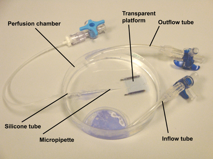
Figure 5: Organ chamber for real-time video microscopy. The chamber was home made. It consisted of a Petri dish of 100 mm diameter with an in- and out-flow tube. The tubes were glued with histoacryl adhesive. Externally oxygenated and carbonated Krebs-Henseleit buffer was circulated by a peristaltic pump via these tubes. One end of a silicone tube was glued to the bottom of the chamber and the other end attached to a three-way stopcock. At the end of the tube, a glass micropipette with a tip of 100 µm was inserted, which served for cannulation of the ophthalmic artery. Via the silicone tube, the ophthalmic circulation was pressurized by filling a silicone tube with Krebs buffer to a level corresponding to 50 mm Hg. Please click here to view a larger version of this figure.
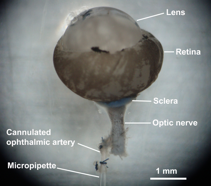
Figure 6: Cannulation of the ophthalmic artery. The ophthalmic artery was cannulated with glass micropipette and sutured with a 10-0 nylon suture. Please click here to view a larger version of this figure.
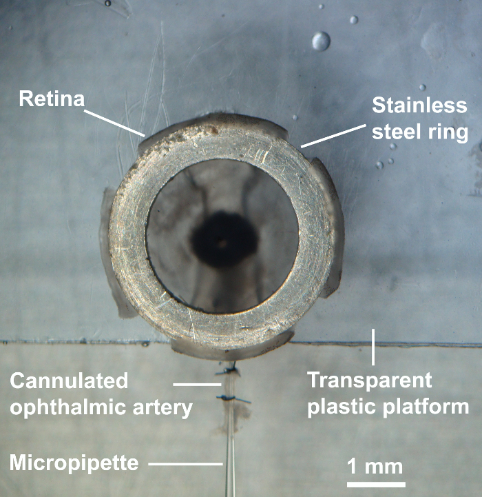
Figure 7: Cannulation of the ophthalmic artery. After placing the retina onto the transparent platform, the lens with the capsular bag was removed, four radial incisions were made into the retina, and a stainless steel of ring of 2.8 mm inner diameter and 4.0 mm outer diameter was placed onto the retina to fix it to the bottom. The retina was then ready for the experiment. Please click here to view a larger version of this figure.
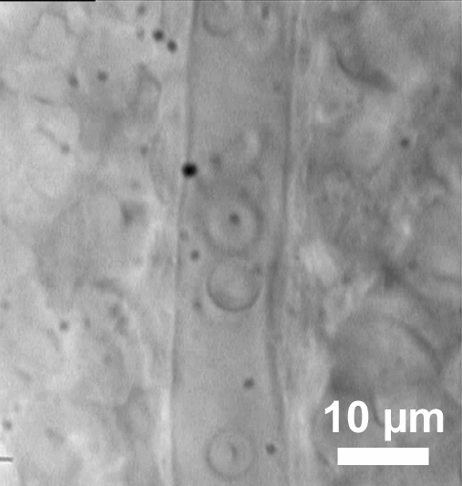
Figure 8: Visualization of retinal blood vessels. An exemplary retinal arteriole with red blood cells inside.
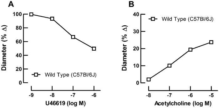
Figure 9: Functional studies using video microscopy. Representative concentration response curves from a single retinal arteriole. (A) Concentration-response curve for the vasoconstrictor, U46619 (10-9 to 10-6 M), and (B) for the endothelium-dependent vasodilator, acetylcholine (10-8 to 10-5 M). Please click here to view a larger version of this figure.
The measurement of vascular responses in the mouse retina is challenging due to the small size of retinal blood vessels. With the presented technique, retinal arterioles are visualized by transmitted light microscopy. This is possible, because the isolated retina is translucent. The advantage of the technique is the high optical resolution. The calculated spatial resolution is 11 px/µm. However, the real resolution for this optical system that uses white light is between 200 and 300 nm, which is explained by the Abbe diffraction limit. Since the first branch of mouse retinal arterioles has an internal diameter of 20 to 30 µm, diameter changes of approximately 1% are detectable with this system. There is no need of additional technical devices or fluorescent dyes as reported in other studies to visualize small retinal arterioles5,6,7. A disadvantage of the technique is the long preparation time, which is between 90 to 120 min by trained investigators. If the preparation time exceeds 180 min, the endothelial function starts to be attenuated.
There are several major critical steps in this technique. First, it is important to thoroughly ligate all retrobulbar arterial branches. If ligation is incomplete, retinal arterioles will not be pressurized due to leakage. Second, the immersion in ethanol for 10 s before opening the eye globe is important to deactivate the smooth muscle reactivity of the ophthalmic artery. We previously demonstrated that immersing for 10 s in 70% ethanol completely deactivates the ophthalmic artery smooth muscle8. In contrast, retinal arterioles are unlikely to be directly affected by ethanol, since they are protected by the bulbar wall. If this step is omitted, diameter changes in the ophthalmic artery may influence the perfusion of the retina. Of note, reactivity of the ophthalmic artery may markedly differ from that of retinal arterioles in response to the same agonist9,10. Third, avoid torsion of the ophthalmic artery. Especially while mounting the retinal preparation onto the platform, the ophthalmic artery may become twisted, resulting in occlusion of the lumen. Also, check carefully that the tip of the glass capillary is not occluded to enable pressurization of retinal arterioles. We suggest not to perform more than three consecutive concentration-response curves in the same retinal preparation, because we observed that responses to acetylcholine became weaker when repeating the concentration-response curves for more than three times. However, there may be differences regarding the repeatability of concentration-response curves depending on the agent applied and, thus, should be tested individually.
We applied pharmacological agents extraluminally in our studies, although intraluminal perfusion using a servo control pump is also possible. Since the technique allows to measure for local mechanisms of vascular reactivity in the retina of small laboratory animals including mice and rats, the advantages of gene-targeting technology by using genetically modified animals can be accessed with this method.
The authors have nothing to disclose.
This work was supported by grants from the Ernst und Berta Grimmke Stiftung and the Deutsche Ophthalmologische Gesellschaft (DOG).
Steel Scissors | Carl Roth GmbH | 3576.1 | 1x 140 mm |
Eye Scissors | Geuder | G-19390 | 1x straight, 10.5 cm |
Precision tweezers, straight with fine tips | Carl Roth GmbH | LH68.1 | 2x type 4 |
Precision tweezers, straight with extra fine tips | Carl Roth GmbH | LH53.1 | 2x type 5 |
Vannas capsulotomy scissors | Geuder | 19760 | 1x straight, 77 mm |
Student Vannas Spring Scissors | Fine Science Tools | 91501-09 | 1x curved, |
Barraquer Needle Holder | Geuder | G-17500 | 1x curved, 120 mm |
Needle | Becton, Dickinson and Company | 305128 | 1x 30 G |
Glass Capillaries (for producing micropipettes) | Drummond Scientific Company | 9-000-1211 | 1x (1.2 x 0.8 mm; outer/inner diameter) |
Nylon Suture | Alcon | 198001 | 1x 10-0 |
Nunclon cell culture dish | Thermo Fisher Scientific | 153066 | 1x 35 mm diameter |
Nunclon cell culture dish | Thermo Fisher Scientific | 172931 | 1x 100 mm diameter |
Discofix C | Braun | 16500C | 10 cm |
Histoacryl adhesive | B. Braun Surgical, S.A. | 1050052 | |
Name | Company | Catalog Number | Comments |
Equipment | |||
Pericyclic pump (CYCLO II) | Carl Roth GmbH | EP76.1 | 1x |
Vertical Pipette Puller Model 700C | David Kopf Instruments | 1x | |
Microscope (Vanox-T AH-2) | Olympus | 1x | |
Water immersion objective LUMPlanFL, 1.0 NA | Olympus | 1x | |
Digital camera (TK-C1381) | JVC | 1x | |
Perfusion chamber | self-made | 1x | |
Name | Company | Catalog Number | Comments |
Drugs and Solutions | |||
Ethanol | Carl Roth GmbH | K928.4 | |
Calcium chloride dihydrate (CaCl2) | Carl Roth GmbH | 5239.1 | |
Kalium chloride (KCl) | Carl Roth GmbH | 6781.1 | |
Kalium dihydrogen phosphate (KH2PO4) | Carl Roth GmbH | 3904.2 | |
Magnesium sulphate (MgSO4) | Carl Roth GmbH | 261.2 | |
Sodium chloride (NaCl) | Carl Roth GmbH | 9265.2 | |
Sodium hydrogen carbonate (NaHCO3) | Carl Roth GmbH | 0965.3 | |
α-(D)-(+)- Glucose monohydrate | Carl Roth GmbH | 6780.1 | |
9,11-dideoxy-9α,11α-methanoepoxy prostaglandin F2α (U-46619) | Cayman Chemical | 16450 | |
Acetylcholine chloride | Sigma-Aldrich | A6625-25G |