We describe a method to investigate the capability of tip-growing plant cells, including pollen tubes, root hairs, and moss protonemata, to elongate through extremely narrow gaps (~1 µm) in a microfluidic device.
In vivo, tip-growing plant cells need to overcome a series of physical barriers; however, researchers lack the methodology to visualize cellular behavior in such restrictive conditions. To address this issue, we have developed growth chambers for tip-growing plant cells that contain a series of narrow, micro-fabricated gaps (~1 µm) in a poly-dimethylsiloxane (PDMS) substrate. This transparent material allows the user to monitor tip elongation processes in individual cells during microgap penetration by time-lapse imaging. Using this experimental platform, we observed morphological changes in pollen tubes as they penetrated the microgap. We captured the dynamic changes in the shape of a fluorescently labeled vegetative nucleus and sperm cells in a pollen tube during this process. Furthermore, we demonstrated the capability of root hairs and moss protonemata to penetrate the 1 µm gap. This in vitro platform can be used to study how individual cells respond to physically constrained spaces and may provide insights into tip-growth mechanisms.
After pollen grains germinate on a stigma, each grain produces a single pollen tube that carries sperm cells to the egg cell and the central cell in the ovule for double fertilization. Pollen tubes elongate through the style and eventually reach the ovule by sensing multiple guidance cues along their way1. During the elongation, pollen tubes encounter a series of physical barriers; the transmitting track is filled with cells, and pollen tubes must enter the minute micropylar opening of the ovule to reach their target (Figure 1A)2. Therefore, pollen tubes must have the ability to penetrate physical obstacles, while tolerating the compressive stress from their surroundings. Root hairs are another type of tip-growing plant cell that must withstand physical obstacles in the environment, in the form of packed soil particles (Figure 1B).
Various mechanical properties of the pollen tube have been studied, including turgor pressure and stiffness of the cell's apical region, which can be measured using the incipient plasmolysis method3,4 and cellular force microscopy (CFM)5,6, respectively. However, these methods alone do not reveal whether pollen tubes are capable of elongating through physical barriers along their growth paths. An alternative technique that allows pollen tube elongation to be monitored in vivo is two photon microscopy7. However, with this method, it is difficult to observe the morphological changes in individual pollen tubes deep inside the ovule tissue. Additionally, root hair growth in soil can be visualized using X-ray computed tomography (CT) and magnetic resonance imaging (MRI)8, albeit with low resolution. Here, we present a method that can be used to acquire high-resolution images of a cell's deformation process on a conventional microscope.
The overall goal of the method described here is to visualize the elongation capability of tip-growing plant cells, including pollen tubes, root hairs, and moss protonemata, in extremely small spaces. As the poly-dimethylsiloxane (PDMS) microdevices presented in this manuscript are optically transparent and air permeable, we can culture living cells inside the device and observe their growth behaviors under a microscope. It is also possible to create micro ~ nanometer scale spaces by the soft lithography technique9 with the use of molds. These features allow us to study the elongation capability of tip-growing plant cells in a physically confined environment.
In this work, we constructed 1 µm wide gaps (4 µm in height) in microfluidic devices and examined the ability of pollen tubes to penetrate these artificial obstacles that are much smaller than the diameter of the cylindrical pollen tube (approximately 8 µm). This experimental platform enables us to visualize the pollen tube's response to microgaps and capture time-lapse images of the response, which track the cell's deformation process. We also developed the microdevices that can be used to investigate the penetration capability of root hairs and moss protonemata. Several microdevices have been reported to date that enable the visualization of plant root10,11,12,13 and moss protonemata14 growth at high resolution. In our device, a series of root hair growth channels are perpendicularly connected to a root growth chamber, and individual root hairs (approximately 7 µm in diameter) are guided to fluidic channels with a 1 µm wide gap. We also cultured moss protonemata (approximately 20 µm in diameter) in a microdevice containing microgaps to examine their responses to these physical barriers. The proposed microfluidic-based approach allows us to explore the capability of various tip-growing plant cells to elongate through extremely small spaces, which cannot be examined by any other currently available method.
1. Fabrication of the PDMS Microdevice to Examine Growing Pollen Tubes and Moss Protonemata
NOTE: We used a maskless photolithography instrument to prepare PDMS molds on silicon wafers. The details regarding the operation of the system are omitted in this manuscript. A standard photolithography technique9 using a photomask may also be used to create the PDMS molds described in this manuscript.
2. Fabrication of the PDMS Microdevice for Root Hairs
3. Preparation of In Vitro Cell Culture Medium for Pollen Tubes (Torenia fournieri)
4. Preparation of In Vitro Cell Culture Medium for Root Hairs (Arabidopsis thaliana)
5. Preparation of In Vitro Cell Culture Medium for Moss Protonemata (Physcomitrella patens)
6. In Vitro Culturing of T. fournieri Pollen Tubes in the Microdevice
7. In Vitro Culturing of A. thaliana Root Hairs in the Microdevice
NOTE: Steps 7.1 – 7.9 (except for 7.3 and 7.5) should be performed in a laminar flow hood.
8. In Vitro Culturing of P. patens (moss) Protonemata in the Microfluidic Device
NOTE: Steps 8.2 – 8.6 (except for 8.3) should be performed in a laminar flow hood.
9. Time-lapse Imaging of T. fournieri Pollen Tube Growth
As illustrated in Figure 1, tip-growing plant cells encounter a series of physical barriers along their growth paths in vivo. The microfluidic in vitro cell culture platforms presented in this study enabled the examination the of tip-growing process in three types of plant cells (pollen tubes, root hairs, and moss protonemata) through 1 µm artificial gaps (Figure 3, Figure 4, Figure 5). For the pollen tube study, live-cell imaging was used to monitor morphological changes in the apical region of pollen tubes as well as the vegetative nucleus and sperm cells in response to encountering an extremely small space (Supplemental Movie 1 and 2).
Although most of the microgaps were successfully fabricated by employing the method described here, we noticed that a few of them were completely closed (Figure 6). Because the 1 µm wide channels created on the silicon mold are fragile, repeated use of this mold might damage the gaps, leading to gap blocking on the PDMS layer. Therefore, before performing the experiment, it is important to check that the PDMS microgaps are intact.
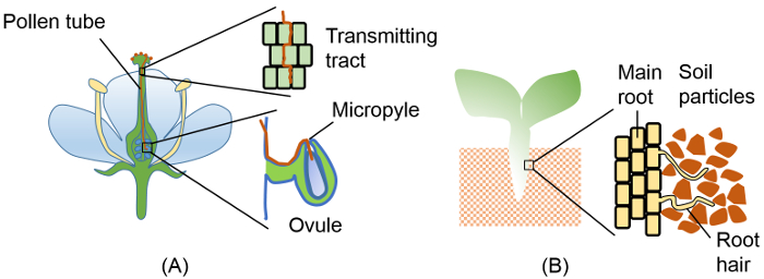
Figure 1: Penetration of tip-growing plant cells into physically constrained spaces. (A) Pollen tube elongating through several physical barriers on its way to an ovule. Barriers include transmitting tract tissue in the style and the integuments surrounding the micropyle. (B) Root hairs penetrating through dense soil. Insets in (A) and (B) show enlargements of the boxed regions. Please click here to view a larger version of this figure.
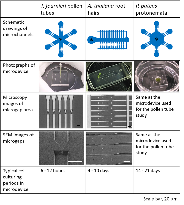
Figure 2: Characteristics of the microdevices used to study T. fournieri pollen tubes, A. thaliana root hairs, and P. patens protonemata. In the schematic drawings of microchannels, a sample is placed at the location indicated by an asterisk mark (*). Microchannel designs, photographs of each device, channel structures, and cell culturing periods are summarized. This figure has been modified with permission from Yanagisawa et al. 201719. Scale bar, 20 µm. Please click here to view a larger version of this figure.
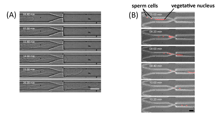
Figure 3: T. fournieri pollen tube elongation through a 1 µm gap. (A) Pollen tube passing through a 1 µm wide and 4 µm high gap. The time-lapse bright field images were captured using an inverted microscope equipped with a spinning-disk confocal system. Scale bar, 20 µm. (B) Time lapse images of a fluorescently labeled vegetative nucleus and sperm cells in a pollen tube of the RPS5Ap::H2B-tdTomato line crossing the microgap. Scale bar, 20 µm. Figure (A) and (B) have been modified with permission from Yanagisawa et al. 201719. Please click here to view a larger version of this figure.
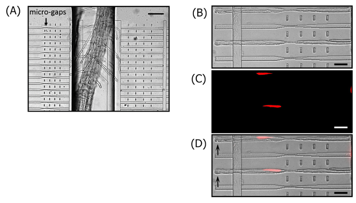
Figure 4. A. thaliana root hair elongation through a 1 µm gap. (A) Root and root hair growth in the microdevice. The microgaps (1 µm in width and 4 µm in height) are located on the left side of the root growth channel. The microchannels on the right side do not contain microgaps and can be used as a control. Scale bar, 100 µm. (B) Bright field, (C) fluorescence, and (D) merged images of root hairs passing through the microgaps. The nuclei in root hairs are fluorescently labeled in the UBQ10pro::H2B-mClover line. The tip of each root hair is indicated by an arrow. Scale bar, 30 µm. These figures have been modified with permission from Yanagisawa et al. 201719. Please click here to view a larger version of this figure.
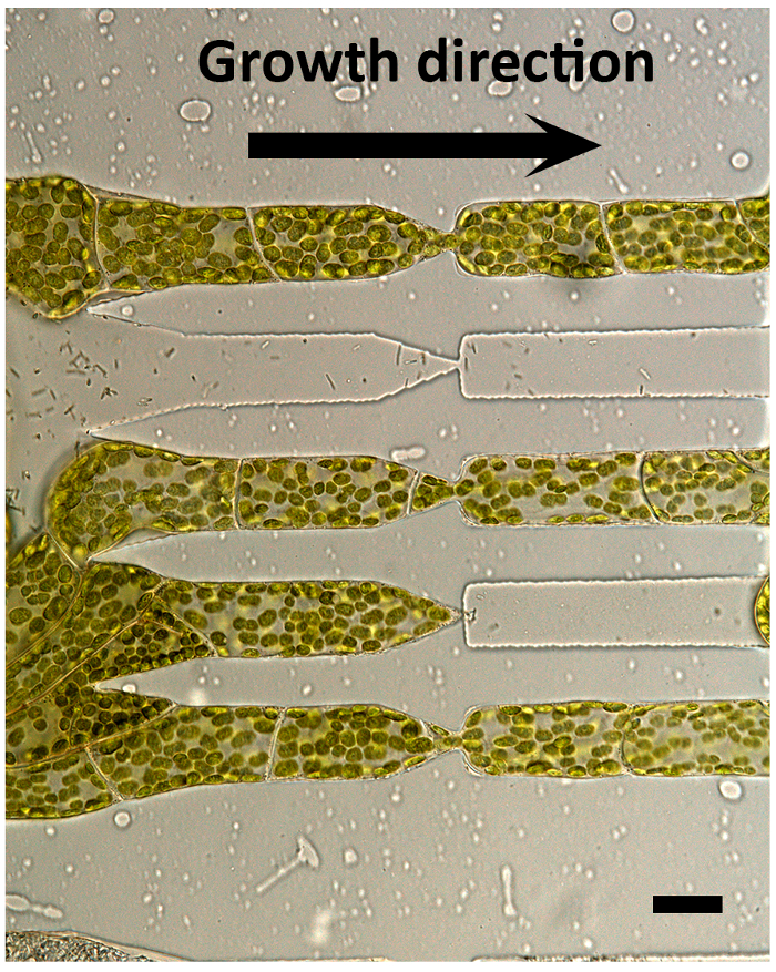
Figure 5. P. patens protonemata elongation through a 1 µm gap. Moss protonemata cells crossing through a 1 µm gap (4 µm in height), which was widened due to turgor pressure of the protonemata. Scale bar, 20 µm. The figure has been modified with permission from Yanagisawa et al. 201719. Please click here to view a larger version of this figure.
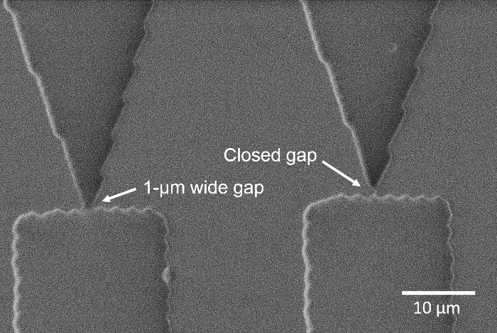
Figure 6. Closed microgaps. SEM image of the 1 µm gap region. The microgap on the right is completely closed. Please click here to view a larger version of this figure.
Supplementary Movie 1. Pollen tube growth through a 1 µm gap.Time-lapse bright field images were captured every 10 s using an inverted microscope equipped with a spinning disk confocal microscope. Scale bar, 20 µm. Please click here to download this file.
Supplementary Movie 2. Penetration of the vegetative nucleus and sperm cells through a 1 µm gap. Time-lapse bright field and fluorescence images were captured every 20 s using an inverted fluorescence microscope. Scale bar, 20 µm. Please click here to download this file.
Several critical steps in the protocol need to be followed precisely to obtain the results presented above. First, the PDMS layer and glass bottom dish surfaces must both be treated with plasma for a sufficient amount of time before bonding. Otherwise, the PDMS layer may locally detach from the glass surface while tip-growing cells are crossing the microgaps. Another crucial step in the root hair and moss protonemata protocol is the sterilization of the microdevice. Normally, root hairs and moss protonemata cells need to be cultured in the microdevice for a couple of weeks. Without sterilizing the device, microorganisms may flourish, which could affect the growth of these tip-growing cells. Sterilization of the pollen tube device is not as critical, since the experiment is concluded within half a day. We have regularly used the pollen tube device without sterilization and did not observe any undesirable effects during the experiment.
The microchannel design should be modified according to the cells of interest. In our work, the channel design used for the A. thaliana root hair experiment was different from the one used for the T. fournieri pollen tube and the P. patens protonemata experiments. Different devices are required because the samples have different dimensions; for instance, root hairs are much shorter than pollen tubes and moss protonemata. Therefore, our root hair device was designed such that the microgap regions were adjacent to the main root channel. In addition to the channel design, the procedure for preparing the root hair device also varies from the procedures used for pollen tubes or moss protonemata. For the pollen tube and moss protonemata studies, we used a microdevice containing a single PDMS layer with the microchannels sealed against a glass bottom dish. However, the device used to study root hairs comprises two PDMS layers: the top layer with a microchannel (depth: 200 µm) for the main root is sealed against the bottom layer, containing microchannels (depth: 4 µm) for root hairs. The standard photolithography technique that was employed to fabricate the silicon mold is limited to an aspect ratio of around 5:1. Therefore, to create a 1 µm wide gap, the channel height will be limited to approximately 5 µm. It is also technically challenging to precisely align a deep channel (e.g., 200 µm) near the 1 µm wide gap on a silicon mold. Therefore, we prepared separate PDMS layers for the main root and the root hairs, and constructed the device by sealing them together.
While this protocol was successfully used to visualize the tip growing process of pollen tubes through the microgaps (Supplementary Movie 1 and 2), time-lapse imaging of root hair and moss protonemata elongation at the microgap region would be more challenging. In the microdevice used for culturing A. thaliana root hairs, the height of the side channels where the microgaps are located (4 µm) is much shallower than that of the root growth chamber (200 µm), so most root hairs are unable to proceed into the side channels. Even if the root hairs do find the side channel entrance, it is hard to predict when they will enter it. To capture time-lapse images of root hair growth at the microgap, we recommend using a microscope equipped with an automated stage that can be programmed to be situated at multiple positions for image acquisition. When such instrumentation is unavailable, we suggest searching for the root hairs that have already entered the side channels but have not yet crossed the microgaps. Since the growth rate of A. thaliana root hairs is much slower (typically 1 – 2.5 µm/min20) than that of T. fournieri pollen tubes (typically 22 – 23 µm/min19), it may be possible to capture time-lapse images of those root hairs that are about to cross the microgaps. On the other hand, moss protonemata observations require a time-lapse system that can be exclusively used for this study, because a prolonged observation period is required due to the slow growth rate.
The microfluidic approach presented here is the first method that allows us to study the capability of tip-growing plant cells to elongate through extremely narrow spaces. These microdevices may be useful for screening assays that investigate the functions of genes associated with cell wall biosynthesis and the integrity of tip-growing cells through their penetration capability. In addition, Denais et al.21 recently demonstrated a nuclear envelope rupture and repair mechanism by squeezing a cancer cell through a confined space prepared in a microfluidic device, and it may be possible to study whether a similar mechanism exists in pollen tubes using our device. This newly developed experimental platform can be used to investigate how individual cells respond to physically constrained spaces and may thus enhance our understanding of tip-growth mechanisms.
The authors have nothing to disclose.
We thank H. Tsutsui and D. Kurihara for providing us with transgenic plants, including the T. fournieriRPS5Ap::H2B-tdTomato line and the A. thaliana UBQ10pro::H2B-mClover line, respectively. This work was supported by the Institute of Transformative Bio-Molecules of Nagoya University and the Japan Advanced Plant Science Network. Financial support for this work was provided by grants from the Japan Science and Technology Agency (ERATO project grant no. JPMJER1004 for T.H.), a Grant-in-Aid for Scientific Research on Innovative Areas (Nos. JP16H06465 and JP16H06464 for T.H.), and Japan Society for the Promotion of Science (JSPS) Grants-in-Aid for challenging Exploratory Research (grant no. 26600061 for N.Y. and grant nos. 25650075 and 15K14542 for Y.S.).
PDMS | Dow Corning Co. | Sylgard184 | |
Murashige & Skoog Medium | Wako Pure Chemical | 392-00591 | |
MES | Dojindo | 345-01625 | |
Sucrose | Wako Pure Chemical | 196-00015 | |
50 mm glass-bottom dish | Matsunami Glass | D210402 | |
35 mm glass-bottom dish | Iwaki | 3971-035 | |
Surgical blade | Feather | No.11 | |
biopsy punches | Harris | Uni-Core | |
Gel loading tips | Bio-Bik | 124-R-204 | |
Inverted Microscope | Olympus | IX83 | |
CSU-W1 | Yokogawa Electric | No Catalog number is avairable for this customized microscope | |
MetaMorph imaging software | Molecular Devices |