Identifying Inhibitors of the HBx-DDB1 Interaction Using a Split Luciferase Assay System
Summary
Here, we present a method for screening anti-hepatitis B viral agents that inhibit the HBx-DDB1 interaction using a split luciferase assay system. This system allows easy detection of protein-protein interactions and is suitable for identifying inhibitors of such interactions.
Abstract
There is an urgent need for novel therapeutic agents for hepatitis B virus (HBV) infection. Although currently available nucleos(t)ide analogs potently inhibit viral replication, they have no direct effect on the expression of viral proteins transcribed from a viral covalently closed circular DNA (cccDNA). As high viral antigen load may play a role in this chronic and HBV-related carcinogenesis, the goal of HBV treatment is to eradicate viral proteins. HBV regulatory protein X (HBx) binds to the host DNA damage-binding protein 1 (DDB1) protein to degrade structural maintenance of chromosomes 5/6 (Smc5/6), resulting in activation of viral transcription from cccDNA. Here, using a split luciferase complementation assay system, we present a comprehensive compound screening system to identify inhibitors of the HBx-DDB1 interaction. Our protocol enables easy detection of interaction dynamics in real time within living cells. This technique may become a key assay to discover novel therapeutic agents for treatment of HBV infection.
Introduction
Hepatitis B virus (HBV) infection is a major public health concern worldwide, with annual estimates of 240 million people chronically infected with HBV and 90,000 deaths due to complications from the infection, including cirrhosis and hepatocellular carcinoma (HCC)1. Although the current anti-HBV therapeutic agents, nucleos(t)ide analogues, sufficiently inhibit viral reverse transcription, they rarely achieve elimination of viral proteins, which is the long-term clinical goal. Their poor effect on viral protein elimination is due to their lack of direct effect on viral transcription from episomal viral covalently closed circular DNA (cccDNA) minichromosomes in the hepatocyte nucleus2.
HBV transcription is activated by HBV regulatory X (HBx) protein3. Recent studies revealed that HBx degrades structural maintenance of chromosomes 5/6 (Smc5/6), a host restriction factor that blocks HBV transcription from cccDNA, via hijacking a DDB1-CUL4-ROC1 E3 ubiquitin ligase complex4,5,6. Therefore, a crucial step in promoting viral transcription from cccDNA is thought to be the HBx-DDB1 interaction. Compounds capable of inhibiting the binding between HBx and DDB1 may block viral transcription, and indeed nitazoxanide was identified as an inhibitor of the HBx-DDB1 interaction via a screening system developed in our laboratory7.
Here, we present our convenient screening system used to identify inhibitors of the HBx-DDB1 interaction, which utilizes a split luciferase complementary assay7,8. Split luciferase subunits are fused to HBx and DDB1, and the HBx-DDB1 interaction brings the subunits into close proximity to form a functional enzyme that generates a bright luminescent signal. As the interaction between the subunits is reversible, this system can detect rapidly dissociating HBx-DDB1 proteins (Figure 1). Using this system, a large compound library can be easily screened, which may result in the discovery of novel compounds capable of efficiently inhibiting the HBx-DDB1 interaction.
Protocol
NOTE: A schematic representation of the split luciferase assay is shown in Figure 1A, and the assay process is outlined in Figure 1B. The interaction dynamics can be measured in real time without cell lysis.
1. Cell Preparation
- Maintain cultured HEK293T cells in Dulbecco's modified Eagle's medium (DMEM) supplemented with 10% v/v fetal bovine serum (FBS), 1x penicillin/streptomycin at 37 °C in 20% O2 and 5% CO2.
- Seed 5 x 10⁶ cells into a 100 mm dish with 10 mL of DMEM and incubate at 37 °C overnight.
- Transiently transfect 1 µg of HBx and DDB1 with split luciferases into the cells according to the following method.
NOTE: The amount of the plasmid DNA transfected may depend on the transfection regent used. The optimal position of the split luciferase fused to the target protein must be determined beforehand. In this case, HBx fused to LgBit at the C-terminus of HBx (HBx-LgBit) and DDB1 fused to SmBit at the N-terminus of DDB1 (SmBit-DDB1) provided the best results (i.e., the brightest luciferase signals). This process has been reported previously in detail7.- Dilute 1 µg of HBx-LgBit expressing DNA plasmid and 1 µg of SmBit-DDB1 expressing DNA plasmid (Table of Materials) in DNA condensation buffer (Table of Materials) to a total volume of 300 µL.
- Add 16 µL of enhancer solution (Table of Materials) and mix by vortexing for 1 s.
- Incubate the sample at room temperature for 3 min.
- Add 60 µL of transfection reagent (Table of Materials) to the sample and mix by vortexing for 10 s.
- Incubate the sample at room temperature for 8 min.
- During incubation, aspirate the culture medium from the dish (prepared in step 1.2), and wash cells with 5 mL of phosphate-buffered saline (PBS). Remove PBS by aspiration and add 7 mL of DMEM.
- Add 3 mL of DMEM to the tube containing the transfection complexes. Mix by pipetting and add the transfection complexes onto the cell in the 10 cm dish.
- Incubate the cells at 37 °C in an incubator under 5% CO2 for 10 h.
- Reseed the cells into a white 96 well plate at 5 x 10⁴ cells/well in 50 µL of medium/well according to the following method.
- Remove the spent cell culture medium and wash cells with 5 mL of PBS.
- Remove PBS by aspiration, add 1 mL of 0.25% trypsin-EDTA, and incubate at 37 °C for 5 min to detach cells.
- Add 4 mL of DMEM and disperse the medium by pipetting over the surface of the cell layer several times. Transfer the cell suspension to a tube.
- Centrifuge cells at 500 x g for 5 min at room temperature.
- Discard supernatant and resuspend the cell pellet in 1 mL of PBS.
- Centrifuge the cell suspension at 500 x g for 5 min at room temperature and discard supernatant.
- Dilute the cell pellet with buffered cell culture medium (Table of Materials) supplemented with 10% FBS to a seeding density of 1.0 x 10⁶ cells/mL.
- Pipette 50 µL of cell suspension into each well of a 96 well plate and return the cells to the incubator.
- Incubate cells at 37 °C under 5% CO2 for 10 h.
2. Compound Screening
- During the incubation, dilute the screening compounds (Table of Materials) and solvent (dimethyl sulfoxide [DMSO]) at 13.5x concentration. For example, if the stock is 10 mM and the screening concentration is 10 µM, add 1 µL of stock solution to 73.1 µL of buffered cell culture medium.
- Add 12.5 µL of luminescent substrate (Table of Materials) to each well and incubate for 5 min at room temperature.
NOTE: As negative controls, the wells at both ends of the plate (i.e., columns 1 and 12) should contain no luminescent substrate. - Measure the baseline luminescence using a luminometer (Table of Materials).
- Immediately after the initial measurement, add 5 µL of compounds and control DMSO diluted in step 2.1 to each well.
NOTE: The final concentration will be 10 µM. - Measure luminescence values every 30 min for 2 h.
NOTE: The plate should be incubated in the dark at room temperature. - Calculate the inhibitory effects by comparison with control DMSO treatment after normalization to the baseline signals.
NOTE: Screening each compound in duplicate or triplicate can reduce variation.
Representative Results
Representative outcomes following the use of this protocol are shown in Figure 2A,B. The signal-to-background ratio was greater than 80 and the Z' factor9 (the gold standard quality index for high-throughput screening) was greater than 0.5, indicating that this assay system was acceptable for high-throughput screening. With the threshold set to >40% inhibition compared with the control (DMSO only), we identified nitazoxanide as a candidate drug7. Using this system, better candidate drugs can be found by screening other, larger compound libraries.
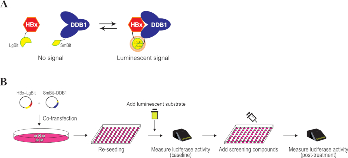
Figure 1: Schematic representation of split luciferase analysis of the HBx-DDB1 interaction. (A) The principle underlying detection of HBx-DDB1 binding using the split luciferase complementary assay system. The separated luciferase subunits, LgBit and SmBit, are fused to HBx and DDB1, respectively. The HBx-DDB1 interaction brings the subunits into close proximity to form a functional enzyme that generates a luminescent signal. The interaction between the subunits is reversible. (B) Split luciferase assay. After co-transfection of plasmids for expression of HBx fused to LgBit and DDB1 fused to SmBit, cells were re-seeded into a 96 well plate. The addition of luminescent substrate enables measurement of luciferase activity without a cell lysis step. Luciferase activities can be measured after adding the screening compounds. Please click here to view a larger version of this figure.
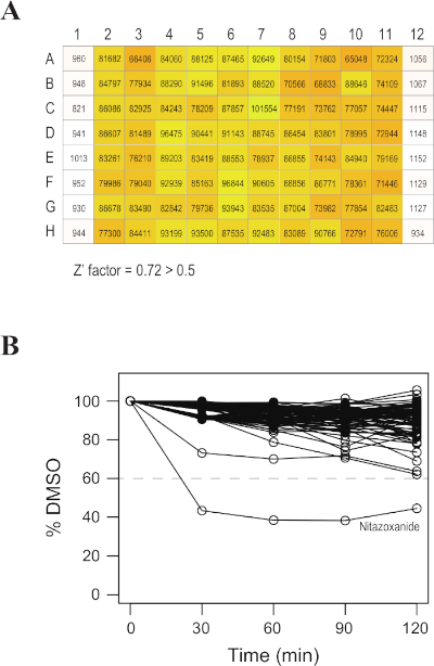
Figure 2: Successful results of the split luciferase assay. (A) Representative baseline luminescent signals from a 96 well plate. Luciferase intensity is represented by numbers and colors. Columns 1 and 12 are controls in which the luminescent substrate was not added. The Z' factor was greater than 0.5. (B) Representative time-series result of relative luciferase activity levels after addition of screening compounds to a 96 well plate. The x-axis represents the inhibitory effects calculated compared to control (DMSO) after standardization to the baseline luciferase activity. The most effective compound was nitazoxanide. Please click here to view a larger version of this figure.
Discussion
We developed a convenient screening method using a split luciferase assay to find HBx-DDB1 binding inhibitors. The interaction dynamics can be detected in real time in living cells without the need for cell lysis. Inhibition of the HBx-DDB1 interaction leads to restoration of Smc5/6, which results in suppression of viral transcription, protein expression, and cccDNA production7. This novel mechanism of antiviral action may overcome the inadequacies of current HBV therapies.
Although a number of methods are available to investigate protein-protein interactions in living cells, examining these interactions remain difficult10. Our procedure is simple and requires only a short time to screen one 96 well plate. Moreover, the screening quality was satisfactory with a high Z' score, the gold standard quality index for high-throughput screening9. Our assay may be suitable for robotic automation11 and is an efficient assay for drug discovery.
While the protocol described here used the HEK293T cell line because its high transfection efficacy and high proliferation ability are suitable for high-throughput screening, this screening method can be performed using other cell lines (e.g., HepG2) without modifications7. As a realistic strategy for screening compounds, HEK293T cells may be used in the first screening followed by HepG2 cells in a second validation screening. Some compounds may not show significant results in different cell lines when the effects are dependent on indirect mechanisms.
As our intention was to develop a high-throughput screening method, subsequent validation studies are necessary to confirm whether the identified compounds function as interaction inhibitors. Decreased levels of luminescent signals in this assay do not always correspond to inhibition of the HBx-DDB1 interaction. Cytotoxicity tests, co-immunoprecipitation studies, and further anti-HBV experiments are important to confirm the effects7.
Although we previously identified nitazoxanide as an inhibitor of the HBx-DDB1 interaction by screening a relatively small-scale compound library7, further studies involving screening of much larger compound libraries can be easily performed to identify novel compounds that are capable of inhibiting protein-protein interactions more efficiently. When performing such further screening, nitazoxanide can be used as a positive control for the assay. Furthermore, the system described here can be applied to other protein-protein interactions. Protein-protein interactions are an important class of drug targets12. Indeed, many other viruses interact with host factors to replicate or express their pathogenicity13,14. The split luciferase-based assay described here, which targets the interactions between viral and host proteins, may provide a new strategy to develop cures for HBV and other infectious diseases.
Disclosures
The authors have nothing to disclose.
Acknowledgements
This work was supported by Grants-in-Aid from the Ministry of Education, Culture, Sports, Science, and Technology, Japan (#19H03430 and #17K09405 to M.O., and #19J11829 to K.S.), by a Grant-in-Aid for Scientific Research on Innovative Areas (#18H05024 to M.O.), by the Research Program on Hepatitis from Japan Agency for Medical Research and Development, AMED (to M.O., #JP19fk021005), by program on the Innovative Development and the Application of New Drugs for Hepatitis B (#JP19fk0310102 to K.K.) from AMED, by grants from the Japan Foundation for Applied Enzymology and from the Kobayashi Foundation for Cancer Research (to M.O.), by GSK Japan Research Grant 2018 (to K.S.), and by a grant from the Miyakawa Memorial Research Foundation (to K.S.).
Materials
| Cell culture microplate, 96 well, PS, F-BOTTOM | Greiner-Bio-One GmbH | 655098 | |
| DMEM | Sigma Aldrich | D6046 | |
| DMSO | Tocris Bioscience | 3176 | |
| Effectene transfection reagent | Qiagen | 301425 | Includes DNA-condensation buffer, enhancer solution and transfection reagent |
| FBS | Nichirei | 175012 | |
| GloMax 96 microplate luminometer | Promega | E6521 | |
| HBx–LgBit expressing DNA plasmid | Our laboratory | Available upon request | |
| HEK293T cells | American Type Culture Collection | CRL-11268 | |
| NanoBiT PPI starter systems | Promega | N2015 | Includes Nano-Glo Live Cell Reagent |
| Opti-MEM | Thermo Fisher Scientific | 11058021 | Described as "buffered cell culture medium" in the manuscript |
| PBS | Takara | T900 | |
| Penicillin-Streptomycin | Sigma Aldrich | P0781 | |
| Screen-Well FDA-approved drug library V2 version 1.0 | Enzo Life Sciences | BML-2841 | Compounds used here were as follows: mequinol, mercaptopurine hydrate, mesna, mestranol, metaproterenol hemisulfate, metaraminol bitartrate, metaxalone, methacholine chloride, methazolamide, methenamine hippurate, methocarbamol, methotrexate, methoxsalen, methscopolamine bromide, methsuximide, methyclothiazide, methyl aminolevulinate·HCl, methylergonovine maleate, metolazone, metyrapone, mexiletine·HCl, micafungin, miconazole, midodrine·HCl, miglitol, milnacipran·HCl, mirtazapine, mitotane, moexipril·HCl, mometasone furoate, mupirocin, nadolol, nafcillin·Na, naftifine·HCl, naratriptan·HCl, natamycin, nebivolol·HCl, nelarabine, nepafenac, nevirapine, niacin, nicotine, nilotinib, nilutamide, nitazoxanide, nitisinone, nitrofurantoin, nizatidine, nortriptyline·HCl, olsalazine·Na, orlistat, oxaprozin, oxtriphylline, oxybutynin Chloride, oxytetracycline·HCl, paliperidone, palonosetron·HCl, paromomycin sulfate, pazopanib·HCl, pemetrexed disodium, pemirolast potassium, penicillamine, penicillin G potassium, pentamidine isethionate, pentostatin, perindopril erbumine, permethrin, perphenazine, phenelzine sulfate, phenylephrine, phytonadione, pimecrolimus, pitavastatin calcium, and podofilox |
| SmBit–DDB1 expressing DNA plasmid | Our laboratory | Available upon request | |
| Trypsin-EDTA | Sigma Aldrich | T4049 |
References
- Tang, L. S. Y., Covert, E., Wilson, E., Kottilil, S. Chronic hepatitis B infection: a review. The Journal of the American Medical Association. 319, 1802-1813 (2018).
- Sekiba, K., et al. Hepatitis B virus pathogenesis: Fresh insights into hepatitis B virus RNA. World Journal of Gastroenterology. 24, 2261-2268 (2018).
- Slagle, B. L., Bouchard, M. J. Hepatitis B virus X and regulation of viral gene expression. Cold Spring Harbor Perspectives in Medicine. 6, a021402 (2016).
- Murphy, C. M., et al. Hepatitis B Virus X Protein Promotes Degradation of SMC5/6 to Enhance HBV Replication. Cell Reports. 16, 2846-2854 (2016).
- Decorsière, A., et al. Hepatitis B virus X protein identifies the Smc5/6 complex as a host restriction factor. Nature. 531, 386-389 (2016).
- Niu, C., et al. The Smc5/6 complex restricts HBV when localized to ND10 without inducing an innate immune response and is counteracted by the HBV X protein shortly after infection. PLoS One. 12, e0169648 (2017).
- Sekiba, K., et al. Inhibition of HBV Transcription From cccDNA With Nitazoxanide by Targeting the HBx-DDB1 Interaction. Cellular and Molecular Gastroenterology and Hepatology. 7, 297-312 (2019).
- Eggers, C. T., et al. NanoLuc Complementation Reporter Optimized for Accurate Measurement of Protein Interactions in Cells. ACS Chemical Biology. 11, 400-408 (2015).
- Zhang, J. H., Chung, T. D. Y., Oldenburg, K. R. A simple statistical parameter for use in evaluation and validation of high throughput screening assays. Journal of Biomolecular Screening. 4, 67-73 (1999).
- Rao, V. S., Srinivas, K., Sujini, G. N., Kumar, G. N. Protein-protein interaction detection: Methods and analysis. International Journal of Proteomics. 2014. 2014, 147648 (2014).
- Michael, S., et al. A Robotic Platform for Quantitative High-Throughput Screening. ASSAY and Drug Development Technologies. 6, 637-657 (2008).
- Skwarczynska, M., Ottmann, C. Protein-protein interactions as drug targets. Future Medicinal Chemistry. 7, 2195-2219 (2015).
- de Chassey, B., Meyniel-Schicklin, L., Vonderscher, J., André, P., Lotteau, V. Virus-host interactomics: New insights and opportunities for antiviral drug discovery. Genome Medicine. 6, 115 (2014).
- Prasad, M., et al. Virus-Host Interactions: New Insights and Advances in Drug Development Against Viral Pathogens. Current Drug Metabolism. 18, 942-970 (2017).
