This manuscript describes a consistent way to quickly perform survival rodent orchiectomies and ovariectomies.
Sex hormone signaling plays a critical role in multiple organ systems as well as in the progression of various diseases, including neurodegenerative disease. The manipulation of sex hormone levels in the murine model system allows for the study of their impact on organs/tissues and within disease progression. Orchiectomy – the surgical removal of the testes – and ovariectomy – the surgical removal of the ovaries – provide a method to deplete the endogenous sex hormones so that the precise hormone levels can be provided through drug or other delivery methods. Here, we provide rapid and minimally invasive methods for both orchiectomy and ovariectomy in the murine model system for the reduction of sex hormones. This protocol details the surgical preparation and excision of the testes through the scrotal sac, and excision of the ovaries via two incisions in the right and left lateral dorsum.
The testes and ovaries are the primary organs responsible for sex hormone production. The cascade of hormonal communication leading to the production of testosterone and estrogen is a well-characterized process that begins in the hypothalamus with the release of gonadotropin-releasing hormone (GnRH)1. The release of GnRH causes the release of luteinizing hormone (LH) and follicle-stimulating hormone (FSH) from the pituitary gland. As these hormones enter the bloodstream, they then affect other tissues in the body. The primary target of LH is the testes (in males) and the ovaries (in females)2. In response to LH, the testes produce and release testosterone3. Similarly, the ovaries produce estrogen4. While the intended effects of these hormones are to prepare the cells and body for fertilization and ensure a functioning reproductive system, many other bodily systems can be affected.
Sex hormones have been linked to several physiological functions. For instance, estrogen helps maintain bone homeostasis by preventing the resorption of bone by osteoclasts. For this reason, ovariectomized mouse models can be used to study the physiology of bone diseases such as osteoporosis5,6,7. Testosterone and estrogen are also research targets for many cardiovascular and neurodegenerative diseases. Recently, elevated testosterone production coupled with high-fat diet has been linked to vascular oxidative stress8. In the brain, changes in LH after ovariectomy have caused alterations in spatial memory9. Reduction of estrogens following ovariectomy has also become a model system for studying cell death in the hippocampus, as this can induce apoptosis, resulting in memory deficits10. Testosterone has also shown a role in the growth of kidneys both in mouse models and humans following kidney transplantation11.
The creation of a hormone-deprived murine model allows for the study of sex hormones and their hormone cascades on various diseases or tissues. This can be accomplished by surgical removal of the testes (orchiectomy) or ovaries (ovariectomy). This procedure can be performed in mice of any strain when they are as young as weaning age (twenty-one days) or any adult age. Ovariectomy is performed in female mice, while orchiectomy is performed in male mice. By removing these organs, the levels of estrogen and testosterone, and many of their derivatives, such as progesterone, can be greatly reduced12,13. The process of performing orchiectomies or ovariectomies in mice can be rapid and minimally invasive with the proper technique. Rapid excision of these organs in a safe and efficient manner can allow for quick surgical processing while keeping mouse numbers minimal by having a 100% survival rate when performed correctly. Here, we detail a protocol for the rapid excision of the testes and ovaries and demonstrate the proper post-surgical monitoring to enable researchers to perform this surgery quickly and safely. We also include visual examples of the sex organs and surrounding tissues to provide the surgeon with anatomical landmarks when performing this procedure.
All animal experiments were approved by the Institutional Animal Care and Use Committee at UTSW (APN#2019-102840).
1. Murine orchiectomy
2. Murine ovariectomy
3. Post-operative care
The procedure presented here is performed in one- to three-month-old mice in the C57BL/6J background. Male mice weighed 16-28 g, and female mice weighed 14-24 g at the time of the procedure. This procedure has been optimized to be applicable for mice of many ages, from weaning through adulthood.
Surgical orchiectomy involves a single skin incision in the ventral scrotal sac, as depicted in Figure 1A. Both testes are removed one at a time and are severed through the vas deferens and spermatic blood vessels, resulting in the removal of the testis and attached epididymis (Figure 1B). The removed contents include the testis, epididymis, and inguinal fat pad, shown in Figure 1C. Successful removal of the testicles can be confirmed by visual observation of removed tissues and ensuring good hemostasis of the tissue stump prior to closing the body cavity. The success of orchiectomy can be measured by observation of well-recovered mice that demonstrate healed incision sites (Figure 1D). Mice that have undergone orchiectomy will also demonstrate a reduction in the level of testosterone measured in mouse serum as early as a week after the procedure (Figure 1E).
Surgical bilateral ovariectomy is done by incisions into the left and right dorsum of female mice (Figure 2A). The lateral incision approach can be used if only one ovary is to be removed. In this approach, the abdominal wall is incised, and the ovarian fat pad is located. The fat pad, ovary, ovarian duct, and distal uterine horn, as depicted in Figure 2B, is exteriorized, followed by severing the distal uterine horn. This technique results in the removal of the ovary and oviduct. The ovary and ovarian duct can be removed without including the distal uterine horn, but there is a risk that any remaining ovarian cells will continue to keep the mouse in estrous cycles. Including the distal uterine horn ensures that the full ovary and duct are removed, there will be a depletion of the sex hormones, and the mice will no longer cycle. Figure 2C depicts an example of the murine female reproductive organs dissected out of the body. It can be observed that the ovaries themselves are much smaller than the surrounding tissues. A dissection scope or loupes aids in the identification of these tissues. Figure 2D demonstrates the ovarian anatomy under a dissecting scope with tissue from the uterine fat pad removed, allowing for better visualization. The success of this procedure can be measured by the observation of well-recovered mice that demonstrate healed incision sites (Figure 2E).
Ovariectomy surgery is safe and efficient when performed rapidly and with minimal disturbance to surrounding tissues. Following the procedure outlined here accomplishes this by ensuring that the skin and abdominal wall incision are positioned correctly, allowing for quick location of the uterine tissues. Identifying the inferior border of the rib cage and the superior border of the leg bones ensures the incision is made near the tissue of interest. Making an incision that is about halfway between the inferior border of the ribs and superior border of the leg bones and is located about 1.5 cm lateral to the spine allows for good positioning of the surgical site when performing a lateral incision approach (Figure 2A).
Confirmation of surgical ovariectomy is shown by observation of uterine atrophy. Figure 2F shows a uterine horn dissected from a 6-month-old mouse, while Figure 2G shows a uterine horn from a 6-month-old mouse that underwent surgical ovariectomy at three months of age. The ovariectomized uterine horn appears thinner and lacks the attachment to the ovary on the distal end. This method of confirmation of the surgical technique effectively shows that the removal of the ovary resulted in atrophy of the uterine tissue.
Once tissues have been successfully identified and excised, the surgery can be completed by closure of the wounds and careful post-operative monitoring. Proper aseptic technique14 is important in ensuring good outcomes when performing survival surgery. As the mouse begins to heal from the incisions, monitoring is performed to look for signs of pain and surgical complications. A possible surgical complication is internal bleeding because of poking or nicking blood vessels while trying to incise or manipulate the tissue. This is denoted by a persistent red, flushed appearance underneath the skin around the surgical site. Avoiding complications such as these is achieved by proper localization of incision sites and aiding the surgeon's vision by use of a surgical microscope or loupes. Visual examination is recommended every 12 hours for the first three days post-surgery. Successful surgery will result in clean, healing wounds (Figure 1D, 2E).
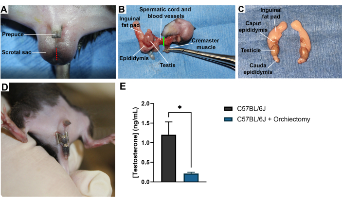
Figure 1: Male murine orchiectomy. (A) A male mouse is shown in the supine position with the location of a surgical incision into the scrotal sac for the removal of the testis, which is shown in red. (B) Image taken during the orchiectomy surgical procedure depicting the point of detachment in green. (C) Dissected-out testis, fat pads, and epididymis from a male mouse. (D) A male mouse with a well-healed wound clip over the scrotal incision following orchiectomy. (E) Mouse serum testosterone concentration in ng/mL as determined by ELISA interpolation. Both groups represent 9-week-old mice. Surgical orchiectomies were performed at 8 weeks of age. N = 3 mice per group. Samples were run in triplicate. Serum samples were collected by cardiac puncture and frozen until the time of analysis. *, p < 0.05. Error bars = SEM. Please click here to view a larger version of this figure.
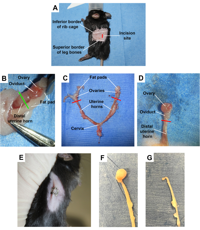
Figure 2: Female murine ovariectomy. (A) A female mouse shown in the lateral position with the surgical incision site for the removal of a single ovary from the dorsal lateral side, shown in red. Other important anatomical landmarks are highlighted. (B) Image taken during surgery depicting the exteriorized ovary, oviduct, and distal uterine horn with the point of detachment shown in green. (C) Dissected uterine horns, ovaries, and fat pads from a female mouse. (D) A close-up view of the dissected ovary, ovarian duct, and distal portion of the uterine horn with fat from the fat pad removed, allowing for better visualization of the ovary. (E) A female mouse with a well-healed wound clip over the incision following ovariectomy. (F) Dissected mouse uterine horn. (G) Dissected mouse uterine horn 3 months post-ovariectomy procedure, showing uterine atrophy. Please click here to view a larger version of this figure.
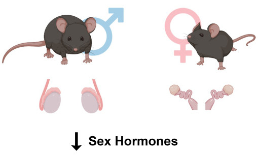
Figure 3: Graphical abstract for murine ovariectomy and orchiectomy. A pictorial representation of the orchiectomy procedure for male mice involving the removal of the testis and ovariectomy procedure involving the removal of the ovaries, both of which result in a sex hormone depleted mouse model. Please click here to view a larger version of this figure.
Surgical removal of testes and ovaries allows for studying murine physiology under controlled hormone deprivation. This technique is important for many fields of science, including neurodegeneration, mineral metabolism, cardiovascular, and reproductive health15,16,17,18,19,20,21. Here, we detail a protocol for the fast, safe, and effective removal of murine ovaries and testis using survival surgery to deplete the sex hormones. When performed by an experienced surgeon, these procedures can take as little as 5 minutes, resulting in a very high survival rate in mice.
Some considerations when planning for a mouse surgery include the best incision site and the physiology of the animal. In the case of orchiectomy, the mouse testis can also be removed through the abdomen by a midline ventral incision13. This technique not only opens the peritoneal cavity, but also relies on the quick identification of the inguinal fat pads. For new surgeons, it is difficult to rapidly perform this technique. When mice are very young, the fat pads are also less developed and, therefore, more difficult to locate. The scrotal approach outlined in this protocol is a suitable option for researchers who are new to mouse surgery, operating on small, young mice, or who wish to minimize the chance of disturbing other organs in the peritoneal cavity by an abdominal incision.
Surgical ovariectomy involves the removal of one or both ovaries. As for most surgical procedures, safe and fast methods usually involve making the incision into the skin and underlying fascia as small as possible. This allows for reduced sutures and wound clips and faster healing. While being able to make the incisions smaller is a skill that comes with practice, being confident about the location of the incision is beneficial. Figure 2A demonstrates an example of how to locate the best incision points. Due to the small mouse anatomy, the ovary will lie near the liver, and on the left side of the mouse, near to the spleen. Accidentally probing and pulling on these organs can result in undesired bleeding or harm. We provide a detailed guide to the correct incision location and identification of the surrounding anatomical landmarks to aid in this technique and reduce the likelihood of adverse complications.
Here, we provide an example of how this procedure can be used to effectively knock down the circulating hormone levels in mice less than a week after the surgical procedure, as well as how the remaining gonadal tissue atrophies over time after the procedure. Previous methods and research detail how these can be done to generate menopause or andropause mouse models, but many of these are performed in rat models rather than mice12,22. While rats and mice have similar anatomy, the murine system has smaller anatomical structures. Here, we provide a method that reliably works in mouse models as young as 3 weeks of age. Overall, these two protocols represent a consistent way to quickly perform survival rodent orchiectomies and ovariectomies, recognize the mouse anatomy, and minimize surgical complications, creating a basis for consistent and effective results.
The authors have nothing to disclose.
We thank the University of Texas Southwestern Medical Center Animal Resource Center for their help in surgical training and protocol review. We thank the Wert Lab support team for their invaluable assistance. This work has been supported by funds from the National Institute of Health (NIH P30EY030413). Biorender.com was used for the creation of cartoon schematics.
1mL Syringe | BD | 309659 | |
30G 1/2" Needle | BD | 305106 | |
AutoClip System | Fine Science Tools | 12020-00 | |
Betadine Solution | Fisher Scientific | NC0158124 | |
Cotton-Tipped Applicators | Fisher Scientific | 10-000-692 | |
Double -ended Micro Spatula | Fine Science Tools | 10091-12 | |
Galilean Loupes | Fine Science Tools | 28050-30 | Optional, can provide better clarity during procedure |
Gauze Sponges, 4"x4" | Fisher Scientific | 13-761-52 | |
Graefe Forceps | Fine Science Tools | 11150-10 | |
High Temp Cautery Kit | Fine Science Tools | 18010-00 | Using the fine tip attachment |
Needle Holders | Fine Science Tools | 12001-13 | |
PGA Absorbable Suture:4-0 / NFS-2 Reverse Cutting 19MM / 30 IN | Covetrus | 29242 | 4-0 or 5-0 Absorbable sutures are best |
Rodent Warming pad | Kent Scientific | RT-0515 | |
Sterile Alcohol Prep Pads | Fisher Scientific | 22-363-750 | |
Straight Locking Micro Needle Holders | Fine Science Tools | 12060-01 | |
Surgical Scissors | Fine Science Tools | 140-60-09 | |
Vannas Spring Scissors – 2.5mm Cutting Edge | Fine Science Tools | 15000-08 | |
Veet Sensitive Hair Remover Gel Cream | Amazon | N/A | |
Wahl Professional Animal Compact Trimmer and Grooming Kit, Blue | Amazon | #9861-900 |