The present study describes methodologies for culturing, freezing, thawing, processing, staining, labeling, and examining entire spheroids and organoids under various microscopes, while they remain intact in a hydrogel within a multipurpose device.
Organoids and spheroids, three-dimensional growing structures in cell culture labs, are becoming increasingly recognized as superior models compared to two-dimensional culture models, since they mimic the human body better and have advantages over animal studies. However, these studies commonly face problems with reproducibility and consistency. During the long experimental processes – with transfers of organoids and spheroids between different cell culture vessels, pipetting, and centrifuging – these susceptible and fragile 3D growing structures are often damaged or lost. Ultimately, the results are significantly affected, since the 3D structures cannot maintain the same characteristics and quality. The methods described here minimize these stressful steps and ensure a safe and consistent environment for organoids and spheroids throughout the processing sequence while they are still in a hydrogel in a multipurpose device. The researchers can grow, freeze, thaw, process, stain, label, and then examine the structure of organoids or spheroids under various high-tech instruments, from confocal to electron microscopes, using a single multipurpose device. This technology improves the studies' reproducibility, reliability, and validity, while maintaining a stable and protective environment for the 3D growing structures during processing. In addition, eliminating stressful steps minimizes handling errors, reduces time taken, and decreases the risk of contamination.
The future of cell research and therapy lies within 3D cell cultures1,2,3. Organoid and spheroid models close the gap between in vitro experiments and animal models by creating better models that mimic human body development, physiology, and diseases4,5,6,7,8,9. However, the reproducibility and repeatability of these models remain challenging. Furthermore, handling, harvesting, transferring, and centrifuging these structures with current technologies results in loss or damage of the organoids and spheroids in many conditions, significantly affecting the results.
Despite many protocols for histological staining, immunohistochemical staining, immunofluorescence labeling, and cryopreservation, there is no universal approach related to standardizing experimental conditions, handling, and processing these delicate structures without losing or damaging them. Current protocols are also incredibly long, alternating from a few days to several weeks, and include complex procedures with various reagents10,11,12,13,14. Additionally, harvesting, pipetting, centrifuging, and transferring 3D growing structures between cell culture vessels and cryovials cause changes in the positioning of the structures and mechanical forces, and ultimately affect the differentiation and maturation of the organoids and spheroids. It has been reported that tissue topology, positioning of the cells, and mechanical forces significantly impact cell differentiation and maturation6,15,16,17.
Therefore, it is desirable to improve the current conventional technologies to generate organoids and spheroids with stable quality. A method/device that will skip the centrifugation and other steps described above and provide the material in a single safe environment from the beginning to the end of the multiple processes would be beneficial to reach the most consistent and reliable data. Additionally, this will reduce time, labor, and cost constraints.
The multipurpose device (MD) described here provides a single safe environment for multiple processes of organoids and spheroids (Supplementary Figure 1). This device and complementing protocols eliminate the harvesting, pipetting, transferring, and centrifuging steps. The organoids and spheroids remain in their in vitro environment during the sequential processes. This environment mainly comprises natural or synthetic extracellular matrix components, like commercially available hydrogels. In other words, the methods described here allow a whole-mount sample of organoids/spheroids to be processed, examined, and frozen while still in a hydrogel drop.
The biocompatible device is resistant to temperatures between 60 °C and -160 °C, which makes it feasible to restore the organoids/steroids in a liquid nitrogen tank at -160 °C or to prepare resin blocks for electron microscopy at 60 °C. The niche in the device has been designed to define a limited space for 3D growing structures and stimulate the formation of spheroids or organoids based on the previous studies18,19,20,21,22,23. That part of the device is transparent and contains a specific plastic that provides high optical quality (refractive index: 1.43; abbe value: 58; thickness: 7.8 mil [0.0078 in or 198 µm]). Both the niche and the surrounding 'side' part cause autofluorescence. The transparent niche in the center has an 80 mm2 area, while the side part is 600 mm2. The depth of the container is 15 mm, and the thickness is 1.5 mm. These features, in addition to the size and design of the device, make it possible to make observations under different types of high-tech microscope and prepare the samples for electron microscopic examinations (Figure 2). The closing system of the device provides two positions, one sealed in the freezer and the other allowing gas flow in the incubator. CCK8 proliferation and cytotoxicity assays demonstrate similar effects on cells compared to the traditional cell culture dishes (Supplementary Figure 2). Trypan blue exclusion test demonstrates high cell viability (94%) during the cell culture in the MD (Figure 3).
The processes that can be performed for one sample in the single device comprise (1) culturing, (2) histological staining, (3) immunostaining, including immunohistochemical and immunofluorescence labeling, (4) freezing, (5) thawing, (6) examining under optical microscopes, such as brightfield, darkfield, fluorescence, confocal, and super-resolution microscopes, (7) coating and examining directly under a scanning electron microscope, or (8) preparing for transmission electron microscopy (Figure 2).
Different methodologies exist for histological staining, immunohistochemical labeling, or fluorescently labeling organoids and spheroids10,11,12,13,14,24,25. Harvesting them from the hydrogel is the first and principal step of the current technology. After this step, some methods allow whole-mount immuno-labeling. The harvested organoids are embedded in paraffin, sectioned, and labeled for staining and immunostaining in others. However, the sections may not present the whole sample and provide only limited data related to the 3D architecture of the structure. Furthermore, damage to these 3D structures and loss of antigenicity are well-known side effects of these technologies.
The complementing new protocols for microscopic examinations in this article allow an analysis of whole-mount samples still in a hydrogel. The protocols described here include two newly developed formulations: solution for immunohistochemistry (S-IHC) and solution for immunofluorescence labeling (S-IF). The methods with these solutions allow researchers to gain more accurate data, since there are no harmful effects of traditional workflows, such as centrifuging, pipetting, and transferring of the delicate structures. The protocol described here also eliminates the need for harvesting, blocking, clearing, and antigen retrieval steps, and shortens the whole procedure to 6-8 h. Furthermore, the methodology allows simultaneously adding one to three antibodies to the same S-IF. Therefore, it is possible to get the results on the same day even after the multiple labeling experiments, which is another advantage of the protocol described here; traditional whole-mount immunofluorescence labeling protocols typically take between 3 days and several weeks10,11,12,13,14.
Paraffin embedding, another harmful step that reduces antigenicity, is also omitted. The 3D structure remains in its in vitro environment from the beginning to the end of the microscopic examination. Since the 3D structure remains in its growing conditions, the protein expression and localization data mimic in vivo conditions better. More accurate results are expected, since the methodology eliminates the steps affecting the sample's antigen expression. Table 1 and Table 2 demonstrate how these new protocols eliminate steps, save time and labor in the lab, and reduce costs and waste products compared to traditional workflows.
In addition to the crucial steps described above, another problem is providing a cryopreservation medium and method to preserve the 3D structure of the sample with higher cell viability rates26,27,28,29,30,31. Cryopreservation is essential to creating a stable model system and enabling the biobanking of organoids and spheroids32,33. Biobanking the entire original 3D structure will allow for a more faithful recapitulation of the natural state of health or disease. The key considerations are the convenience and reliability of cryopreservation and the thawing of organoids/spheroids. Post-thaw organoid recovery is very low in most current technologies, often less than 50%. However, recent studies have shown promising results with improved survival rates26,27,28,29. Lee et al. demonstrated that 78% of spheroid cells survived after cryopreservation when they used the University of Wisconsin solution containing 15% DMSO28. The cell survival ratio increased to 83% in the study of Arai et al.29. However, the results after cryopreservation are significantly affected since the 3D structures cannot maintain the same characteristics and quality. In addition, serum-free reagents are required for good manufacturing practice in pharmaceutical and diagnostic settings. Traditional workflows use a medium containing fetal bovine serum (FBS) and dimethyl sulfoxide (DMSO) for the slow-freezing method, both of which are associated with handicaps. FBS is an animal-derived product and can have batch variations. DMSO is a very successful cryoprotectant, but long-term exposure, especially during thawing, might cause cytotoxic effects30,31.
This article also describes the freezing/thawing methodology of entire organoids or spheroids while still in a hydrogel. Two formulas for freezing organoids and spheroids are used in the study: (1) 10% DMSO containing traditional freezing solution (FS) and (2) a serum- and DMSO-free cryopreservation medium. This cryopreservation medium contains extracellular matrix components, which are different from current formulas. The extracellular matrix comprises two main classes of macromolecules, proteoglycans, and fibrous proteins, which are essential for physical scaffolding for the cellular constituents, but also initiate processes required for tissue morphogenesis, differentiation, and homeostasis34,35,36,37,38,39,40. Collagens provide tensile strength, regulate cell adhesion, support chemotaxis and migration, and direct tissue development37. In addition, elastin fibers provide recoil to tissues that undergo repeated stretch38. A third fibrous protein, fibronectin, directs the organization of the interstitial extracellular matrix and has a crucial role in mediating cell attachment and functions as an extracellular mechano-regulator39. Du et al. have demonstrated the cryoprotective effect of chicken collagen hydrolysate on the natural actomyosin model system41. Their results suggest that collagen hydrolysate can inhibit ice crystal growth, reduce protein freeze-denaturation and oxidation similarly to commercial cryoprotectants, and provide a better gel structure after freeze-thaw cycles. Therefore, adding extracellular matrix components to the cryopreservation media provides a more secure and protective environment for the sample and supports the living structures to heal after freezing-thawing.
Additionally, the present study describes a straightforward protocol to label cytoplasmic membranes and nuclei of live organoids and spheroids while they are still in the hydrogel.
1. Culturing organoids and spheroids
2. Hematoxylin and Eosin staining of whole-mount organoids/spheroids in a hydrogel
3. Immunohistochemistry of whole-mount organoids/spheroids in a hydrogel
4. Immunofluorescence labeling of whole-mount organoids/spheroids in a hydrogel
5. Plasma membrane and nucleus labeling of living organoids and spheroids in a hydrogel
6. Freezing and thawing of whole-mount organoids/spheroids in a hydrogel
7. Scanning electron microscopy of whole-mount organoids/spheroids
8. Transmission electron microscopy of whole-mount organoids/spheroids in a hydrogel
The present article represents a multipurpose device (MD) and complementing methodologies for culturing, freezing, thawing, histological staining, immunohistochemical staining, immunofluorescence labeling, coating, and processing of entire organoids or spheroids while still in a hydrogel in a single uniquely designed environment. The current study was designed to prepare HepG2 liver cancer spheroids in 35 hydrogel drops in 35 MDs. Experiments were conducted in triplicate to ensure accuracy. Additionally, lung organoids in MDs were immunofluorescent labeled as an example to demonstrate the results of the current methodologies for organoid studies.
Figure 1 shows a close look at the device containing a hydrogel dome. The niche's size and shape are designed to provide a protective environment for the hydrogel containing the organoids/spheroids and save the reagents used during various processes. In addition, the side part of the device that surrounds the niche can be used as the humidity chamber during immunostaining experiments. The medium to feed the sample may vary between 100-200 µL, depending on the height of the hydrogel drop. The current study focuses on the dome-based method since many hydrogels, commercial extracellular matrix components, and basement membrane extracts allow the user to prepare drops. However, it is also possible to fill the niche of the MD with the hydrogel and then seed the cells inside it to generate organoids/spheroids. That method might be preferred if the viscosity of the matrix does not allow the preparation of domes or if the experiment includes large volumes of organoids. The hydrogel type and the hydrogel/medium ratio for the preparation of drops may vary according to the cell type, the experimental design, and the medium. Figure 1 also represents the device's design that makes it possible to investigate the organoids/spheroids under brightfield, confocal, and scanning electron microscopes while organoids/spheroids are still in their native in vitro environment.
Figure 2 presents how to seed cells in a hydrogel and examine the development of spheroids or organoids. The design and dimensions of the multipurpose device have also been presented in that figure. Video 1 demonstrates the location of 3D growing spheroids in a hydrogel. Figure 3 represents live images of growing spheroids from day 3 to day 21. The time for the formation of spheroids and organoids may vary according to the nature of the sample. For example, spheroids from HepG2 cells and HEK cells formed within 3 days, while the formation of liver and biliary organoids lasted 2 weeks during the experiments.
Hematoxylin and Eosin-stained spheroids are seen in Figure 4. The image represents well-preserved and homogenously stained spheroids at different sizes and fusions of spheroids. Live cells in the center of the spheroids are noteworthy. The image also demonstrates the delicate connections between the cells from different spheroids. These fragile connections could not be visualized after traditional workflows since transferring, pipetting, or centrifuging would damage them. Figure 5 demonstrates immunostaining of spheroids with the antibody specific for Arginase, one of the most common markers for the diagnosis and prognosis of hepatocellular carcinoma42,43. The micrographs reveal differentiated and undifferentiated liver cancer cells in the same spheroids. This figure contains images with and without counterstaining with Hematoxylin. Researchers might choose to omit counterstaining with Hematoxylin in cases in which it would be hard to differentiate labeled areas in a 3D structure.
Figure 6 represents another straightforward protocol for whole-mount visualization of living organoids/spheroids in a hydrogel: live cell membrane and nucleus staining. The methodology allows the same labeling density in the periphery and the center, showing complete reagent penetration. Figure 7, Figure 8, and Figure 9 show representative images of spheroids labeled with one, two, or three antibodies, respectively. One to three primary antibodies are diluted in S-IF simultaneously. Similarly, one to three matching secondary antibodies suitable for the experiment are diluted simultaneously in S-IF. The immunolabeling protocol allows researchers to mark the 3D structures within 4-6 h without losing or damaging them. The background is transparent, and the technology used here does not require additional clearing, antigen retrieval, or blocking methods/solutions. The method also allows researchers to label the specimen with multiple antibodies in a single step. In other words, the user prepares one solution containing one to three primary antibodies and another solution matching with one to three secondary antibodies. The protocol described here eliminates sequential labeling steps with different antibodies in conventional methodologies.
Figure 10 represents scanning and transmission electron microscopic images of the spheroids. The first row demonstrates pictures of entire spheroids in the MD under a scanning electron microscope. The second row contains transmission electron microscopic images of spheroids after preparation of the resin blocks containing whole-mount spheroids in the MD, as well as images of sectioned and stained spheroids. Well-preserved cytoplasmic organelles and other ultrastructural features of the cells demonstrate the effectiveness of this straightforward protocol, which protects the 3D structure of the whole sample. The MD also allows the researchers to freeze and thaw the whole-mount samples in a hydrogel. Figure 11 demonstrates hydrogel domes and the spheroids at a higher magnification before and after freezing. The irregularity at the boundaries of the hydrogel dome is notable. However, the roundness of the cryopreserved spheroids is nearly stable after thawing compared to the spheroids before freezing. Live cell membrane and nucleus labeling methods are also applied 48 h after thawing to demonstrate how the freezing/thawing procedure affected the 3D architecture, the cell membrane, and the cell viability. The traditional freezing solution containing dimethyl sulfoxide and the current newly formulated solution, SF, demonstrate similar results. More than 75% of the 3D structures could survive in this protocol. However, further experiments are needed to reveal the long-term side effects of each formulation on organoids and spheroids.
Table 1 and Table 2 compare the traditional workflows with those described here based on step number, duration, and waste production (i.e., the total number of plastic gloves, pipette tips, serological pipettes, centrifuge tubes, microcentrifuge tubes, cell culture vessels, cryovials, etc. for each workflow).
Supplementary Figure 1 represents sequential steps that can be performed in a single MD schematically. Supplementary Figure 2 demonstrates the results of the experiments designed to compare the cell viability, toxicity, and proliferation rates of HepG2 cells in an MD or a traditional glass-bottomed dish. Supplementary Figure 3 shows the foam boxes that have been used for freezing the samples in MDs. Supplementary Figure 4 summarizes the steps to take out a resin block from an MD. Supplementary Figure 5 includes images of airway organoids that were immunofluorescent labeled and then visualized while they were still in MDs. Similarly, Video 2 demonstrates two immunofluorescent labeled airway organoids in an MD. Finally, Supplementary Figure 6 is a graph comparing the mean intensity of cell membrane labeling intensity in live spheroids before and after freezing using traditional workflow and the workflow described here. Image J software has been used for analysis.
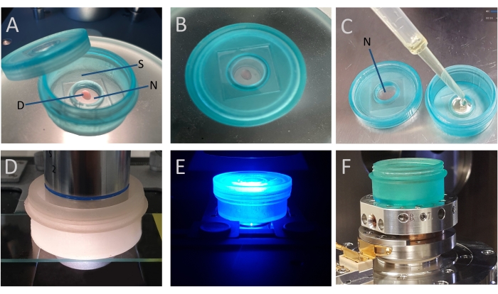
Figure 1: The multipurpose cell culture device (MD). (A) The niche (N), the central part of the device, is designed to create a protective environment for the organoids/spheroids grown in a hydrogel drop (D) during sequential processes. The side (S), the surrounding part of the niche, can be used as a humidifier chamber during immunostaining experiments. (B) The transparent center in the lid allows the user to observe the organoids/spheroids when the device is closed. (C) The hydrogel dome containing organoids in the MD can be stained or embedded in a resin block for transmission electron microscopy. The lid also includes the niche (N) for hanging drop methodology. The size and the design of the device allow the user to examine the organoids/spheroids under (D) brightfield, (E) confocal, and (F) scanning electron microscopes. Please click here to view a larger version of this figure.
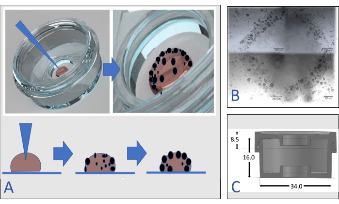
Figure 2: Seeding cells inside a hydrogel. (A) The cell pellet in the pipette is inserted into the top of the dome. After 3-5 days, spheroids become visible in the dome, especially at the periphery of the drop. (B) Four live images of growing spheroids from each quarter in a dome were captured and merged. Scale bar = 200 µm. (C)The design and dimensions (in centimeters) of the multipurpose device (MD). Please click here to view a larger version of this figure.
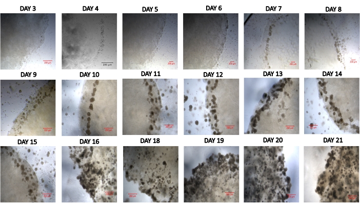
Figure 3: Developing spheroids in a hydrogel drop from day 3 to day 21. Scale bar = 200 µm. Please click here to view a larger version of this figure.
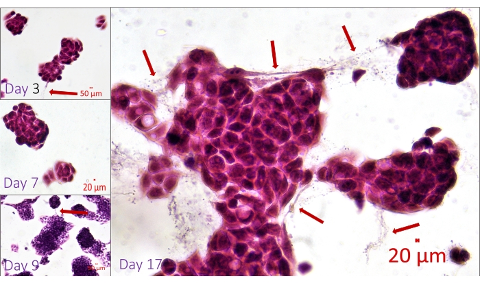
Figure 4: Hematoxylin and Eosin-stained whole-mount spheroids within the MD. Visualization of the connections between the cells located in the adjacent or fusing spheroids. The delicate processes between the cells (arrows) are visible since the whole-mount sample in the hydrogel is fixed, stained, and examined without damaging the 3D growing structures. Scale bar: day 3, day 9 = 50 µm; day 7, day 17 = 20 µm. Please click here to view a larger version of this figure.
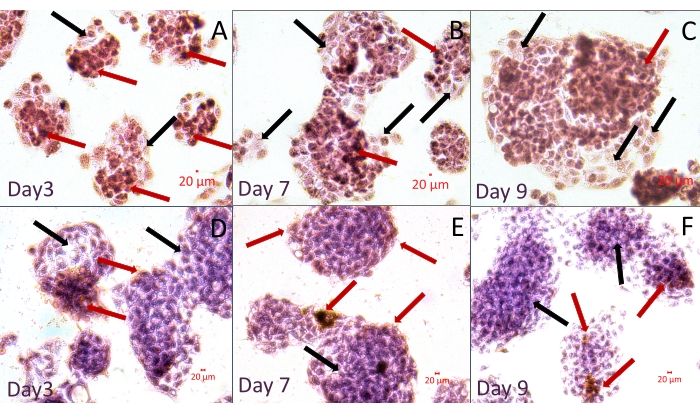
Figure 5: Immunohistochemical staining of whole-mount spheroids in the hydrogel within the MD. The samples were immunostained with an antibody specific to Arginase. Arginase positive (red arrows) and negative (black arrows) cells are seen in the spheroids. Images are from two experiments: (A–C) without and (D–F) with counterstaining with Hematoxylin. Scale bar = 20 µm. Please click here to view a larger version of this figure.
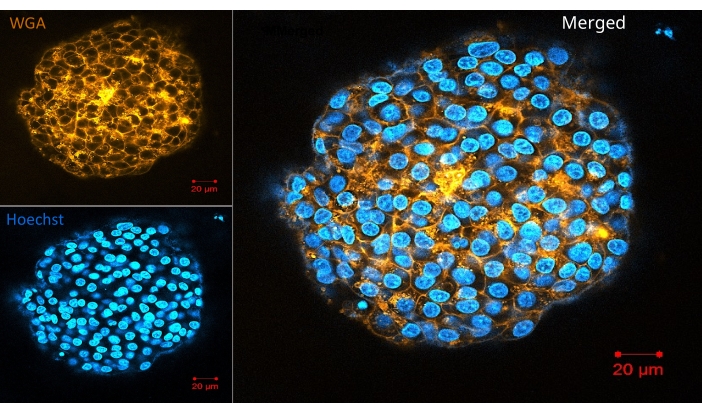
Figure 6: Confocal image of a live organoid in the hydrogel. The living organoids were labeled with the live cell membrane (WGA) and nucleus stains (Hoechst). Scale bar = 20 µm. Please click here to view a larger version of this figure.
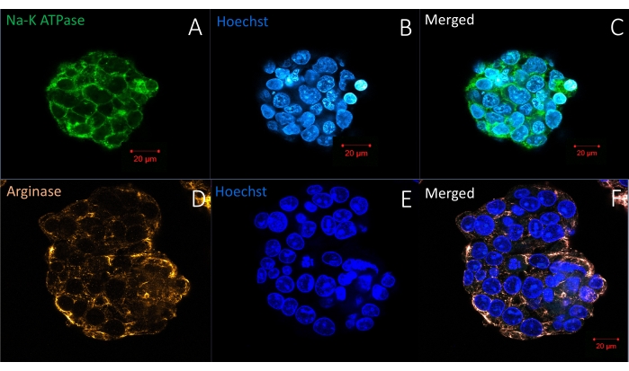
Figure 7: Immunofluorescence labeling of whole-mount spheroids in a hydrogel with one antibody and a nuclear stain (Hoechst). (A–C) The upper panel shows a spheroid labeled with an antibody specific for Na-K ATPase in the cell membrane. (D–F) The bottom panel demonstrates the location of another antibody specific for Arginase, a cytosolic protein specific for hepatocellular carcinoma, in liver cancer spheroids. Duration of staining protocol: 6 h. Scale bar = 20 µm. Please click here to view a larger version of this figure.
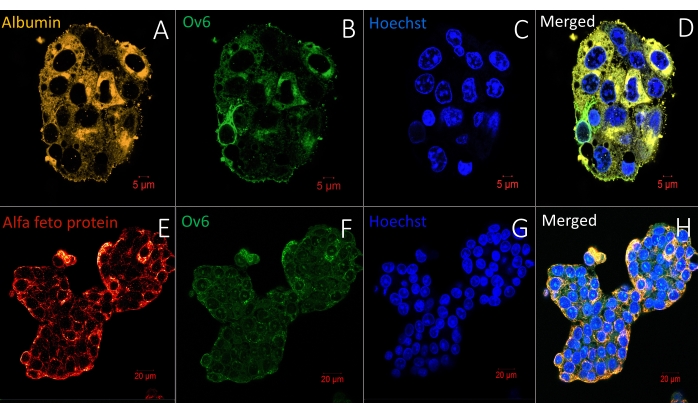
Figure 8: Immunofluorescence labeling of whole-mount spheroids in a hydrogel with two antibodies and a nuclear stain (Hoechst). (A–C) The upper panel shows a spheroid labeled with two antibodies specific for albumin and Ov6. Scale bar = 5 µm. (D–F) The bottom panel demonstrates the location of alpha feto protein and Ov6 in liver cancer spheroids. Scale bar = 20 µm. Duration of staining protocol: 6 h. Please click here to view a larger version of this figure.
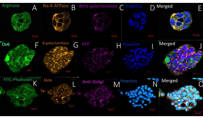
Figure 9: Immunofluorescence labeling of whole-mount spheroids in a hydrogel with three antibodies and a nuclear stain (Hoechst). (A–E) The spheroid in the upper panel is labeled with antibodies specific for Arginase, Na-K ATPase, and beta-galactosidase. Scale bar = 10 µm. (F–J) The middle panel demonstrates a spheroid labeled with Ov6, beta-galactosidase, and alpha-feto protein. Scale bar = 20 µm. (K–O) The spheroid in the lower panel has been labeled with FITC-phalloidin, anti-mitochondrial antibody, and anti-Golgi antibody. Scale bar = 20 µm. Note the high labeling specificity and reduced background. Duration of staining protocol: 6 h. Please click here to view a larger version of this figure.
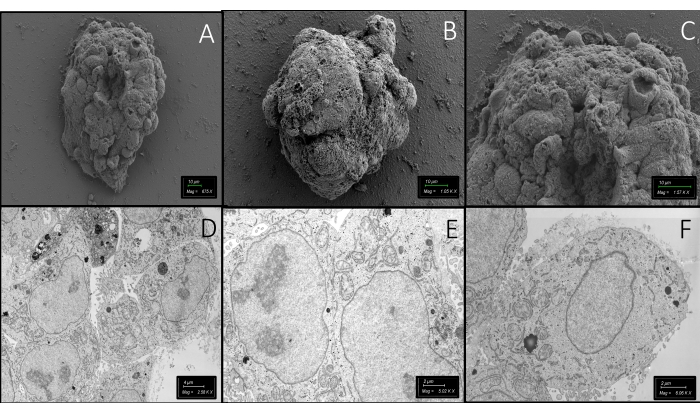
Figure 10: Electron microscopy images. (A–C) Scanning electron microscopic images of entire spheroids in the hydrogel in the MD. Scale bar = 10 µm. (D–F) Ultrastructural features of the cells in a spheroid have also been visualized under a transmission electron microscope. Scale bars: (D) = 4 µm; (E,F) = 2 µm. Please click here to view a larger version of this figure.
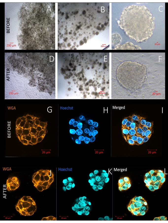
Figure 11: Live images of spheroids before and after freezing. (A–F) The irregularity of the borders of the hydrogel domes is evident after freezing. However, the size and roundness of spheroids remain similar. (F) Cell migration on the culture surface can be seen after thawing. (G–L) The live cell membrane (WGA) and nucleus staining (Hoechst) demonstrate similar cell viability and intact cell membranes before and after freezing the entire spheroids in a hydrogel in the MD. Scale basr: (A,B,D,E) = 200 µm; (C,F,G–L) = 20 µm. Please click here to view a larger version of this figure.
Tradional workflow | Workflow with multipurpose device (MD) | |
Steps | 22 | 6 |
Time | 9 days | 4 days |
Waste Products | 26 | 9 |
Table 1: Comparison of step number, time, and waste production between the traditional and new workflows during a short-term experiment.
Traditional workflow | Workflow with Solution for immunofluorescence (S-IF) | |
Steps | 25 | 7 |
Time | 120 h | 6-8 h |
Waste Products | 28 | 10 |
Table 2: Comparison of the conventional immunolabeling and the new immunolabeling protocol.
Video 1: Time series of images at different levels of a hydrogel containing spheroids under an inverted phase contrast microscope. Please click here to download this Video.
Video 2: 3D structure of two airway organoids labeled with antibodies for Cytokeratin 5 and DAPI. Please click here to download this Video.
Supplementary Figure 1: Overview of the experiment. This schematic shows the sequential experimental steps for growing and examining whole-mount organoids in a hydrogel. The technology described here makes it possible to grow, freeze, thaw, label, and examine the organoids under different microscopes. Please click here to download this File.
Supplementary Figure 2: Comparison of the cell viability, toxicity, and proliferation rates of HepG2 cells in a traditional glass-bottomed dish or an MD. HepG2 cells were grown on(A) a traditional glass-bottomed cell culture dish or (B) in an MD to compare cell viability and toxicity. (C) Trypan blue exclusion test was used to demonstrate the viability. Scale bar = 20 µm. The results were compared using (D–E) CCK8 cytotoxicity and (F) cell proliferation assays. Please click here to download this File.
Supplementary Figure 3: Arrangement of the two foam boxes. One foam box is placed inside the other during the slow freezing process of the entire organoids or spheroids in the MD. *denotes the two boxes. Please click here to download this File.
Supplementary Figure 4: Steps demonstrating how to remove the resin block from the MD after polymerization of the resin. (A) The resin block surrounding the spheroids or organoids is prepared inside the MD niche and then polymerized. (B–D) Then, with an appropriate thin rod, the resin block is pushed to remove it from the niche. (E) Next, the resin block, with the plastic below, is removed. (F–G) Finally, a pair of tweezers is used to separate them if the block is still attached to the plastic. (H) The block is ready for thin sectioning. The whole-mount organoids or spheroids in the resin block (*) are ready for sectioning for transmission electron microscopy. Please click here to download this File.
Supplementary Figure 5: Airway organoids that were immunofluorescent labeled and then visualized while they were still in MDs. (A–C) The upper panel shows circular airway organoids with a central lumen. Green represents airway progenitor cells expressing Cytokeratin 5 in the outermost ring of the organoid, and blue represents the nuclei. Scale bar = 50 µm. (D–F) The bottom panel shows the three-dimensional image of the two side-by-side organoids in the (D) DAPI and (E) Alexa 488 filters, as well as the (F) merged image. Scale bar = 100 µm. Please click here to download this File.
Supplementary Figure 6: Comparison of the labeling density on the live cell membranes of the cells. The graph compares the spheroids before and after freezing using the traditional and presently adopted workflow. Analysis was performed using the Image J image analysis software. Abbreviations: IntDen = Integrated Density (fluorescence intensity in the selected spheroids). CTCF = Corrected total cell fluorescence. Please click here to download this File.
The MD, complementing formulations and protocols described here, facilitates rapid and spontaneous 3D growth of organoids and spheroids in a more controlled environment and continues the experiment in the same conditions. The specimen stays in the same environment during the entire process, and nearly 100% of the 3D growing architectures remain intact in the container. This improves the homogeneity during the sequential experiments and allows for an extended culture period. In addition, the number of steps during organoids and spheroid processing is drastically decreased compared to traditional workflows (Figure 4), which in turn minimizes handling time and human errors and reduces contamination risk. Furthermore, this improves reliability and validity since experimental conditions remain stable while protecting the 3D growing structures in a single environment. It helps to save time and labor in the lab while improving accuracy, reliability, and validity. In addition, it allows tracking of individual 3D growing structures during the entire sequential process. Examination of the size and position changes during the culture and their response to different compounds is possible in the device. This provides an excellent in vitro experimental model for recapitulation of the development of the human body and diseases. Researchers can investigate the stages during the fusion of spheroids and organoids, an essential mechanism for migration in diseases and normal development44,45,46,47. Recent studies aiming at the modeling of fusion in the lab prefer using organoid and spheroid models44,45,46,47.
This device also allows researchers to stop the investigation in an urgent situation by freezing them in their current state and then continuing without any loss when the conditions allow new experiments. For example, because of the COVID pandemic, researchers needed to stop experiments immediately, losing their precious materials and data. There is no current solution to secure an experiment if there is a sudden interruption of the studies due to a change of circumstances.
Limitations of the technique
The techniques described here have been developed to examine the spontaneous development of organoids and spheroids, a model to understand the evolution of the normal human body and diseases. Therefore, the current device and methodologies cannot be used for developing uniformly sized spheroids or organoids that have been preferred for drug screening tests. Another limitation is related to the size of the spheroids and organoids. Full organoids and spheroids can be very dense, with several cell layers requiring reagents that preserve or label the samples to penetrate their core. However, incubation periods for all protocols, including staining, labeling, and freezing, vary according to the sample's size and the microenvironment gel surrounding the sample. For example, larger organoids and stiffer gels require more extended incubation periods to allow for penetration into the center of the sample.
Future applications
The device and these methodologies allow long-term examination of 3D growing organoids and spheroids to understand organogenesis, tissue morphogenesis, and diseases while improving reproducibility, reliability, and accuracy. The organoids or spheroids labeled in MDs can be examined using high-tech microscopy technologies, including multiphoton laser scanning and light-sheet fluorescence microscopy. Additionally, researchers can examine the same sample in one single device under both a confocal and scanning electron microscope to make a correlative study, CLEM (Correlative light electron microscopy). The device and methodologies can also be used for biobanking organoids and spheroids while protecting 3D architectures.
The authors have nothing to disclose.
We are grateful to Dale Mertes from the University of Chicago for the preparation of diagrams, to Dr. Mehmet Serif Aydin for his technical support at Istanbul Medipol University Research Institute for Health Sciences and Technologies, and to Dr. Rana Kazemi from Maltepe University for editing the manuscript.
Absolute Ethanol (EtOH) | Merck | 8187602500 | Dilute in dH2O to make 30%, 50%, 70%, 80%, 90% and 96% solution and store at RT |
Acetone | Merck | 8222512500 | Store at RT |
Alexa fluor wheat germ agglutinin and Hoechst in Hank's balanced salt solution (HBSS) | Invitrogen | I34406 | Image-IT LIVE Plasma Membrane and Nuclear Labeling Kit, Store at -20 °C |
Alpha-1-Fetoprotein (AFP) Concentrated and Prediluted Polyclonal Antibody | Biocare Medical | CP 028 A | Store at +4 °C |
Anti-albumin antibody | Abcam | EPR20195 | Store at +4 °C, Dilution: 1:50 |
Anti-beta galactosidase antibody, Chicken polyclonal | Abcam | 134435 | Store at +4 °C, Dilution: 1:25 |
Anti-cytokeratin 5 | Abcam | 53121 | Store at +4 °C, Dilution: 1:100 |
Arginase-1 Concentrated and Prediluted Rabbit Monoclonal Antibody | Biocare Medical | ACI 3058 A, B | Store at +4 °C, Dilution: 1:50 |
Calcium chloride (CaCl2) | Sigma | C1016-500G | Dissolve in Karnovsky's fixative to make 2 mM CaCl2; store at RT |
Cell Counting Kit 8 (WST-8 / CCK8) | Abcam | ab228554 | |
Centrifuge tubes, 15 mL | Nest | 601051 | |
Centrifuge tubes, 50 mL | Nest | 602052 | |
Class II Microbiological Safety Cabinet Bio II Advance Plus | Telstar | EN12469 | |
CO2 Incubator | Panasonic | KM-CC17RU2 | |
Copper Grids | Electron Microscopy Sciences | G100-Cu | Ultra-thin sections put on the grids; 100 lines/inch square mesh |
Critical Point Dryer | Leica | EM CPD300 | For drying biological samples for SEM applications in absolute acetone |
DAB/AEC chromogen solution mixture | Sigma Aldrich | AEC101 | Store at +4 °C |
Diamond knife | Diatome | Ultra 45°, 40-US | Use for ultra-thin sections for TEM |
Dimethyl sulfoxide for molecular biology | Biofroxx | 67-68-5 | |
Disposable Plastic Pasteur Pippettes | Nest | ||
DMEM – Dulbecco's Modified Eagle Medium | Gibco | 41966-029 | Store at +4 °C |
Eosin Y Solution Alcoholic | Bright Slide | 2.BS01-105-1000 | |
Epon resin | Sigma | 45359-1EA-F | Epoxy Embedding Medium kit, Store at +4 °C |
Fetal Bovine Serum with Additive Fortifier | Pan Biotech | P30-3304 | Store at +4 °C |
Freezing Solution (FS) | Cellorama | CellO-F | Store at +4 °C |
Glass knife maker | Leica | EM KMR3 | For make glass knives in 8 mm thickness |
Glass knife strips (Size 8 mm x 25.4 mm x 400 mm) | Leica | 7890-08 | Use for ultra- or semi-thin sections for TEM |
Glutaraldehyde Aqueous Solution, EM grade, 25% | Electron Microscopy Sciences | 16210 | Dilute in dH2O to make 2.5% solution and store at +4 °C |
Glycerol solution | Sigma Aldrich | 56-81-5 | Store at -20 C, Dilution :1:100 |
Goat anti-chicken IgY (H+L) Secondary Antibody,Alexa, 647 | Invitrogen | A32933 | Store at RT |
Goat anti-Mouse IgG (H+L) Secondary Antibody, DyLight, 488 | Invitrogen | 35502 | Store at +4 °C, Dilution :1:50 |
Goat anti-Mouse IgG (H+L) Secondary Antibody, DyLight, 550 | Invitrogen | 84540 | Store at +4 °C, Dilution :1:50 |
Goat anti-Rabbit IgG (H+L) Secondary Antibody, DyLight, 488 | Invitrogen | 35552 | Store at +4 °C, Dilution :1:50 |
Goat anti-Rabbit IgG (H+L) Secondary Antibody, DyLight, 550 | Invitrogen | 84541 | Store at +4 °C |
Hematoxylin Harris | Bright Slide | 2.BS01-104-1000 | |
HepG2 cells | ATCC | HB-8065 | Store in nitrogen tank |
Human/Rat OV-6 Antibody Monoclonal Mouse IgG1 Clone # OV-6 | R&D Systems | MAB2020 | Store at -20 °C |
Hydrogel | Corning | 354248 | Matrigel, Basement Membrane Matrix High Concentration (HC), LDEV-free, 10 mL, Store at -20 °C |
Hydrogel | Corning | 354234 | Matrigel, Basement Membrane Matrix, LDEV-free, 10 mL, Store at -20 °C |
Hydrogel | ThermoFischer Scientific | A1413201 | Geltrex, LDEV-Free Reduced Growth Factor Basement Membrane Matrix |
Hydrogel | Biotechne, R&D Systems | BME001-01 | Cultrex Ultramatrix RGF BME, Store at -20 °C |
Karnovsky's fixative | %2 PFA, %2.5 Glutaraldehyde in 0.15 M Cacodylate Buffer, 2 mM CaCl2; prepare fresh; use for TEM & SEM samples | ||
L-Aspartic acid | Sigma | 11189-100G | Store at RT |
Lead aspartate solution | Dissolve 40 mg aspartic acid in 10 mL ddH2O and add 66 mg lead nitrate. Solution stabilize at 60 °C and adjust pH to 5; prepare fresh | ||
Lead nitrate | Electron Microscopy Sciences | 17900 | Store at RT |
Leica Confocal Microscope | Leica | DMi8 | |
LSM 700 Laser Scanning Confocal Microscope | Zeiss | ||
Microplate reader | Biotek Synergy | ||
Multipurpose Device (MD) | Cellorama | CellO-M | |
Nuclear-DNA stain | Invitrogen | H3569 | Hoechst 33258, Pentahydrate (bis-Benzimide) – 10 mg/mL Solution in Water, Store at +4 °C |
Nuclear-DNA stain | ThermoFischer Scientific | 62248 | DAPI solution, Store at +4 °C |
Osmium Tetroxide (OsO4) ,4% | Electron Microscopy Sciences | 19190 | Dilute in dH2O to make 2% solution; store at +4 °C and in airtight container; protect light |
Ov6 antibody | R&D systems | MAB2020 | Store at +4 °C |
Paraformaldehyde (PFA) solution, 4% | Sigma | 1.04005.1000 | Dissolve 4% PFA in dH2O and boil, cool and aliquot; store at -20 °C |
Paraformaldehyde solution 4% in PBS, 1 L | Santa Cruz Biotechnology | sc-281692 | Store at +4 °C |
Phosphate Buffered Saline (PBS), tablets | MP Biomedicals, LLC | 2810305 | |
Post-fixative solution | %2 OsO4, %2.5 Potassium Ferrocyanide in dH2O; prepare fresh | ||
Potassium Ferrocyanide aqueous solution, 5% | Electron Microscopy Sciences | 26603-01 | Store at RT |
Primovert – Inverted Bright Field Microscope – ZEISS | Zeiss | Item no.: 491206-0001-000 | |
Round bottom microcentrifuge tubes, 2 mL | Nest | 620611 | |
Scanning Electron Microscopy with STEM attachment | Zeiss | GeminiSEM 500 | We use Inlens Secondary Electron (SE) detector at 2-3 kV for scanning electron micrographs and aSTEM detector at 30 kV for transmission electron micrographs. |
SensiTek HRP Anti-Polyvalent Lab Pack | ScyTek Laboratories | SHP125 | Store at +4 °C |
Sodium Cacodylate Buffer, 0.4 M, pH 7.2 | Electron Microscopy Sciences | 11655 | Dilute in dH2O to make 0.2 M and store at +4 °C |
Sodium/Potassium ATPase alpha 1 antibody [M7-PB-E9] | GeneTex | GTX22871 | Store at -20 °C |
Solution for Immunofluorescence Labeling (S-IF) | Cellorama | CellO-IF | Store at +4 °C |
Solution for Immunohistochemistry (S-IHC) | Cellorama | CellO-P | Store at +4 °C |
Specimen trimming device | Leica | EM TRIM2 | For prepare epon sample block to ultramicrotome |
Sputter coater | Leica | EM ACE200 | Coat the SEM samples with 6 nm gold/palladium for 90 s |
Thiocarbohydrazide (TCH) | Sigma | 223220-5G | Dilute in dH2O to make 0.5% solution and filter with 0.22 µm membrane filter; store at RT; prepare fresh |
Trypan Blue Solution, 0.4% | Gibco | 15250061 | |
Ultra gel super glue | Pattex | PSG2C | For glue polymerized epon block with sample to holder epon block |
Ultramicrotome | Leica | EM UC7 | For prepare high-quality ultra- or semi-thin sections for transmission electron microscopy (TEM) |
Universal Pipette Tips, 10 µL | Nest | 171215-1101 | |
Universal Pipette Tips, 1000 µL | Isolab | L-002 | |
Universal Pipette Tips, 200 µL | Nest | 110919HA01 | |
Uranyl Acetate | Electron Microscopy Sciences | 22400 | Dilute in dH2O to make 2% solution and filter with 0.22 µm membrane filter; keep tightly closed container store at RT |