To improve the drainage of patients with necrotizing pancreatitis complicated with peripancreatic abscess and reduce the mortality of patients with necrotizing pancreatitis, we adopted a retroperitoneal approach for peripancreatic abscess debridement and drainage by laparoscopy.
In patients with severe necrotizing pancreatitis, pancreatic necrosis and secondary infection of surrounding tissues can quickly spread to the whole retroperitoneal space. Treatment of pancreatic abscess complicating necrotizing pancreatitis is difficult and has a high mortality rate. The well-accepted treatment strategy is early debridement of necrotic tissues, drainage, and postoperative continuous retroperitoneal lavage. However, traditional open surgery has several disadvantages, such as severe trauma, interference with abdominal organs, a high rate of postoperative infection and adhesion, and hardness with repeated debridement. The retroperitoneal laparoscopic approach has the advantages of minimal invasion, a better drainage route, convenient repeated debridement, and avoidance of the spread of retroperitoneal infection to the abdominal cavity. In addition, retroperitoneal drainage leads to fewer drainage tube problems, including miscounting, displacement, or siphon. The debridement and drainage of pancreatic abscess tissue via the retroperitoneal laparoscopic approach plays an increasingly irreplaceable role in improving patient prognosis and saving healthcare resources and costs. The main procedures described here include laying the patient on the right side, raising the lumbar bridge and then arranging the trocar; establishing the pneumoperitoneum and cleaning the pararenal fat tissues; opening the lateral pyramidal fascia and the perirenal fascia outside the peritoneal reflections; opening the anterior renal fascia and entering the anterior pararenal space from the rear; clearing the necrotic tissue and accumulating fluid; and placing drainage tubes and performing postoperative continuous retroperitoneal lavage.
The treatment of severe acute pancreatitis (SAP) complicated by peripancreatic infection and necrosis has long been a difficult problem to solve1,2. Pancreatic infection and necrosis are serious complications of severe acute pancreatitis3. Reducing abdominal and retroperitoneal pressure, removing necrotic tissue as much as possible, and reducing the absorption of toxic substances are the main surgical principles for successfully treating SAP4. Traditional open surgery and continuous postoperative lavage have saved the lives of many SAP patients. However, the transabdominal approach requires perforation of the gastrocolic ligament, entry into the minor omental sac, and subsequent invasion of all segments of the pancreas, which inevitably interferes with various abdominal organs and might introduce retroperitoneal bacteria into the abdominal cavity, increasing the risk of abdominal infection. In addition, the drainage tube is drained from the retroperitoneal cavity to the outside of the abdominal wall. Owing to poor drainage, compression of the intestinal tube may also cause intestinal fistula and abdominal bleeding. In 2013, the Evidence-based Guidelines for the Treatment of Acute Pancreatitis issued by the International Society of Pancreatology and the American Society of Pancreatology noted that minimally invasive debridement of necrotic tissue was superior to open debridement for patients with symptomatic infectious necrosis5. Chen et al.6 used peritoneal laparoscopy to reach the peritoneum, remove necrotic pancreatic tissue, and place the drainage tube, achieving satisfactory results. However, theoretically, problems such as bacterial displacement and peritoneal infection caused by communication between the peritoneum and retroperitoneum are inevitable. Sileikis et al.7 used three-hole laparoscopic retroperitoneal resection of pancreatic necrotic tissue and catheter drainage from 2007 to 2009 and cured 8 SAP patients.
Since 2016, we have adopted retroperitoneal laparoscopy to remove necrotic pancreatic tissue through a retroperitoneal approach. Compared to other minimally invasive surgeries, this method is more direct and safer. It provides direct access to the retroperitoneal space, allowing direct visualization and removal of necrotic tissue in the renal capsule. The field of view is clear, bleeding is easily controlled, and a drainage tube can be placed as needed, ensuring effective drainage. The drainage tube does not pass through the abdominal cavity, causing minimal disturbance to the cavity and reducing the risk of abdominal infection. The necrosis and infection of SAP occur mostly in the tail of the pancreas. There is often necrosis of the peripancreatic fatty tissue, which sometimes extends into the left colonic sulcus. Therefore, we utilized a left-sided approach in all patients, and typically, a single surgery was sufficient to thoroughly remove the necrotic tissue. We believe that retroperitoneal laparoscopic surgery via a left-sided approach is particularly suitable for patients with necrosis of the pancreatic body and tail combined with massive effusion.
The protocol was conducted in accordance with the Declaration of Helsinki and was approved by the Ethics Committee of Meizhou People's Hospital. (MS-2023-C-95). Informed consent was obtained from patients for this study.
1. Patient selection
2. Preoperative preparation
3. Surgical procedure
4. Postoperative management
5. Follow-up procedures
A total of six patients received this surgical treatment (Table 1). Preoperatively, all patients were assessed based on PaO2/FiO2, systolic blood pressure, and creatinine levels and were assigned modified Marshall score10 and APACHE II scores (Table 2). All patients underwent surgery smoothly, and the duration of surgery ranged from 75 to 100 min, with an average of 84 ± 6.7 min. Preoperative bacterial cultures obtained through PCD tubes revealed Escherichia coli, Enterococcus faecalis, Pseudomonas aeruginosa, Burkholderia cepacia, and Acinetobacter baumannii. Postoperatively, systemic toxicity symptoms rapidly improved, and CT scans performed 1 week later showed a substantial reduction in necrotic tissue and peripancreatic fluid accumulation (Figure 4). Preoperatively, the blood white blood cell count, neutrophil ratio, and PCT were elevated in all six patients, and their values significantly decreased 1 week after surgery. Four patients had elevated blood amylase levels preoperatively, and 1 week after surgery, all patients had normal levels (Table 3).
A single patient experienced recurrent high fever without resolution at 2 weeks post-surgery, with unobstructed drainage. Repeat CT indicated increased peripancreatic necrosis and fluid compared to observations 1 week earlier. A repeat laparoscopic procedure was performed on the right side with the patient in the left lateral position using the same technique as the initial left-sided retroperitoneoscopic surgery. Symptoms were rapidly ameliorated after the second surgery. Complications occurred in two patients. One patient developed a fever shortly after surgery and exhibited drainage fluid containing yellowish fluid with fecal-like debris after 2 weeks. Considering the possibility of a colonic fistula, conservative treatment was initiated for 1 week. Since the drainage fluid still contained fecal-like material (approximately 10-50 mL daily), the patient underwent subsequent surgery for terminal ileostomy, after which the drainage fluid gradually cleared. Another patient exhibited bloody drainage through the drainage tube 1 week post-surgery, accompanied by progressively decreasing hemoglobin levels in blood tests, indicating intraperitoneal bleeding. The patient received transfusions of red blood cells, fresh frozen plasma, and cryoprecipitate, followed by enhanced hemostatic treatment, which successfully stopped further bleeding. The other four patients experienced no postoperative complications.
During the 1-year follow-up period, all patients achieved satisfactory clinical outcomes, with almost complete disappearance of the peripancreatic fluid and necrotic tissue.
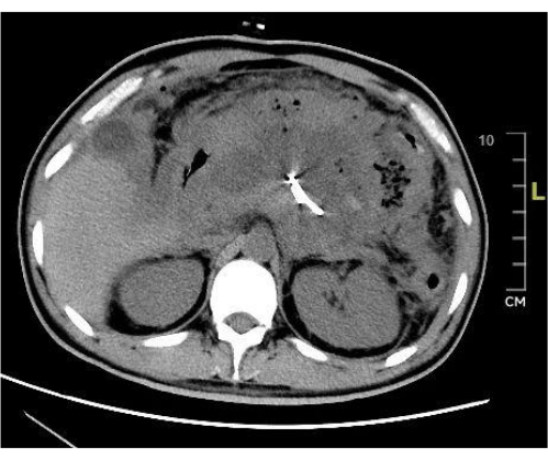
Figure 1: Preoperative CT scan. The preoperative CT scan shows abundant peripancreatic fluid accumulation and necrotic tissue. Please click here to view a larger version of this figure.
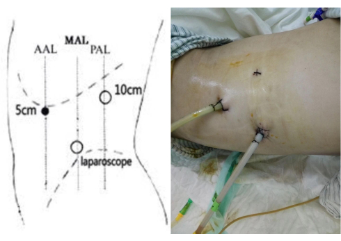
Figure 2: Trocar set up. Three-port arrangement of trocars in the retroperitoneoscopic adrenal surgery procedure and the postoperative placement of drainage tubes. Please click here to view a larger version of this figure.
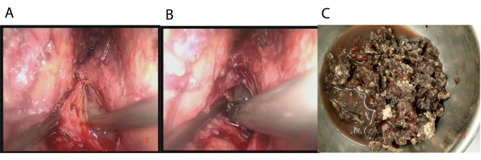
Figure 3: Surgical procedure. (A) Pus was discovered immediately after the aspirator entered the preperitoneal space adjacent to the kidney. (B) Careful extraction of black necrotic tissue using tissue forceps. (C) Excised pancreatic necrotic tissue during surgery. Please click here to view a larger version of this figure.
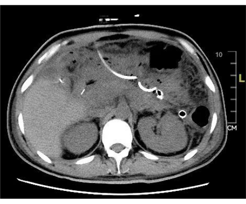
Figure 4: Postoperative CT scan. The postoperative CT scan shows a significant reduction in peripancreatic fluid accumulation and necrotic tissue after surgery. Please click here to view a larger version of this figure.
Table 1: Clinical data showing identified bacterial strains in patients receiving surgical treatment. Please click here to download this Table.
Table 2: APACHE II scores for patients. Please click here to download this Table.
Table 3: Clinical parameters of patients receiving surgical treatment before and after treatment. Please click here to download this Table.
The optimal timing of surgical intervention for SAP has been a subject of ongoing debate. In the past, surgical intervention was assumed to be performed immediately upon the occurrence of pancreatic infection-related necrosis. However, since 2000, an increasing number of experts have suggested that the timing of surgical intervention for SAP should be postponed as much as possible11,12,13. Aseptic peripancreatic necrosis may not require immediate treatment. When infected necrosis develops, PCD can be considered initially to alleviate systemic toxic symptoms. Surgical removal of necrotic tissue can be delayed until approximately 4 weeks later14. At this point, patients have generally passed the acute inflammatory and multiorgan failure stages, and their systemic condition has stabilized. The necrotic tissue in the peripancreatic region becomes more localized and encapsulated, which helps reduce the risk of colon injury and intra-abdominal bleeding during surgery. The median surgical duration in this group was 38.5 (range: 11-63) days. In one patient who underwent early surgery, during the removal of necrotic tissue with forceps, there was a risk of tearing small, nonorganized blood vessels enveloped within the necrotic tissue, leading to bleeding and making thorough clearance of necrotic tissue challenging.
Reports suggest that endoscopic treatment of infected necrotizing pancreatitis is associated with decreased surgical complications15. However, this approach has strict surgical indications, and the clearance of necrotic tissue in the retroperitoneum remains challenging. The percutaneous nephroscopic pancreatic abscess clearance procedure is simpler but limited by restricted visualization, making effective removal of necrotic tissue challenging and often requiring multiple repetitive operations. This method is more direct, and postoperative septic symptoms were rapidly relieved in all six patients. The drainage tubes were left in place for 35-66 days and remained unobstructed. The extended use of drainage tubes is connected with the natural course of SAP, which is known for its prolonged and progressively deteriorating nature. In SAP, the pancreatic necrosis process is gradual and can continue for a long duration. Complete and sufficient drainage is the key to treatment. Most cases of SAP with pancreatic infection and necrosis occur in the tail of the pancreas and often involve necrosis of peripancreatic fat tissue that sometimes extends into the left colonic gutter. In this cohort, all patients were treated using a left-sided approach, and typically, necrotic tissue could be removed during a single operation. Since only one patient underwent bilateral surgery, the trauma associated with repeated debridement procedures during treatment was reduced overall, providing greater alignment with the concept of minimally invasive surgery16. The three-port approach is characterized by its high safety, direct access to the preperitoneal space adjacent to the kidney, direct visual guidance for necrotic tissue removal, excellent visualization, manageable bleeding, and flexible placement of drainage tubes for more effective drainage. The drainage tubes are placed around the abdominal cavity, minimizing disruption and decreasing the chances of retroperitoneal fluid accumulation and abdominal cavity contamination, ultimately reducing the risk of intra-abdominal infection.
For beginners, locating and identifying the retroperitoneal space is a crucial step for surgical success. In patients with pancreatitis, the retroperitoneal tissues are often swollen, necessitating thorough removal of retroperitoneal fat tissue. In this scenario, the preoperative placement of a PCD tube is particularly important. We injected approximately 1000 mL of normal saline through the PCD tube to expand the preperitoneal space further, aiding in its identification during surgery. The anterior renal space is an avascular zone, and post pancreatitis congestion and edema cause the anatomical structures to be unclear. In most cases, blunt dissection of this space using a suction device is recommended. Sharp dissection with energy platforms such as ultrasonic scalpels should be avoided to prevent unnecessary collateral damage. The renal hilum vascular structures often serve as anatomical landmarks during surgery. By analyzing CT scans before the procedure, the primary distribution of fluid and necrotic tissue can be located, and this anatomical landmark can be used as a guide to enter the preperitoneal space adjacent to the kidney.
Compared to traditional open or laparoscopic procedures, this method involves a smaller operational space, making intraoperative bleeding more challenging to manage. When using tissue forceps to grasp necrotic tissue, it is preferable to use a gentle and repetitive approach to avoid forceful tearing of large segments of necrotic tissue. In case of oozing/bleeding, rinsing with hydrogen peroxide and applying pressure can be effective for hemostasis. When there is fresh tissue on the wound and no loose necrotic tissue, surgery should be promptly performed. To avoid unnecessary bleeding, tissue forceps should not be used excessively to clamp tightly adhered necrotic tissue at this point. Inherently, SAP often leads to ongoing pancreatic necrosis and accumulation of peripancreatic fluid after surgery. Therefore, ensuring unobstructed drainage and thorough postoperative irrigation is particularly important. The use of larger diameter drainage tubes with irrigation capability and multiple side holes, coupled with irrigation and drainage, is essential.
Several limitations should be noted. First, the surgical removal and drainage of necrotic tissue and fluid in the pancreatic head region and the ventral side of the pancreas are challenging, which limits the applicability of this procedure to a relatively smaller subset of patients. Second, this procedure merely represents our initial exploration of retroperitoneoscopic surgery, with a small sample size and a relatively short follow-up period. Further research with multicenter collaborations and a larger sample size is necessary.
In conclusion, laparoscopic removal and drainage of pancreatic necrotic tissue for SAP with concurrent peripancreatic infection is safe and effective. The left-sided approach is commonly employed, and adherence to standardized procedures during surgery can reduce the occurrence of complications. Nevertheless, further research involving multicenter collaboration and a larger sample size is needed.
The authors have nothing to disclose.
This research was not supported by any grants.
Dissecting forceps | KANGJI | ZP485RB 330mm×Φ5 | |
Drainage tube | Kang-Li | QH-F28 | |
Grasping forceps | KANGJI | ZP531RB 330mm×Φ5 | |
Laparoscope | STORZ | 26003BA | |
Normal Saline 3000ml | Ke-Lun | 3000ml:27g | |
Suction and Irrigation | KANGJI | Φ5/Φ10×330mm | |
Suture | ETHICON | SA86G | |
Trocar | KANGJI | ZP015RN Φ5.5/ ZP018RN Φ12.5 | |
Ultrasonic knife | An-He | AH-600 | |
Veress needle | KANGJI | ZP001RN Φ2.5×100mm |
.