The method presented here can evaluate the effect of reagents on angiogenesis or vascular permeability in vivo without staining. The method uses dextran-FITC injection via the tail vein to visualize neo-vessels or vascular leakage.
Several models have been developed to investigate angiogenesis in vivo. However, most of these models are complex and expensive, require specialized equipment, or are hard to perform for subsequent quantitative analysis. Here we present a modified matrix gel plug assay to evaluate angiogenesis in vivo. In this protocol, vascular cells were mixed with matrix gel in the presence or absence of pro-angiogenic or anti-angiogenic reagents, and then subcutaneously injected into the back of recipient mice. After 7 days, phosphate buffer saline containing dextran-FITC is injected via the tail vein and circulated in vessels for 30 min. Matrix gel plugs are collected and embedded with tissue embedding gel, then 12 µm sections are cut for fluorescence detection without staining. In this assay, dextran-FITC with high molecular weight (~150,000 Da) can be used to indicate functional vessels for detecting their length, while dextran-FITC with low molecular weight (~4,400 Da) can be used to indicate the permeability of neo-vessels. In conclusion, this protocol can provide a reliable and convenient method for the quantitative study of angiogenesis in vivo.
Angiogenesis, the process of formation of neo-vessels from pre-existing vessels, plays a critical role in many physiological and pathological processes, such as embryonic development, wound healing, atherosclerosis, tumor development, etc.1,2,3,4,5. This dynamic process involves several steps, including the degradation of the matrix, vascular cell proliferation, migration and self-organization to form tubular structures and the stabilization of the neo-vessels6. Promoting angiogenesis has been demonstrated to be critical in the treatment of myocardial infarction, stroke and other kinds of ischemic diseases7 while inhibiting angiogenesis has been considered a promising strategy in the treatment of cancers8 and rheumatoid diseases9. Angiogenesis has been considered an organizing principle for drug discovery10. Thus, the construction of a reliable and convenient method to assess the extent of angiogenesis is critical for mechanical research or drug discovery in angiogenesis-dependent diseases.
Several in vitro and in vivo models have been developed to evaluate angiogenesis11. Among these, two-dimensional (2-D) models, like matrix gel tube formation assay12, cannot form functional tubular structures. The animal models, such as the hind limb ischemia model13,14, can reproduce the angiogenesis process but are complex and require a laser speckle blood flow imaging system. 3D models of vascular morphogenesis, like matrix gel plug assay, provide a simple platform that can mimic the process of angiogenesis in vivo15, but the detection of angiogenesis requires immunohistochemistry or immunofluorescence staining16,17,18, which are variable and poorly visualized.
Here, we describe a protocol for a modified matrix gel plug assay where vascular cells were mixed with matrix gel and subcutaneously injected into the back of mice to form a plug. In the plug, vascular cells need to degrade the matrix, proliferate, migrate, and self-organize to finally form functional vessels with blood flow in the internal environment. Thereafter, fluorescent-labeled dextran is injected via the tail vein, to flow through the plug, and the label is visualized to indicate neo-vessels. The content of angiogenesis can be quantitatively evaluated by the length of the vessels. This method can form functional vessels that cannot be produced in 2-D angiogenesis models12, and does not need complex stain process as in ordinary matrix gel plug assay11. It also does not require expensive specific instruments like laser speckle blood flow imaging system in hind limb ischemia model13,14,19. This method is versatile, low-cost, quantifiable, and easy to perform, and can be used to determine the pro- or anti-angiogenic capability of drugs or be used in mechanical research involved in angiogenesis.
All procedures involving animal subjects were approved by the Institutional Animal Care and Use Committee (IACUC) of Wenzhou Medical University (XMSQ2021-0057, July 19th, 2021). All reagents and consumables are listed in the Table of Materials.
1. Culture medium preparation
2. Vascular cell preparation
3. Matrix gel preparation
4. Mouse preparation
5. Matrix gel mixture injection
6. Dextran-FITC injection through the tail vein
7. Matrix gel plug collection
8. Embedding matrix gel plug and section preparation
9. Quantification of angiogenesis (Figure 3)
10. Quantification of vascular permeability (Figure 4)
Figure 1 is the flowchart depicting how to prepare the mixture of matrix gel, vascular cells, culture medium and reagent. The mixture was then subcutaneously injected into the back of Nu/Nu mice and heated using a heating pad to accelerate its coagulation to finally form gel plug.
Figure 2A is the flowchart to indicate vessels with fluorescent labeled dextran. Fluorescent labeled dextran was injected via the tail vein and circle for 30 min, so it can enter the functional vessel in the gel plug. Thereafter, the gel plugs were collected, embedded using tissue embedding gel (the orientation of matrix gel when embedded was indicated in Figure 2B). A 12 µm of thick section was sliced from the plugs (5 slices from each side) and fluorescent pictures were taken for quantitative analysis of angiogenesis and/or vascular permeability.
Figure 3 is the influence of anti-angiogenic reagent palmitate and pro-angiogenic reagent fibroblast growth factor 1 (FGF1) on angiogenesis in the gel plug. Figure 3A is the appearance of gel plugs with vehicle, palmitate or FGF1. Blood can enter the functional neo-vessel in the gel plug, which makes the plug red to varying degrees. Figure 3B is the fluorescent image of matrix gel plugs, in which the functional vessels were visualized by dextran-FITC with high molecular weight. Figure 3C is the quantitative result of vessel length of different group. Palmitate can significantly decrease the length of neo-vessels, while FGF1 treatment can obviously increase the length.
Figure 4 compares the vascular permeability in gel plugs with or without vascular endothelial growth factor (VEGF) treatment. Figure 4A is the fluorescent image of matrix gel plugs, in which the leakage area is visualized by dextran-FITC with low molecular weight and indicated by arrows. Figure 4B is the quantitative result of leakage area. The increased leakage area in VEGF treatment group revealed that VEGF can increase vascular permeability.
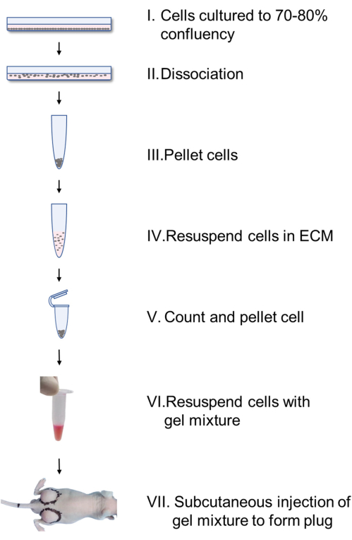
Figure 1. Flowchart showing formation of a gel plug in mice. Vascular cells cultured in monolayer culture are dissociated, pelleted, resuspended with endothelial cell medium (ECM), and counted (I-IV). After pelleting and resuspension in matrix gel mixture (containing 8.8 volume matrix gel, 1 volume 10x M199 supplemented with 10% FBS and 0.2 volume reagent), 300 µL of gel mixture was subcutaneously injected into the back of the mice (V-VII) to form plugs. Please click here to view a larger version of this figure.
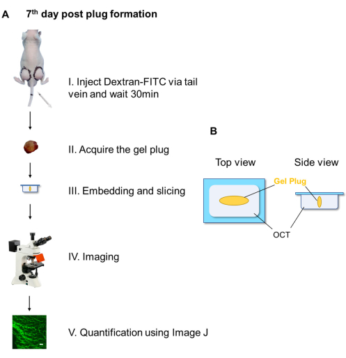
Figure 2. Flowchart showing dextran-FITC injection and quantification of angiogenesis. (A) Dextran-FITC was injected via the tail vein (I). After 30 min, gel plug was acquired, embedded and sections were sliced using freezing microtome (II, III). Thereafter, fluorescence images were taken using fluorescence microscope (IV) and angiogenesis was quantitatively analyzed using Image J software (V). (B) The diagram of matrix gel plug orientation when embedded in tissue embedding cassette. Abbreviations: OCT = optimum cutting temperature compound. Please click here to view a larger version of this figure.
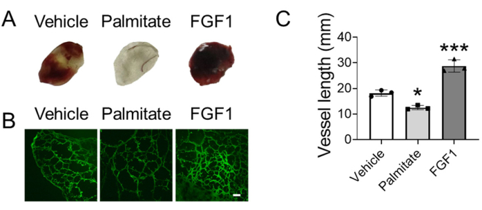
Figure 3. Evaluating angiogenesis using modified matrix gel plug assay. (A) Appearance of representative matrix gel plugs. (B) The fluorescent picture of functional vessels in matrix gel plugs indicated by Dextran-FITC. (C) The quantitative analysis of the length of functional vessels in matrix gel plugs using one-way ANOVA. *p<0.05 versus vehicle; ***p<0.001 versus vehicle. The scale bar is 100 μm. Abbreviations: FGF1 = Fibroblast growth factor 1. Please click here to view a larger version of this figure.
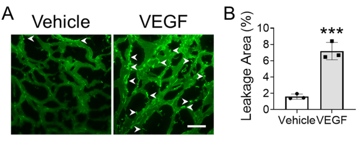
Figure 4. Evaluating vascular permeability using modified matrix gel plug assay. (A) The representative fluorescence image of vessels in gel plug, the arrows indicate leakage site. (B) The quantitative analysis of leakage area to evaluate vascular permeability using Student's t-test. ***p<0.001. The scale bar is 100 μm. Abbreviations: VEGF = vascular endothelial growth factor. Please click here to view a larger version of this figure.
We present a reliable and convenient method for the quantitative evaluation of angiogenesis in vivo without staining. In this protocol, vascular cells were mixed with matrix gel in the presence of pro-angiogenic or anti-angiogenic reagents, and then subcutaneously injected into the back of Nu/Nu mice to form gel plug (Figure 1). After 7 days of gel plug formation, dextran-FITC was intravenously injected and circulated for 30 min. The gel plug was collected and embedded with tissue embedding gel, and 12 µm sections were sliced for photography (Figure 2). Dextran-FITC can indicate functional vessels without staining, and the length of neo-vessels can be used to quantitatively evaluate the pro-angiogenic or anti-angiogenic function of reagents. In the example presented in this protocol, palmitate treatment reduced length of neo-vessels, while FGF1 increased length of neo-vessels (Figure 3), which indicated that palmitate is anti-angiogenic, while FGF1 is pro-angiogenic. Besides that, this protocol can also be used in the evaluation of vascular permeability (Figure 4).
Caution should be exercised on the selection of the recipient mice, handling and injecting the matrix gel, collecting gel plug, and detecting the length of the vessels. Immunodeficient mice aged 6-8 weeks are recommended, although C57BL/6 mice are also workable. Considering the variability in the plug, 3-5 plugs in each group is recommended. The number of vascular cells should be even between different groups. Matrix gel should be carefully handled according to the instructions and should not be used if solidified or containing particulate matter or numerous bubbles. In the matrix gel preparation step, the final concentration of matrix gel should not be less than 80%, and vortex is not allowed as bubbles may be formed. It should be noted that the shape of gel plug may influence the reproducibility of the results, so heating pad was used to accelerate the solidification of matrix gel plug. A 37 °C animal incubator can also be used. Besides that, during gel plug embedding and section preparation, plugs and sections should be protected against bright light, and photos should be taken under a fluorescence microscope as soon as possible.
Among the various in vivo angiogenesis assays, the matrix gel plug assay is one of the most widely used methods because it is versatile, less costly, and easy to perform11. However, the staining process in ordinary matrix gel plug assay is error prone. The plug is fragile and with high water content. The dehydration and fixation steps included in the staining process may distort the gel plug, and further influence the evaluation of the length of the vessels. Besides that, the scattered vascular cells in the plug may be stained and recognized as functional vessels in ordinary matrix gel plug assay. One of the most important advantages of this modified matrix gel plug assay is that the functional vessels can be visualized without staining. FITC-labeled dextran was used to indicate vessels, and only functional vessels that have blood flow can be indicated, which contributes to the convenience and reliability of this assay.
Besides the length of neo-vessels, the permeability also affects the function of neo-vessels20. Fluorescent-labeled dextran with low molecular weight (~4,400 Da) can pass through the gap in vessels and can be used to indicate the permeability of neo-vessels. If needed, researchers can evaluate both angiogenesis and vascular permeability in the same gel plug by using both fluorescent-labeled dextran with low molecular weight and that with high molecular weight21.
Angiogenesis can be affected by the in vivo environment22, such as diabetes13,14,19, etc. This in vivo environment is hard to reproduce in these in vitro models. However, in this modified protocol, the in vivo environment can be easily reproduced by choosing proper recipient mice. Besides that, the interaction between different vascular cells also affects the process of angiogenesis. Different kinds of vascular cells, including endothelial cells, smooth muscle cells and pericytes, can be added to form neo-vessels in the plug in this protocol to investigate the contribution of different vascular cells in angiogenesis.
However, there are also some limitations for this method. The formation of neo-vessels can be affected by the shape of gel plug. Moreover, the neo-vessels formed in the gel plug is not even. There are more functional vessels at the edge of gel plug than that at the center (Figure 2A). Thus, the results may be affected by the proficiency of performer.
The authors have nothing to disclose.
This work was funded by Natural Science Foundation of Zhejiang Province (LY22H020005), and National Natural Science Foundation of China (81873466).
Adhesion Microscope Slides | CITOTEST | 188105 | |
Anesthesia System | RWD | R640-S1 | |
Cell Counter | Invitrogen | AMQAX1000 | |
Cell Culture Dish | Corning | 430167 | |
Cryoslicer | Thermo Fisher | CryoStar NX50 | |
Dextrans-FITC-150kDa | WEIHUA BIO | WH007N07 | |
Dextrans-FITC-4kDa | WEIHUA BIO | WH007N0705 | |
Embedding Cassettes | CITOTEST | 80203-0007 | |
Endothelial Cell Medium | ScienCell | 35809 | |
Endothelial Growth Supplements | ScienCell | 1025 | |
Fetal Bovine Serum | Gibco | 10100147C | |
Fibroblast Growth Factor 1 | AtaGenix | 9043p-082318-A01 | FGF1 |
Fluorescence Microscope | Nikon | ECLIPSE Ni | |
Heating Pad | Boruida | 30-50-30 | |
Insulin Syringe | BD | 300841 | |
Isoflurane | RWD | R510-22-10 | |
Laboratory Balance | Sartorius | BSA124S-CW | |
Matrigel | Corning | 356234 | Matrix gel |
Medium 199 powder | Gibco | 31100-035 | |
Microtubes | Axygen | MCT-150-C | |
Optimal Cutting Temperature (OCT) Compound | SUKURA | 4583 | Tissue embedding gel |
Palmitate Acid | KunChuang | KC001 | |
Penicillin-Streptomycin Liquid | Solarbio | P1400 | |
Phosphate Buffer Saline | Solarbio | P1022 | |
Surgical Instruments | RWD | RWD | |
Tail Vein Injection Instrument | KEW BASIS | KW-XXY | |
Trypsin-EDTA Solution | Solarbio | T1320 | |
Ultra-Low Temperature Freezer | eppendorf | U410 | |
Vascular Endothelial Growth Factor | CHAMOT | CM058-5HP | VEGF |