Summary

Evisceration of Mouse Vitreous and Retina for Proteomic Analyses

Published: April 03, 2011
doi:

Summary

The dissection technique illustrates evisceration of the vitreous, retina, and lens from the mouse eye, separation by centrifugation, and characterization with protein assays.

Abstract

While the mouse retina has emerged as an important genetic model for inherited retinal disease, the mouse vitreous remains to be explored. The vitreous is a highly aqueous extracellular matrix overlying the retina where intraocular as well as extraocular proteins accumulate during disease.1-3 Abnormal interactions between vitreous and retina underlie several diseases such as retinal detachment, proliferative diabetic retinopathy, uveitis, and proliferative vitreoretinopathy.1,4 The relative mouse vitreous volume is significantly smaller than the human vitreous (Figure 1), since the mouse lens occupies nearly 75% of its eye.5 This has made biochemical studies of mouse vitreous challenging. In this video article, we present a technique to dissect and isolate the mouse vitreous from the retina, which will allow use of transgenic mouse models to more clearly define the role of this extracellular matrix in the development of vitreoretinal diseases.

Protocol

1. Anterior segment dissection.

  1. Scleral tissue posterior to the limbus is grasped with a 0.22 forcep and the globe (eye ball) is stabilized.
  2. A microsurgical blade is used to make a linear incision in the cornea from limbus to limbus.
  3. A 0.12 Colibri forcep is then used to grasp the cornea at the incision.
  4. The anterior chamber fluid is then absorbed with a Weck-Cel surgical spear.

2. Lens evisceration.

  1. A 0.12 Colibri forcep grasping the cornea is used to stabilize the globe.
  2. A fine curved needle holder (or curved dressing forceps) is inserted behind the lens toward the posterior aspect of the globe. The needle holder is partially closed, and pulled forward. Pressure is applied using the forceps on the external surface of the sclera, which pushes the lens forward through the corneal incision while leaving the eye wall intact. The vitreous appears as a translucent gel and is partially adherent to the lens.
  3. The lens-vitreous tissue is then placed into a filtered centrifugation tube containing 20 microliters of protease inhibitor cocktail (Roche) dissolved in PBS.

3. Retina evisceration.

  1. Continue to stabilize the globe with 0.12 Colibri forceps grasping the corneal incision.
  2. A fine curved needle holder (or curved dressing forceps) is placed as far posterior to the globe as possible, near the optic nerve. The needle holder is partially closed, and pulled forward. Pressure is applied using the forceps on the external surface of the sclera, which pushes the retina forward through the corneal incision. The vitreous appears as a translucent gel adherent to the retina. The retina is visualized as a yellow, vascularized tissue. Any strips of pigmented tissue can be peeled away with forceps.
  3. The retina-vitreous tissue is then placed into the filtered centrifuge tube containing the lens-vitreous tissue.

4. Filtered centrifugation.

  1. The filtered centrifuge tube is placed in a benchtop centrifuge. Spin at 14,000 x G for 12 minutes.
  2. Aspirate the eluant from the lower chamber, which is the vitreous. The retina remains in the upper chamber. These samples can be utilized for protein studies.

5. Representative Results

Vitreous samples were analyzed by SDS-PAGE (Figure 2). Samples were also used for an enzymatic activity assay (Figure 3).

Figure 1
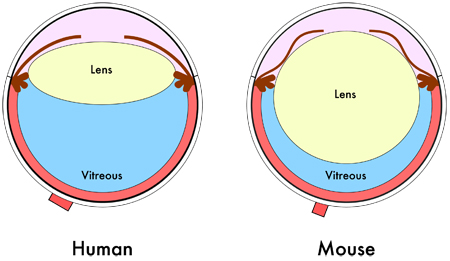
Figure 1. Mouse eye diagram. Cross-sectional schematic of the human and mouse eye shows the relative smaller vitreous volume in the mouse eye due to its larger lens.

Figure 2
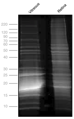
Figure 2. One-dimensional SDS-PAGE of mouse vitreous and retina. Protein profiles of mouse vitreous, 13.6 mg/mL (lane 1), and retina, 11.35 mg/mL (lane 2). Gel electrophoresis was performed at 150 kV for 45 minutes, stained with Flamingo fluorescent gel stain and visualized using a VersaDoc Imaging system (BioRad, Hercules, CA).

Figure 3
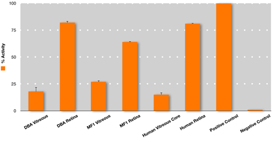
Figure 3. Superoxide dismutase (SOD) enzyme activity in the mouse and human vitreous. Measurement of total SOD activity (SOD activity colorimetric aasay kit, #AB65354, Abcam, Inc, Cambridge, MA) was performed on vitreous and retina collected by evisceration from DBA and albino mice. This was compared to SOD activity in human vitreous core and human retina collected from post-mortem human donor eyes to determine relative activity. Undiluted vitreous core samples were manually aspirated using a 23-gauge needle, 11 and retina was collected by flowering the eye and cutting the tissue away from the RPE/choroid complex. Although the assay does not distinguish between SOD isoforms, the vitreous SOD activity is likely to reflect SOD3, since it is the only extracellular isoform.12 The retina SOD activity is likely to reflect all three SOD isoforms: intracellular SOD1, mitochondrial SOD2, and extracellular SOD3. 12 For highly sensitive enzymatic assays, including this SOD assay, the possibility of cross-contamination of proteins is possible and requires further study. Error bars represent SEM.

Discussion

Transgenic mice are an important model for investigating retinal and vitreoretinal disease.6-10 The mouse vitreous body, however, comprises a significantly smaller proportion of the eye in comparison to the human vitreous due to the large lens in the mouse eye.5 This makes it difficult to isolate and purify the mouse vitreous. Understanding the protein changes associated with the vitreous during diseases such as proliferative diabetic retinopathy, age-related macular degeneration, uveitis, and retinal detachment will give insight into the mechanisms by which they progress. This visualized experimental protocol provides a means to obtain and purify the mouse vitreous body, while maximizing the protein yield for enzymatic and proteomic analyses.

Disclosures

The authors have nothing to disclose.

Acknowledgements

Funding was provided by Fight for Sight and Research to Prevent Blindness.

Materials

Name Company Catalog Number Comments
15°, BD Beaver Microsurgical Blade Becton-Dickinson 374881  
PBS, pH 7.4 Invitrogen 70011-044  
0.22 Fine-Castroviejo Suturing Forceps Storz Ophthalmics E1805  
0.12 Colibri forceps Storz Ophthalmics 2/132  
Microcon Centrifugal Filter Ultracel YM-100 or YM-50 Millipore 42412, 42415  
SOD Activity Colorimetric Assay Kit Abcam Ab65354  
Weck-Cel surgical spears Medtronic 0008680  
Protease inhibitor cocktail Roche 11 836 170 001  
Flamingo fluorescent gel stain Bio-Rad 161-04910  

References

  1. Bishop, P. N. Structural macromolecules and supramolecular organisation of the vitreous gel. Prog. Retin. Eye Res. 19, 323-344 (2000).
  2. Gao, B. B., Chen, X., Timothy, N., Aiello, L. P. &. a. m. p. ;. a. m. p., Feener, E. P. Characterization of the vitreous proteome in diabetes without diabetic retinopathy and diabetes with proliferative diabetic retinopathy. J. Proteome Res. 7, 2516-2525 (2008).
  3. Izuta, H. Extracellular SOD and VEGF are increased in vitreous bodies from proliferative diabetic retinopathy patients. Mol. Vis. 15, 2663-2672 (2009).
  4. Sebag, J. Molecular biology of pharmacologic vitreolysis. Trans. Am. Ophthalmol. Soc. 103, 473-494 (2005).
  5. Smith, R. S., Smith, R. S., John, S. W. M., Nishina, P. M., Sundberg, J. P. . Systematic evaluation of the mouse eye: Anatomy, pathology, and biomethods. , 161-195 (2002).
  6. Ihanamaki, T., Metsaranta, M., Rintala, M., Vuorio, E., Sandberg-Lall, M. Ocular abnormalities in transgenic mice harboring mutations in the type II collagen gene. Eur. J. Ophthalmol. 6, 427-435 (1996).
  7. Kaarniranta, K. A mouse model for Stickler’s syndrome: ocular phenotype of mice carrying a targeted heterozygous inactivation of type II (pro)collagen gene (Col2a1. Exp. Eye Res. 83, 297-303 (2006).
  8. Marneros, A. G. &. a. m. p. ;. a. m. p., Olsen, B. R. Age-dependent iris abnormalities in collagen XVIII/endostatin deficient mice with similarities to human pigment dispersion syndrome. Invest. Ophthalmol. Vis. Sci. 44, 2367-2372 (2003).
  9. Martin, A. C. Pathogenesis of persistent hyperplastic primary vitreous in mice lacking the arf tumor suppressor gene. Invest. Ophthalmol. Vis. Sci. 45, 3387-3396 (2004).
  10. Song, B. J., Tsang, S. H., Lin, C. S. Genetic models of retinal degeneration and targets for gene therapy. Gene. Ther. Mol. Biol. 11B, 229-262 (2007).
  11. Skeie, J. M., Mahajan, V. B. Dissection of Human Vitreous Body Elements for Proteomic Analysis. J Vis Exp. , (2011).
  12. Ciechanowski, K. Impaired synthesis is not the reason for decreased activity of extracellular superoxide dismutase in patients with diabetes. Arch Med Res. 36, 148-153 (2005).
check_url/cn/2795?article_type=t

Play Video

Cite This Article
Skeie, J. M., Tsang, S. H., Mahajan, V. B. Evisceration of Mouse Vitreous and Retina for Proteomic Analyses. J. Vis. Exp. (50), e2795, doi:10.3791/2795 (2011).

View Video