Collagen is a core component of the ECM, and provides essential cues for several cellular processes ranging from migration to differentiation and proliferation. Provided here is a protocol for embedding cells within 3D collagen hydrogels, and a more advanced technique for generating randomized or aligned collagen matrices using PDMS microchannels.
Historically, most cellular processes have been studied in only 2 dimensions. While these studies have been informative about general cell signaling mechanisms, they neglect important cellular cues received from the structural and mechanical properties of the local microenvironment and extracellular matrix (ECM). To understand how cells interact within a physiological ECM, it is important to study them in the context of 3 dimensional assays. Cell migration, cell differentiation, and cell proliferation are only a few processes that have been shown to be impacted by local changes in the mechanical properties of a 3-dimensional ECM. Collagen I, a core fibrillar component of the ECM, is more than a simple structural element of a tissue. Under normal conditions, mechanical cues from the collagen network direct morphogenesis and maintain cellular structures. In diseased microenvironments, such as the tumor microenvironment, the collagen network is often dramatically remodeled, demonstrating altered composition, enhanced deposition and altered fiber organization. In breast cancer, the degree of fiber alignment is important, as an increase in aligned fibers perpendicular to the tumor boundary has been correlated to poorer patient prognosis1. Aligned collagen matrices result in increased dissemination of tumor cells via persistent migration2,3. The following is a simple protocol for embedding cells within a 3-dimensional, fibrillar collagen hydrogel. This protocol is readily adaptable to many platforms, and can reproducibly generate both aligned and random collagen matrices for investigation of cell migration, cell division, and other cellular processes in a tunable, 3-dimensional, physiological microenvironment.
Many cellular processes have been extensively studied in 2 dimensions, thereby forming a collective knowledge of basic cell signaling mechanisms. These studies, however, neglect important cellular cues received from the structural and mechanical properties of the local cellular microenvironment and extracellular matrix (ECM). To better understand how cells interact within a physiological context, it is important to study them in 3-dimensional (3D) assays. The ECM for these 3D assays can either be cell-derived or reconstituted from purified proteins. Regardless of the source of the ECM, 3D matrix assays have proven to be invaluable for understanding how cells navigate and interact within the physiological world. For example, cells grown in 3D matrices display distinct modes of locomotion that depend on the mechanical nature of their surrounding ECM which are not observed in 2D experiments4-6. Moreover, cells cultured in 3D also have fewer and less pronounced stress fibers and focal adhesions than their counterparts grown on hard surfaces such as glass or plastic7.
The importance of contextual 3D assays is not limited to cell migration, however. Some other cell signaling events can only be investigated through the use of 3D assays. During tissue and cell differentiation, the stiffness of the extracellular environment and ECM provides signals that can influence morphogenic events. For example, mammary epithelial tubulogenesis only occurs in low stiffness 3D matrices, but not in stiff matrices nor on 2D substrata8,9. When cultured within stiff 3D matrices, these same epithelial cells take on an aberrant phenotype with increased proliferation and cell membrane protrusions driven through altered FAK and ERK signaling10. Many other signaling pathways and cellular processes are known to be similarly affected by the stiffness of the local cellular environment, and these signaling cascades highlight the importance of investigating signaling events and cellular phenotype in the context of appropriate local mechanical properties of a 3D ECM.
Collagen I is a particularly relevant protein to use for in vitro studies as it is the most abundant component of the ECM and is responsible for many of the mechanical properties of the cellular microenvironment. While it was originally thought of as merely a structural protein, its role is now known to be much more complex. Collagen fiber composition, architecture, orientation, density, and stiffness all provide a concentrated milieu of signaling information5. During the progression of certain diseases, such as chronic inflammation and tumorigenesis, the collagen network is dramatically remodeled2,11. More specifically in breast cancers, increased collagen deposition and tissue stiffness accompany and likely contribute to tumor progression. In these early tumors, the stiffened collagen network appears strained and more aligned, such that most of the fibers encapsulate the growing tumor2. As the tumor progresses, the collagen continues to reorganize, and regions of the fibrillar network become orientated perpendicular to the tumor boundary2,12. Perpendicular alignment serves as a prognostic biomarker where these patients have a poorer disease free progression and overall survival1. One explanation for this correlation is that the poor outcomes are a consequence of increased dissemination of tumor cells via persistent cell migration in aligned collagen networks3.
To understand how cells specifically respond to alignment and organization that is observed in tumor progression, it is necessary to generate both random and aligned 3D collagen matrices for experimentation. There are three basic methodologies to induce alignment within fibrillar networks. The first technique utilizes a strain-inducing device where the collagen between two points is contracted or stretched to generate alignment. Fibers parallel to the axis of force are pulled taut while fibers perpendicular to the axis are compressed and buckled. While strain-induced techniques typically offer superb alignment, this approach requires bulky equipment that is not easily adaptable to many platforms3,13. Alternatively, cell-induced strain can be created by placing localized plugs of cells that subsequently contract and align the collagen13. This method has the problem of being variable, as many parameters may be subject to change. The second method utilizes magnetic beads and a magnetic field during polymerization to induce collagen alignment13,14. Good results can be obtained from this method with unsophisticated equipment, but it does require the use of antibodies or some other method to magnetize the polymer. Therefore, it can be somewhat expensive to use, and the stiffness of the collagen gel is potentially modified by the increased connections in the network. Moreover, the magnetic beads used in this process are often autofluorescent, which is problematic for imaging experiments. Lastly, alignment can be generated by PDMS microfluidic channels3,15,16. In this method, collagen alignment is achieved by flowing polymerizing collagen through small microfluidic channels. These microfluidic channels can be made in a multitude of designs, and are easily adaptable to many platforms. Moreover, they are very economical as very small quantities of collagen and other reagents are used due to their diminutive sizes.
Provided here is a simple protocol for embedding cells within a 3-dimensional, fibrillar collagen hydrogel. In addition, a more advanced technique, wherein PDMS microfluidic channels are used to control the organization and alignment of the collagen matrix is also provided. This protocol is readily adaptable to many platforms, and can reproducibly generate both aligned and random collagen matrices for investigation of cell migration, cell division, and other cellular processes in a 3-dimensional, physiological microenvironment.
1. Neutralization, Dilution and Polymerization of Collagen Solutions for 3D Investigation and Cellular Contraction Assays
2. Western Blotting, Cell Morphogenesis and Gel Gontractility Assay
3. Generation of PDMS Microchannels for Collagen Fiber Alignment
Note: To generate aligned collagen matrices, a mold for PDMS microchannels (Figure 2A) requires a SU-8 silicon master made via soft-lithography15.
4. Prepping PDMS Microchannels for Use
5. Collagen Preparation for Use in Microchannels
6. Pouring Aligned and Random Collagen Microchannels
While 3D assays can be done within the same stiffness of collagen gel, varying the gel stiffness can be used to determine how the cells will respond to mechanical changes in their cellular microenvironment. A stiff collagen hydrogel is defined as a gel where the embedded cells are unable to locally contract the surrounding collagen. The intrinsic contractility of different cell types is unique, and thus it is best to begin with a simple contractility curve to establish the collagen concentration that will be sensed as compliant and stiff.
Begin by holding the cell numbers constant and increasing the concentration of the collagen gel. The stiffness of the gel will scale exponentially with concentration, as shown previously10,20. A representative example of a contractility assay is shown in Figure 1. Here, 50,000 4T1 cells are embedded in either a 2 mg/ml or 3.5 mg/ml collagen gel for 5 days. The 4T1 cell line is a highly contractile, metastatic cancer line, but is only able to contract the high density gel by approximately 10% (Figure 1a, c). This low level of contraction is typical for a stiff gel classification. Comparable low density gels (2 mg/ml, Figure 1b, c) are contracted 57% (n = 4) during the same time frame. It is important to note that increasing the amount of collagen ligand can impact cell signaling pathways, therefore a second means to vary the stiffness is to hold the collagen concentration constant and compare gels that are restrained (stiff) by being left attached to the bottom of the dish or contractile (compliant) because they have been released to float freely in the cell medium. This can serve as a good control to determine whether changes in cellular signaling result from increased collagen ligand or the stiffness of the matrix.
While collagen gel contraction assays are straightforward, there are a few items to be aware of when using these types of assays. For one, under high density conditions it is important to verify that there is not a loss of cell viability. We have observed that there is a ceiling for the stiffness of the microenvironment that a cell will tolerate, and have observed increased apoptosis or cell death in some cell lines, which can cause subsequent results to be misinterpreted. There should be no appreciable loss of cell viability between high and low stiffness conditions. This can be determined by Western blotting for caspase activity or some other assay for measuring cell viability. Secondly, collagen gels can also be contracted too much thereby creating a stiffer environment, and diminishing the differences between high and low stiffness conditions. We recommend not exceeding contractions of 60 – 70%. To maximize reproducibility, it is important to optimize the timeframe of the experiment, the initial cell seeding density, and the stiffness of the matrix to the contractility of the cell type in use.
Once mastery of embedding cells within 3D matrices is achieved, assays can be modified to alter the organization and orientation of the collagen fibers. This is achieved by the use of PDMS microfluidic channels (Figure 2). For the purpose of this manuscript, two similar conformations were used, a narrow and a wide channel, with 3 openings or ports (Figure 2b). In a typical experiment to determine the cellular response to random compared to aligned matrices, unpolymerized collagen is drawn through the microfluidic channel from port A to port C with cells being added to port B. The degree of collagen alignment is modulated by the rate of collagen flow such that higher collagen flow rates yield better alignment. The use of narrower channels coupled with higher vacuum pressures enhances the alignment of the collagen network, while a wider channel used in conjunction with low vacuum pressures generates random matrices. The organization of the fibrillar network can be visualized by second harmonic generation, as shown in representative images (Figure 2c, d). In these images, the collagen fibers (indicated by the arrowheads and pseudocolored in purple) align more consistently with the axis of collagen flow in the narrow channels (Figure 2d). The collagen fibers in wide channels have a variety of orientations, and have a more random distribution (Figure 2c).
The difference between wide and narrow channels is discernable to the human eye, but can also be analyzed via software developed specifically for fiber analysis. CT-FIRE, an open source and freely available software, utilizes a curvelet-based algorithm for pre-processing followed by a fiber extraction algorithm to quantify fiber angle, thickness, length, and straightness21. For each condition, more than 500 images were randomly sampled from different locations within the channels, and more than 125,000 fibers were extracted and analyzed by CT-FIRE. The subsequent fiber angle histogram generated for the narrow channels (Figure 2f) shows a clear enrichment of fibers oriented parallel to the direction of flow. In contrast, wide channels have a more equivalent fiber angle distribution (Figure 2e) which is consistent with a more random matrix.
Importantly, the use of microfluidic devices, as a technique to control the organization and orientation of collagen fibers, allows for assessment of various cellular responses to altered matrix conditions. For example, Sung et al. determined that viability of human mammary fibrobasts is dependent on collagen fiber.15 Additionally, recent work from our laboratory demonstrates that breast carcinoma cells migrate more persistently on aligned collagen fibers compared to random collagen matrices.3 These results help to better define the role of the microenvironment within the context of mammary biology and breast cancer. Moving forward, the ability to precisely control the 3D cellular microenvironment will allow for continued investigation of important cellular responses within the context of many different disease states.
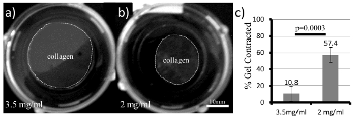
Figure 1. 3D Collagen Gels for Measuring Cell Contractility and Cellular Responses to ECM Stiffness. 4T1 cells were plated in both (a) 3.5 mg/ml and (b) 2 mg/ml collagen gels and allowed to grow for 5 days. Over time, the collagen gel (Gray area within dotted line) will contract and the diameter is measured and quantified in (c, Student's t-test, error bars +/- SD). Please click here to view a larger version of this figure.
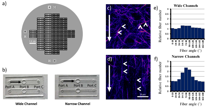
Figure 2. Generating Random and Aligned Collagen Matrices Using PDMS Microchannels. A six inch silicon wafer (a) was used as a template for both wide and narrow PDMS microchannels (b). Identical collagen concentrations were drawn through both types of channels, and individual fibers (arrowheads, c, d) align with the direction of flow (arrows) in narrow microchannels (d). Wide microchannels yielded more random matrices. Fiber angles with respect to the bottom of image were counted and shown in (e) and (f). Scale bar = 26 µm. Please click here to view a larger version of this figure.
3D collagen gels are a valuable addition to our toolbox to understand how cells interpret and respond to their local microenvironment. This manuscript has provided a very basic protocol for embedding cells within a 3D collagen matrix, and to reproducibly generate matrices with random or aligned collagen fibers. Both protocols work as adaptable platforms where different collagen isoforms, crosslinkers, or other matrix proteins could potentially be added at the time of polymerization. It is also easy to modify the platforms to assess different timeframes and endpoints for gene expression and protein production. Imaging, assessing protein and RNA levels, and reading metabolic profiles are all very compatible with these 3D techniques.
As is important with all techniques, it is important to assess the quality of the gel and assay. A sign of a good quality collagen gel is one that can handle manual manipulation. For example, a 1 ml [1 mg/ml] collagen gel poured into a six well plate should be easily picked up with a forceps and manipulated. If the gel tears or fragments, the quality of the gel is suboptimal, and steps need to be taken to improve the collagen polymerization. Temperature and pH are the most important parameters required for good fiber formation and gelation. Begin the troubleshooting process by making sure all components are kept on ice until ready for gelation to occur. It is also very important to make sure that the neutralization buffer and collagen stock solution are being mixed thoroughly. Next, verify that the pH of the neutralization buffer is at pH 7.4. A HEPES buffered PBS is recommended because it is very compatible to tissue culture, has a broad buffering range, and stock solutions tend to be very stable at 4 ˚C for long periods of time. It also has the added benefit of producing longer, more curved fibers which are more similar to the physiological state15. NaOH is another commonly used neutralizing solution, but needs to be titered more closely. It also appears to have the effect of producing shorter, thicker fibers15. Regardless, the pH of the neutralized collagen should be at a pH of 7.4. If all those conditions are being satisfied, and the integrity of the gels is still not satisfactory, then the problem lies with the stock collagen solution. Although the stock collagen solution has a listed shelf life of 6 months at 4 ˚C, we have found that it is generally shorter than commercial sources claim when used for 3D gels. Discard the old bottle, and order from a new lot. It is not unusual for the quality of collagen from the manufacturer to vary significantly from lot to lot.
In addition to identifying quality collagen gels, it is also important to identify the steps that are critical for the generation of good alignment within the PDMS microchannel. The underlying principle of this technique is that the rate of collagen flow through the microchannel subsequently induces filament alignment. Therefore, the most critical step in controlling the degree of fiber alignment is proper modulation of the vacuum pressure. For random collagen alignment in a microchannel, the vacuum pressure must be low, and the collagen must flow slowly down the channel. If the collagen flows through too quickly, the fibers will begin to align. A wider channel makes this proposition easier. Conversely, for good collagen alignment, higher vacuum pressures must be used and the collagen solution must be pulled through more quickly. A second critical step in the generation of aligned matrices is the temperature. It is very important to make sure all components and solutions are chilled on ice prior to use. If the channels are not chilled prior to use, the collagen in the channels will polymerize too quickly, and will result in thinner, smaller fibers. If the neutralized collagen is chilled at 4 ˚C and not on ice, the enhanced nucleation will give rise to larger fiber diameters which are more difficult to align by flow. It is also important to chill the microchannel itself. Without prior chilling, the small volume of collagen will warm very rapidly yielding poor results.
The collagen microchannels are a very useful technique, but there are certain limitations. For one, the alignment in a flow-induced microchannel is never perfect, and will never equal a strain-induced alignment technique. There will always be some fibers oriented perpendicular to the direction of flow, but the reproducibility, adaptability, and ease of use outweigh the small variations in alignment within the channel. More importantly, cells have been shown to effectively sense small changes in alignment, and respond with more persistent migration in these channels3,15. The cells will also enhance the alignment of the channel as their numbers increase and they begin to migrate. If greater alignment is required, the PDMS microchannels can be easily modified to narrower dimensions or to have small constrictions to enhance alignment. Both have been shown to be effective, but have other associated trade-offs16. Another limitation to the microchannel technique is that the PDMS channel has little permeability to drugs or fluorescent dyes. This means that these compounds need to diffuse down the channel from the ports, which is surprisingly slow, and can pose difficulty in washing out treatments.
Although there are certain limitations to this microchannel platform, it does offer several advantages over existing techniques. It is more experimentally accessible as it does not require bulky instruments3,13 or small populations of cells for generating alignment. It also does not require exogenous magnetic beads13,14 which tend to be autofluorescent and could artificially crosslink the collagen network. The collagen microchannel is also simply more cost-effective and economical. In addition to the current advantages, it also has numerous benefits that could be leveraged in future applications. For one, the collagen microchannel can be designed or modified in almost any configuration or experiment. Advanced designs allow the channels to create hollow collagen tubes or 3D structures to mimic the architecture of simplified organs or tumors22. Moreover, the channels can be made of materials other than PDMS. This potentially could convey different mechanical properties to the embedded collagen networks or matrices. Lastly, the small amount of reagents used for this assay might also make it attractive and economical for smaller high throughput screens.
The authors have nothing to disclose.
The authors would like to acknowledge grant numbers UO1CA143069, R01CA142833, R01CA114462, RO1CA179556, T32-AG000213-24, and T32-GM008692-18 for funding this work. We also acknowledge Jeremy Bredfelt and Yuming Liu of LOCI for the development of and assistance with the CT-FIRE analysis.
High Concentration Rat tail Collagen | Corning | 354249 | |
SylGard184 elastomer kit | Corning | NC9285739 | Elastomer for PDMS channels |
HEPES | Fisher | BP310 | For HEPES neutralization buffer |
KCl | Fisher | BP366 | For HEPES neutralization buffer |
KH2PO4 | Fisher | BP362 | For HEPES neutralization buffer |
Na2HPO4 | Fisher | S374 | For HEPES neutralization buffer |
NaCl | Fisher | BP358 | For HEPES neutralization buffer |
Levy Improved Neubauer Hemacytometer | Fisher | 15170-208 | cell counting |
6-well non-tissue culture plate | Corning | 351146 | |
50 mm glass bottom dish | MatTek | P50g-1.5-30-f | |
Bel-Art Plastic Vacuum Desiccator | Bel-Art | F4200-2021 | Degassing chamber for PDMS |
transparency film | 3M | pp2950 | Plastic film for pouring pdms channels |
ThermoScientific CimaRec | ThermoScientific | HP141925 | Hot plate for curing PDMS microchannels |
Vacuum regulator | Precision Medical | PM3100 | Vacuum regulator for collagen microchannels |
8" X 8" rubber sheet | Amazon – Rubber-Cal | Silicone – 60A | rubber sheet for pouring PDMS microchannel |
8" X 8" X .125" acrylic sheet | Amazon | Plexiglass sheets | for pouring PDMS microchannels |
10 lb weights | Amazon | CAP Barbell | for pouring PDMS microchannels |
15 ml Conical tubes | Fisher | 352097 | |
50 ml Conical tubes | Fisher | 352098 | |
Plastic pipets | Dot Scientific | 229202B, 229206B, and 667225B | 2ml, 5ml, and 25ml |
70%EtOH | Fisher | NC9663244 |