Here the protocol describes arthrocentesis of the knee, a procedure in which a needle is inserted into the knee joint, and synovial fluid is aspirated. Synovial fluid may be removed for testing to determine the nature of the knee effusion. Arthrocentesis of the knee is typically performed with the patient supine.
Arthrocentesis of the knee is a procedure in which a needle is inserted into the knee joint, and synovial fluid is aspirated. An arthrocentesis can be diagnostic or therapeutic. Synovial fluid may be removed for testing to determine the nature of the knee effusion. If septic arthritis is suspected, urgent arthrocentesis before initiation of antibiotic treatment is indicated. Moreover, arthrocentesis can also aid in diagnosing crystal-induced arthritis such as gout or pseudogout, or non-inflammatory arthritis such as osteoarthritis. Identifying the cause of the knee effusion can guide treatment. Furthermore, removing fluid from a knee can reduce intraarticular pressure to decrease pain and improve range of motion. There is no absolute contraindication to performing this procedure, but in selecting the needle entry site, an area of skin that is infected should be avoided. Therefore, caution should be exercised when a patient presents with suspected cellulitis over the knee joint to avoid the potential risk of causing iatrogenic septic arthritis. A knee that has undergone arthroplasty should be assessed for arthrocentesis by an orthopedic surgeon. Arthrocentesis of the knee is typically performed with the patient supine. The site for needle insertion is marked, and then the skin is disinfected. After a local anesthetic is administered, a needle is inserted along the pathway that was anesthetized. Synovial fluid is aspirated, and then the needle is withdrawn. Pressure is applied until any bleeding stops. The synovial fluid can be analyzed for infection and inflammation but cannot directly confirm a diagnosis of internal derangement or autoimmune causes of arthritis. In addition to the history and physical examination, laboratory findings and imaging can clarify the etiology of a knee effusion.
Arthrocentesis is performed to successfully aspirate synovial fluid from a joint such as a knee, shoulder, elbow, wrist, or ankle. A patient with a newly detected knee effusion can undergo a diagnostic arthrocentesis to determine the nature of the effusion. Before proceeding to attempt an arthrocentesis, knee swelling by history must be confirmed on physical examination to assess whether an effusion exists. With the patient supine, the knees can be compared on inspection to see if the swelling is unilateral. The knee with the effusion may appear larger than the other knee. With a large effusion (at least 20 mL), convexity can be seen proximal to the patella. With a small effusion (5-10 mL), pressing the fluid superolaterally with one hand can allow the other hand to palpate a fluid bulge. Palpating the fluid can help decide if a successful arthrocentesis is probable. In addition to the supine position, arthrocentesis of the knee can also be done on a patient in the sitting position, but there is a higher chance that less synovial fluid would be aspirated1. Synovial fluid from the knee can be aspirated from a medial or lateral approach, but the latter is preferred in complicated circumstances2. A knee effusion is not always tender on exam and thus does not necessarily cause an antalgic gait. An urgent arthrocentesis before antibiotic treatment is indicated if septic arthritis is suspected. An orthopedic surgeon can perform a knee joint aspiration to diagnose a prosthetic joint infection in a patient who had a knee arthroplasty.
In addition to evaluating for infection, arthrocentesis can assist in identifying diagnoses, such as crystal-induced arthritis (gout or pseudogout), rheumatoid arthritis, spondyloarthritis, reactive arthritis, psoriatic arthritis, hemarthrosis, or osteoarthritis. The findings on synovial fluid analysis can lead to the appropriate treatment. In a patient with pain and restricted movement of the knee due to an effusion, aspirating the fluid can improve these symptoms. Furthermore, arthrocentesis of a knee prior to an intra-articular steroid injection has been shown to reduce the risk for arthritis relapse in rheumatoid arthritis3. There is no absolute contraindication to arthrocentesis of a knee, but the needle should be inserted away from any cellulitis to not introduce any infection into the joint. Moreover, arthrocentesis has been shown to be generally safe in patients on anticoagulation with warfarin or direct oral anticoagulants4,5,6,7. With the proper technique and clinical indication, a patient can undergo this procedure with minimal risks.
This protocol follows the guidelines at BronxCare Health System. A written informed consent is necessary from the patient.
1. Identifying anatomical structures
2. Skin sterilization
3. Anesthetizing
4. Arthrocentesis needle insertion
5. Needle removal
A prospective randomized study compared complete aspiration of synovial fluid from the knee and intra-articular injection with corticosteroid alone. It demonstrated that aspirating as much synovial fluid as possible can reduce the risk for recurrence of arthritic symptoms when treating rheumatoid arthritis patients with intra-articular corticosteroids. Figure 1 shows the reduction in the proportion of relapses in the arthrocentesis group3.
A retrospective chart review of arthrocenteses and joint injections compared the incidence of clinically significant bleeding in patients receiving warfarin with an international normalized ratio ≥2.0 and those whose anticoagulation was adjusted to an international normalized ratio <2.0. Table 1 shows no statistically significant difference in bleeding between the two groups6.
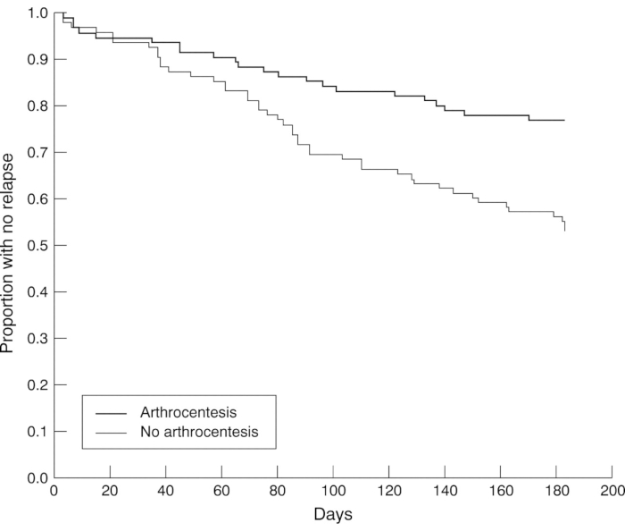
Figure 1: Proportion of arthritis relapse with or without arthrocentesis. The proportion of relapses in the arthrocentesis group was significantly reduced (p = 0.0009) and at the end of 6 months, there were 23% relapses in the arthrocentesis group and 47% in the no arthrocentesis group (p=0.001). Ninety-five knees were randomized to arthrocentesis and 96 had no arthrocentesis before triamcinolone hexacetanoide (20 mg) was injected into the inflamed knee joint. This figure is adapted with permission from Weitoft et al.3. Please click here to view a larger version of this figure.
Complications | Group A (INR ≥ 2) (n = 456) | Group B (INR < 2) (n = 184) | P Value |
Clinically significant bleeding (early) | 1 (0.2%) | 0 | NS |
Clinically significant bleeding (late) | 0 | 0 | NS |
Infection of joint (late) | 1 (0.2%) | 0 | NS |
Pain of joint causing physician visit | 3 (0.7%) | 0 | NS |
INR=international normalized ratio; NS=not significant. |
Table 1: Early and late complications between two groups. Procedure-related complications were defined as early (within 24 h) and late (within 30 days) clinically significant bleeding in or around a joint, infection of joint related to the arthrocentesis, and pain requiring a physician visit excluding chronic pain. Four patients accounted for 5 complications. This table has been modified with permission from Ahmed et al.6.
Knee arthrocentesis is a bedside or clinic procedure in which a needle is inserted into the joint capsule, and synovial fluid is aspirated. Before attempting an arthrocentesis, knee swelling by history should be confirmed to be an effusion on physical examination. A knee x-ray can reveal an effusion but is not necessary prior to aspiration. If body habitus complicates the physical examination, ultrasonography can be used to confirm effusion size and to direct insertion of the needle for aspiration8. If a patient has a swollen, painful, and warm knee with restricted movement, then a diagnostic arthrocentesis should be performed promptly to evaluate for septic arthritis. Arthrocentesis is usually performed with the patient supine and the knee extended as fully as it can be to increase intraarticular pressure. Mechanical compression with a knee brace has been shown to improve the success of knee arthrocentesis and fluid yield in rheumatoid arthritis and osteoarthritis9. A fully extended knee can be difficult if the patient has pain and restricted movement. It is possible, though, to aspirate fluid from a flexed knee, especially with an external compression brace. A study of 35 flexed knees with mechanical compression to the superior knee led to successful arthrocentesis fluid yields identical to the extended knee position10. Obvious unilateral knee swelling still should be palpated to detect the presence of fluid. Moderate to large volume knee effusions are easier to detect than small effusions. In a patient with morbid obesity, quadriceps hypertrophy, or osteoarthritic bony changes of the knee, it may be difficult to identify an effusion and so ultrasound can be used to confirm the presence of synovial fluid.
A skin marker is preferred over a pen because pen ink may be erased by iodine or chlorhexidine. An alternative to marking an "X" is to impress the skin with the tip of a ballpoint retractable pen or the sterile end of a needle sheath. Instead of soaking three gauze sponges with iodine solution, three iodine swabs or three chlorhexidine applying devices can be used to sterilize the skin for arthrocentesis. In a study of 166 specimens from native knees disinfected with chlorhexidine and then iodine, there were no false-positive cultures11. This suggests that a positive culture detected from an arthrocentesis performed under sterile conditions is highly suspicious for septic arthritis. Antibiotics can alter the synovial fluid analysis. Among 81 patients with septic arthritis, the average synovial leukocyte count was lower in the group who had received antibiotics prior to arthrocentesis compared to those who had not received antibiotics12. This highlights the importance of aspirating synovial fluid before antibiotics. Ethyl chloride spray is labeled as nonsterile, but a study of 15 healthy adults prepared for mock bilateral shoulder and bilateral knee injections showed that the percentage of positive cultures from the skin did not significantly increase after the application of ethyl chloride13. The burst of ethyl chloride spray to cool the skin as a topical anesthetic is optional. Subcutaneous lidocaine as a local anesthetic can be sufficient prior to an arthrocentesis. One disadvantage of this local anesthesia is the antibacterial property of lidocaine 2% in vitro14. Thus, this theoretically could affect the results of the synovial fluid culture.
The larger bore needle for aspiration should be inserted at the same location as the infiltration of lidocaine. The entire length of this needle need not be inserted into the synovial space. The syringe size for arthrocentesis should be at least 3 mL, but typically a 5 mL syringe would be the minimum to send synovial fluid analysis for cell count, crystal examination, gram stain, and culture. Only a drop of the synovial fluid is required for the wet mount, but an adequate amount of fluid is preferred to extract sediment of a centrifuged specimen for crystal examination and Gram stain15,16,17,18. A knee effusion can contain more than 100 mL of synovial fluid, and so if a large knee effusion is detected, a 50 mL syringe can be used to aspirate.
Even before an arthrocentesis, data gathered from the patient's history, physical examination, laboratory findings, and imaging can suggest a particular etiology of a knee effusion. Acute onset of knee pain and swelling can be seen in septic arthritis or a gout flare. Chronic knee swelling in an older, obese patient can be due to osteoarthritis. Trauma to the knee can cause hemarthrosis. Physical examination findings such as malar rash or psoriasis can lead one to suspect lupus arthritis or psoriatic arthritis, respectively. Laboratory tests such as an elevator rheumatoid factor and an anti-cyclic citrullinated peptide antibody can be seen in rheumatoid arthritis. The radiographic finding of chondrocalcinosis in the knee can be seen in pseudogout. The diagnostic arthrocentesis can provide key information to supplement the suspected diagnosis for a knee effusion. The arthrocentesis can also be therapeutic by decreasing pain and improving the range of motion of the knee. Arthrocentesis of the knee is a useful procedure in which synovial fluid is aspirated through a needle from the knee joint.
The authors have nothing to disclose.
The authors have no acknowledgments.
Alcohol prep pad | Medline | MDS090670Z | sterile 2-ply pad |
Eclipse needle | BD | DGW60702 | 25G x 5/8" |
Ethyl Chloride instant topical anesthetic spray | Gebauer's | P/N 0386-0008-03 | non-flammable |
Lidocaine HCl injection | Fresenius Kabi Usa, Llc | NDC 63323-492-27 | 1% single dose vial |
Plastic bandage | Curad | CUR02278RB | 4-sided seal |
Plastipak 3 mL syringe | BD | 309651 | sterile |
Plastipak 5 mL syringe | BD | 309649 | sterile |
Povidone iodine topical solution | Major | NDC 0904-1103-09 | topical antiseptic |
Precision glide needle | BD | 305196 | 18G x 1 1/2" |
Sterile gauze sponge | CARING | PRM2208 | 2 in. x 2 in. |
Sterile regular tip surgical skin marker | MEDLINE | DYNJSM01 | |
Surgical gloves | TRIUMPH | MSG2265 | sterile & powder-free |