This study demonstrates a reproducible heterotopic abdominal heart transplantation technique in rats that beginners can learn and perform. Additionally, a novel aortic regurgitation model in rats is generated by performing heterotopic abdominal heart transplantation and damaging the donor’s aortic valve using a guidewire after harvesting.
Over the past 50 years, many researchers have reported heterotopic abdominal heart transplantation in mice and rats, with some variations in the surgical technique. Modifying the transplantation procedure to strengthen the myocardial protection could prolong the ischemia time while preserving the donor's cardiac function. This technique's key points are as follows: transecting the donor's abdominal aorta before harvesting to unload the donor's heart; perfusing the donor's coronary arteries with a cold cardioplegic solution; and topical cooling of the donor's heart during the anastomosis procedure. Consequently, since this procedure prolongs the acceptable ischemia time, beginners can easily perform it and achieve a high success rate.
Moreover, a new aortic regurgitation (AR) model was established in this work using a technique different from the existing one, which is created by inserting a catheter from the right carotid artery and puncturing the native aortic valve under continuous echocardiographic guidance. A heterotopic abdominal heart transplantation was performed using the novel AR model. In the protocol, after the donor's heart is harvested, a stiff guidewire is inserted into the donor's brachiocephalic artery and advanced toward the aortic root. The aortic valve is punctured by pushing the guidewire further even after the resistance is felt, thus inducing AR. It is easier to damage the aortic valve using this method than with the procedure described in the conventional AR model. Additionally, this novel AR model does not contribute to the recipient's circulation; therefore, this method is expected to produce a more severe AR model than the conventional procedure.
Heterotopic abdominal heart transplantation in rats was first reported in 1964 by Abbott et al.1and has been used to study acute and chronic allograft rejection, cardiac allograft vasculopathy, ischemia-reperfusion injury, and cardiac remodeling2,3,4,5,6,7,8,9,10,11. Some modifications have been added to the procedure over the past 50 years. The fundamentals of the current procedure are as follows. The donor's ascending aorta and pulmonary artery (PA) are end-to-side anastomosed to the recipient's abdominal aorta and inferior vena cava, respectively. Although the donor's left atrium and ventricle do not receive any intracavitary flow, blood flows to the donor's coronary system; therefore, the donor's heart starts beating again after de-clamping.
Some experts with experience in hundreds or thousands of operations have reported a high success rate with short ischemia time for heterotopic abdominal heart transplantation2,3,4,5; however, it is difficult for beginners to achieve the short ischemia time from the outset. Sufficient cardioprotection is an important factor for obtaining good cardiac contraction of the donor's heart. Insufficient myocardial protection can stiffen the donor's heart. Therefore, we modified the transplantation procedure to strengthen the protection of the donor's heart. One of the aims of this study is to demonstrate a reproducible heterotopic abdominal heart transplantation procedure that beginners can easily perform since it prolongs the acceptable ischemia time.
Additionally, some researchers have reported an aortic regurgitation (AR) model in rats, which has been used to examine the effects of agents on left ventricular (LV) remodeling12,13,14,15. The conventional procedure includes the following: (1) a right lateral neck incision is made to expose the right carotid artery after anesthesia; (2) a catheter is cannulated from this vessel and advanced toward the aortic root; and (3) AR is induced by puncturing the native aortic valve under continuous echocardiographic guidance.
However, puncturing the aortic valve while holding the echocardiography probe and obtaining a good view of the ascending aorta, the aortic valve, and the catheter with an echocardiogram is challenging. Furthermore, cardiac failure following acute AR is another complication. Therefore, a novel AR model, which can be easily created and does not contribute to the recipient's circulation, has been established in this work to solve these challenges. The other aim of this study is to create an AR model by using heterotopic abdominal heart transplantation and damaging the donor's aortic valve using a guidewire after harvesting.
All the animal procedures were conducted in accordance with "An Outline of the Act on Welfare and Management of Animals" and "Standards Relating to the Care and Keeping and Reducing Pain of Laboratory Animals" by the Ministry of the Environment, Government of Japan and the "Guidelines for Proper Conduct of Animal Experiment" by the Science Council of Japan16,17,18. The animal protocols were reviewed and approved by the Institutional Animal Care and Use Committee of the University of Tokyo (M-P19-065).
1. Heterotopic abdominal heart transplantation in rats
NOTE: Heterotopic abdominal heart transplantations were conducted in male Jcl:Wistar rats aged 7-9 weeks old. A microscope with 6.7x to 45x magnification was used to perform the procedure. The surgical instruments were autoclaved for sterilization.
2. Novel AR model using heterotopic abdominal heart transplantation in rats
NOTE: A novel AR model using heterotopic abdominal heart transplantation was generated using male Jcl:Wistar rats aged 7-9 weeks old. A microscope with 6.7x to 45x magnification was used to perform the procedure. The surgical instruments were autoclaved for sterilization.
Regarding the normal model, good LV contraction was successfully established after de-clamping. The ischemia time of the transplanted heart and the recipient's anesthesia time were approximately 60 min and 130 min, respectively (Table 1).
Good LV contraction was also obtained after de-clamping in the new AR model. The ischemic time of the transplanted heart and the recipient's anesthesia time in the AR model were approximately 5 min and 10 min longer than the times of the normal model (Table 2). The AR model showed significantly larger LV dimensions and a thinner LV wall than the normal model (Table 3), and postoperative echocardiography detected an AR jet flow in the AR model (Figure 3). Macroscopic examination showed LV dilation and endocardial thickening (Figure 4), and the Masson's Trichrome-stained samples demonstrated fibrotic changes in the myocardium and endocardium (Figure 5). In contrast, these fibrotic changes were not found in the normal model.
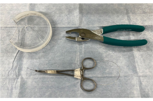
Figure 1: Surgical instruments and materials for creating an aortic regurgitation model using heterotopic abdominal heart transplantation. 1, a modified Petri dish with a hole in the center; 2, pliers; and 3, a stiff guidewire Please click here to view a larger version of this figure.
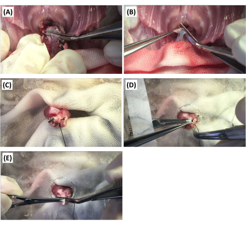
Figure 2: Surgical procedure for the creation of the aortic regurgitation model. (A) The donor's pulmonary artery is transected using Potts scissors. (B) The donor's ascending aorta is transected distally to the brachiocephalic artery with Potts scissors. (C) The donor's heart is fixed with pliers. (D) After the donor's ascending aorta is fixed with a vascular clip, the aortic valve is punctured with a stiff guidewire. (E) The aorta is transected proximally to the brachiocephalic artery using Potts scissors. Please click here to view a larger version of this figure.
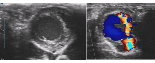
Figure 3: Postoperative echocardiography for the aortic regurgitation model. Left ventricular dilatation and a severe aortic regurgitation jet flow were detected. Please click here to view a larger version of this figure.
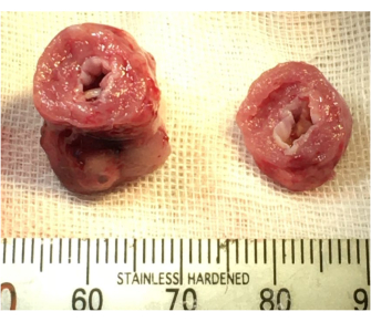
Figure 4: Macroscopic findings of the aortic regurgitation model. Left ventricular dilatation and endocardial thickening were confirmed. Please click here to view a larger version of this figure.
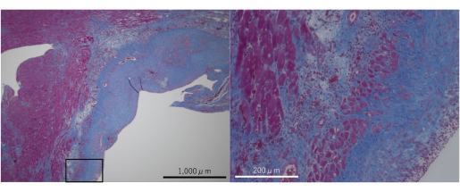
Figure 5: Masson's Trichrome-stained microphotographs of the aortic regurgitation model. Fibrotic changes were confirmed in the myocardium and endocardium. Please click here to view a larger version of this figure.
Variables | Number | Variables | Number |
Donor weight (g) | 236.0±40.6 | PA anastomosis time (min) | 18.8±2.7 |
Recipient weight (g) | 294.6±43.6 | Ischemia time (min) | 59.7±4.8 |
Clamp time (min) | 48.5±3.0 | ||
Harvest time (min) | 16.5±2.0 | Anesthesia time (min) | 132.7±8.6 |
Ao anastomosis time (min) | 26.9±2.7 | Extubation time (min) | 39.0±19.2 |
Table 1: Operative records of the normal model generated using heterotopic abdominal heart transplantation in rats (n = 19). Continuous variables are expressed as the mean ± standard deviation. Abbreviations: Ao = aorta; PA = pulmonary artery
Variables | Number | Variables | Number |
Donor’s weight (g) | 211.5±46.9 | PA anastomosis time (min) | 18.8±2.1 |
Recipient’s weight (g) | 261.2±42.0 | Ischemia time (min) | 65.7±7.2 |
Clamp time (min) | 49.3±4.9 | ||
Harvest time (min) | 17.3±2.2 | Anesthesia time (min) | 143.7±14.6 |
Ao anastomosis time (min) | 28.2±3.6 | Extubation time (min) | 28.0±14.5 |
Table 2: Operative records of the aortic regurgitation model generated using heterotopic abdominal heart transplantation in rats (n = 40). Continuous variables are expressed as the mean ± standard deviation. Abbreviations: Ao = aorta; PA = pulmonary artery
Variables | Normal model | AR model | P value |
LV wall (mm) | 3.05±0.50 | 2.19±0.57 | 0.002 |
LVDd (mm) | 2.23±0.55 | 4.56±2.13 | 0.003 |
LVDs (mm) | 1.32±0.34 | 3.30±1.79 | 0.003 |
LV-FS (%) | 40.49±9.41 | 29.06±8.24 | 0.008 |
Table 3: Postoperative echocardiography data of the normal and AR models created using heterotopic abdominal heart transplantation in rats. Continuous variables are expressed as the mean ± standard deviation. A Student's t-test was used to compare the differences between the two groups (P < 0.05). Abbreviations: AR = aortic regurgitation; FS = fractional shortening; LV = left ventricular; LVDd = left ventricular end-diastolic diameter; LVDs = left ventricular end-systolic diameter.
Key steps were discovered to prevent the donor heart from stiffening during implantation. First, it is vital to transect the donor's abdominal aorta before harvesting to unload the donor's heart4,7. If the donor's surgical procedure is performed without endotracheal intubation, breathing ceases after the thoracotomy, which obstructs the donor's pulmonary circulation. Consequently, the donor's heart becomes overloaded, preventing good contraction of the donor's heart after de-clamping. Second, perfusing the donor's coronary arteries with the cardioplegic solution is crucial8. Therefore, the cardioplegic solution should be perfused into the coronary arteries by clamping the ostium of the ascending aorta using tweezers after harvesting the donor's heart until the red color of the coronary arteries is diminished. Third, topical cooling of the donor's heart with slushed ice and cold normal saline is necessary. The donor's heart easily becomes warm and stiff when touching the bowel. By placing the donor's heart on a small plate, slushed ice can be placed around it. Additionally, the donor's heart can be submerged in cold normal saline during the anastomosis. Furthermore, the amount of slushed ice and cold normal saline needed can be minimized, thus preventing excessive cooling of the recipient. Moreover, using side-biting forceps to clamp the abdominal aorta and inferior vena cava is convenient7. These side-biting clamp forceps enable simultaneous clamping of the branches of both vessels and prevent backflow from these branches, thus simplifying the anastomosis of the donor's heart.
Experts with abundant experience in performing such operations can perform heterotopic abdominal heart transplantation with a short ischemia time. Plenter et al. reported a minimum ischemia time of approximately 35-45 min2. In the hands of Niimi, the ischemia time was consistently under 35 min3. Additionally, Westhofen et al. showed that the cold/warm ischemia time improved from 45 min/100 min to 10 min/20 min4. They performed heterotopic abdominal heart transplantation using mice rather than rats; however, their ischemia times were short. Therefore, completing the transplantation procedure within this short ischemia time appears difficult for beginners. This study's transplantation procedure showed approximately 60 min of ischemia time (Table 1), and all cases had good LV contraction after de-clamping because of the strengthened myocardial protection. Therefore, beginners can perform this study's procedure and achieve a high success rate.
Nevertheless, dozens of operations are required for the success of heterotopic abdominal heart transplantation. In this study, 62 operations were required to establish the transplant procedure and achieve a high success rate. Additionally, whether beginners can perform the transplant procedure easily by watching this study's video is yet to be proven. It would be useful and positive if the process could assist beginners.
Additionally, in this work, a novel AR model in rats was established using heterotopic abdominal heart transplantation and by damaging the donor's aortic valve using a guidewire after harvesting the donor's heart. Only two studies by Shimada et al. (the first author of both studies is a co-author of the present study) have reported AR models using heterotopic heart transplantation in rats19,20. Some important aspects should be considered when generating the AR model. First, the tool to puncture the aortic valve is important. The aortic valve of adult rats (approximately 200 g) is relatively sturdy; therefore, in our work, it was difficult to damage the aortic valve using a soft guidewire. In contrast, the aortic valve was easily pierced using a 23 G needle, although the risk of injury to other tissues was high, and few recipients could survive after the transplantation procedure because of bleeding. Therefore, a stiff guidewire was selected (Figure 1 and Figure 2). Second, immobilizing the donor's heart and the ascending aorta is important. Initially, the aortic valve was punctured while the ascending aorta wall was grasped using micro tweezers. However, damage to the ascending aorta wall was frequently observed due to excessive traction. Therefore, the donor's heart and ascending aorta were immobilized using a modified Petri dish with a hole in the center, pliers, and a vascular clip. (Figure 1 and Figure 2). Notably, the risk of injury to the ascending aortic wall was reduced after introducing this method.
This study's novel AR model has some benefits. First, with this model, it was possible to puncture the aortic valve more easily and in a shorter time compared to using the traditional AR model. The ischemia time in the AR model was only approximately 5 min longer than in the normal model (Table 1 and Table 2). Moreover, this method can produce various models with different degrees of AR by changing the number of punctures. Since this model does not contribute to the recipient's circulation, the recipient can survive even when the donor's heart shows severe AR (Figure 3).
Notably, the severe AR model showed fibrotic changes in the myocardium and endocardium due to severe AR jet flow (Figure 4 and Figure 5). Therefore, this model may contribute to studies on the pathomechanisms of myocardium and endocardium fibrosis and the evaluation of anti-fibrotic agents.
The authors have nothing to disclose.
We would like to thank Editage (www.editage.com) for the English language editing.
Antisedan (atipamezole) | Nippon Zenyaku Kogyo Co., Ltd. | ||
Domitor (medetomidine) | Nippon Zenyaku Kogyo Co., Ltd. | ||
Dormicum (midazolam) | Maruishi Pharmaceutical Co., Ltd. | ||
heparin | AY Pharmaceuticals Co.,Ltd. | ||
Jcl:Wistar rats | CLEA Japan, Inc. | ||
microscope | Orinpas Co., Ltd. | SZ61 | |
modified Krebs-Henseleit cardioplegic solution | Merck KGaA | ||
sevoflurane | FUJIFILM Wako Pure Chemical Corporation | ||
SURGICEL FIBRILLAR | Johnson & Johnson K.K. | ||
Vetorphale (butorphanol) | Meiji Animal Health Co., Ltd. |