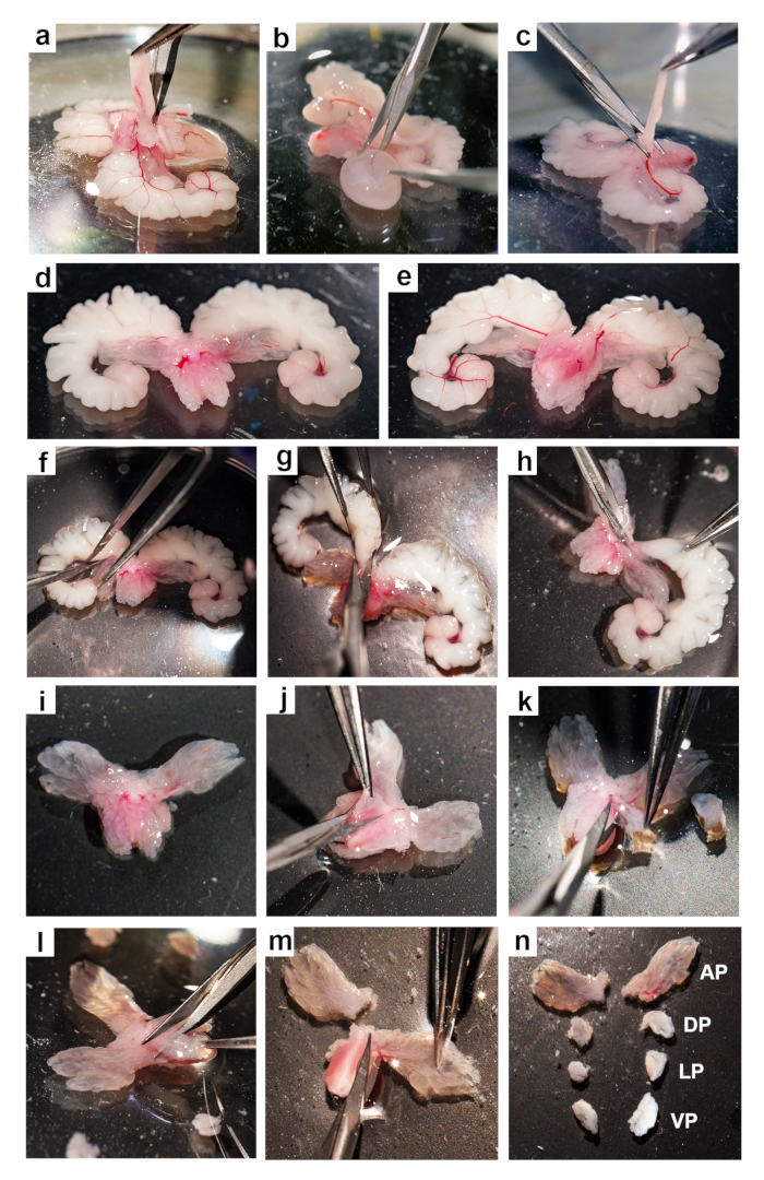
Figure 1: Dissection of the mouse prostate. Step-by-step images for the dissection of the mouse prostate from the UGS: (a) removing fat, (b) removing the urinary bladder, (c) removing the vas deferens, (d) ventral view of the prostate with the urethra, (e) dorsal view of the prostate with the urethra, (f-h) removing the seminal vesicles, (i) ventral view of the prostate, (j) dissecting a dorsal lobe, (k) dissecting a lateral lobe, (l) dissecting a ventral lobe, (m) dissecting an anterior lobe, and (n) the dissected prostate lobes: anterior = AP, dorsal = DP, lateral = LP and ventral =VP.
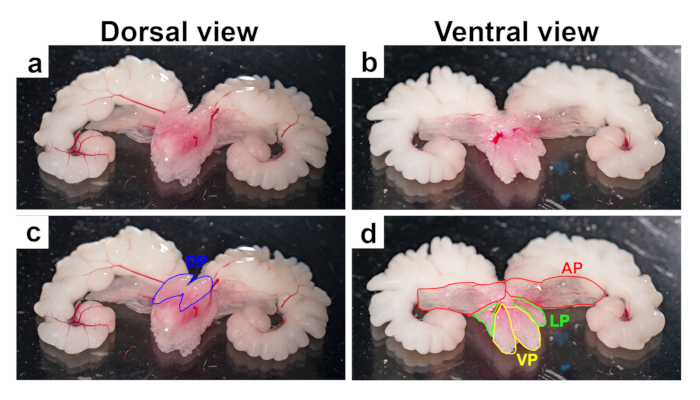
Figure 2: Anatomy of the mouse prostate lobes. Representative images from 3-month-old mouse UGS after removal of the fat, bladder, and vas deferens. Images show prostate lobes with seminal vesicles and urethra, with dorsal (a and c) and ventral (b and d) views. Top panels (a and b) show untouched images, while bottom panels (c and d) show the lobes outlined in blue (dorsal), red (anterior), yellow (ventral), and green (lateral).
