Calvarial Window Generation in Mouse Model: A Surgical Procedure to Create Window in the Top Part of the Mouse Skull

Published: April 30, 2023

Abstract

Source: Geisler, J. A. et al. Modeling Brain Metastases Through Intracranial Injection and Magnetic Resonance Imaging. J. Vis. Exp. (2020)

In this video, we describe the surgical procedure to create a calvarial window for accessing the brain of a mouse model.

Protocol

All procedures involving animal models have been reviewed by the local institutional animal care committee and the JoVE veterinary review board.

1. Prepare Stereotactic Units for Surgery

  1. Turn on all anesthesia machines, digital Vernier scales, and digital injectors.
    NOTE: All surgical tools should be adequately cleaned and sterilized prior to surgery.
  2. Utilize anesthetic machines with an induction chamber attachment in a biological safety cabinet (Figure 1A).
  3. Ensure all tubes from anesthesia machines are connected to the stereotactic frames (Figure 1Binset 1C) and clamps on the tubes are open for all units being used. Close any clamps on tubes going to stereotactic frames that will not be used for surgery.
  4. Set anesthesia machines to manufacturer recommended nose cone flow rate based on the mouse weight (e.g., 25 g animal weight: nose cone flow rate 34 mL/min).
    NOTE: The head holder included in this stereotactic setup is recommended only for adult mice. Ensure that the manufacturer recommendations included with the stereotactic setup are followed.
  5. Ensure that the appropriate anesthetic (e.g., isoflurane) prefilled in the syringe is attached to the anesthesia machine (Figure 1B).
    NOTE: Over-priming the syringe can cause too much anesthesia to be delivered to the mice during surgery and result in an anesthetic overdose.
  6. Prepare the drills by twisting the stage lock, inserting a drill bit adaptor and a 1 mm drill bit into each drill and lock the drill by manually tightening the bit-lock.
  7. Attach drills onto the stereotactic frames (Figure 1C).

2. Place Mice on Stereotactic Frames

  1. Anesthetize mice (e.g., isoflurane) using the aforementioned induction chamber.
  2. Maintain mice throughout the procedure at a deep anesthetic plane. The % anesthesia administered by the machine depends on a number of factors including: flow rate, degree of stimulation, and body temperature. Monitor the mice frequently throughout the procedure for depth of anesthesia by evaluating for rhythmic respirations (animal is not gasping); lack of palpebral reflex (fluttering of the eyelids when stimulated with a cotton tip applicator); and lack of toe pinch (withdrawal of limb upon the noxious stimulus of pinching the toes).
    NOTE: Different strains of mice will have a different response to anesthesia.
  3. After mice are at an appropriate, deep anesthetic plane, transfer the mice to the stereotactic unit. Use a heating pad while the mouse is on the stereotactic machine to maintain body temperature and an appropriate anesthetic plane.
    NOTE: To achieve low profile we use air-activated hand warmers placed within an inverted pipette tip box (mouse elevating box in Figure 1D).
  4. Open the mouse's mouth and place teeth in the trough of the mouth bar located on the nose piece on the stereotactic frame (Figure 2A). Slide the nose cone over the mouse's nose/mouth (Figure 2B).
    1. Place mice with their heads level to the mouth bar. Gently open the mouth with the blunt end of a cotton tip applicator and slide into place. Ensure the nose cone is fully over the mouse's nose or anesthesia will not be delivered properly. The nose cone will not engage with the nose if the teeth are not seated within this groove (Inset Figure 2A).
  5. Using a sterile cotton swab, place eye lubricant on each of the mouse's eyes. Application of eye lubricant mitigates drying of the cornea and reduces the chance of corneal damage and postoperative complications related to corneal trauma.
  6. Stabilize the mouse's skull by depressing the left ear bar up against the medial canthus of the left ear, locking it in place using the screw on the stereotactic frame. Then, slide the right ear bar against the medial canthus of the right ear and screw tight on the stereotactic frame (Figure 2B).
    NOTE: Make sure the mouse's head is level and straight when placing ear bars. If the head is crooked or angled, the injection will be in the incorrect place in the brain. The ear bars should be tightened ONLY until the skull is immobilized upon stimulation with moderate manual probing.

3. Make a Calvarial Window

  1. Prepare and clean the scalp with 3x alternating passes each of a betadine solution and 70% ethanol. Due to the close proximity of the surgical site to the eyes, use the betadine solution over the surgical scrub.
  2. Using a sterile scalpel, make a 3 mm incision through the skin along the central median aspect of the cranium (following the sagittal suture line). Bleeding at the incision should be minimal. Should it occur, apply consistent, firm pressure at the site of bleeding with a sterile cotton tip applicator for >30 seconds.
  3. Identify and orient the drill perpendicular to the bregma (Figure 2C), making sure to reset the digital Vernier scale to zero.
  4. Move the drill 2 mm lateral to the sagittal suture and 1 mm anterior to the coronal suture (Figure 2C). For reproducibility, ensure the location to the left or right of the sagittal suture line remains consistent for all animals.
  5. Turn the drill onto its highest speed. Ensure skin is moved away from drill to avoid tissue damage caused by the rotating bit and carefully initiate the drill onto the skull. Drill a hole roughly 0.5 mm deep through the calvaria, resulting in the calvarial window. Be careful not to lower the drill too far or it will drill into the brain. Dropping sterile saline at the drill site while the drill is in motion can offset any heat generated by the machine that may cause thermal damage to the surrounding tissue.
  6. Once the calvarial window is made, carefully raise the drill and remove it from the stereotactic frame.
  7. Clean the drill bits using 70% ethanol and set aside if being used again. If not, remove drill bits and submerge in 70% ethanol.

Representative Results

Figure 1
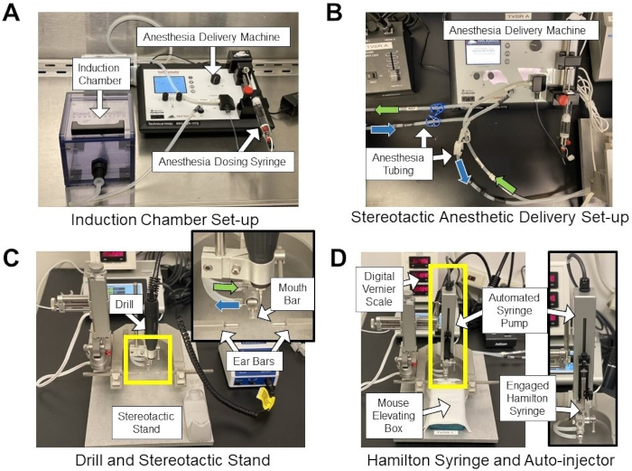
Figure 1: Stereotactic and anesthesia systems. (A) Anesthesia induction chamber setup within a biological safety cabinet. (B) Stereotactic anesthetic delivery setup highlighting anesthesia tubing from the anesthetic machine to the nose cone on the stereotactic apparatus (see inset in (C)). Green arrows indicate delivered anesthetic gas tubing, and blue arrows indicate scavenged gas tubing. (C) Stereotactic stand with drill attachment. Inset shows anesthetic tubing (green and blue arrows), mouth bar, and ear bars. (D) Stereotactic stand with automated syringe pump and mouse elevating box. The box is an inverted pipette tip box containing hand warmers used to elevate the mouse to the appropriate height and maintain body temperature. Inset shows the orientation of the Hamilton syringe in the automated injection apparatus.

Figure 2
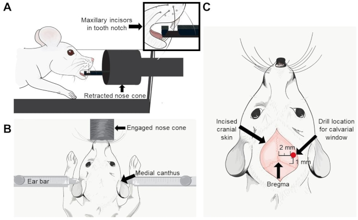
Figure 2: Pictorial representation of the tooth placement on a stereotactic device, location of ear bars, and calvarial window relative to bregma. (A) The pictorial of the maxillary incisors in the tooth notch on the nose cone. (B) The location of the left and right ear bars within the medial canthus of the respective ears. (C) An arrow indicates the bregma and a red dot indicates the location where the calvarial window should be made (2 mm lateral to the sagittal suture and 1 mm anterior to the coronal suture).

Disclosures

The authors have nothing to disclose.

Materials

Surgical Materials
Betadine Purdue Products 19-027132 Povidone-iodine, 7.5%
Bone Wax Surgical Specialities 903 Sterile and malleable beeswax and isopropyl palmitate
Buponorphine SR-Lab ZooPharm N/A Long acting injectable analgesic 5 mL (0.5 mg/mL) polymetric formulation
Cotton tip applicators Puritan 25-806 10WC Sterile long stemmed cotton tip applicators
Eye Ointment Puralube 17033-211-38 Lubricating petrolatum and mineral oil based ophthalmic ointment
Handwarmers  Hothands  HH2 Air-activated heat packs
Ibuprofen Up & Up 094-01-0245 100 mg per 5 mL in liquid suspension
Isoflurane Henry Schein INC 1182097 Liquid anesthetic for use in anesthetic vaporizer
Scalpels Integra Miltex 4-410 #10 disposable scalpel blade
Suture Covidien SP5686G 45 cm swedged 5-0 monofilament polypropylene suture
Stereotaxic Unit
High Speed Drill (Foredom) Kopf Model 1474 Max of 38,000 RPM
Mouse Gas Anesthesia Head Holder Kopf Model 923-B Mouth bar with teeth hole and nosecone
Non-Rupture Ear Bars Kopf Model 922 Ear bars suitable for mouse applications
Stereotaxic Instrument Kopf Model 940 Base plate, frame and linear scale assembly with digital readout monitor
Anesthesia Machine
SomnoSuite Low-Flow Digital Vaporizer Kent Scientific SS-01 Digital anesthesia machine
SomnoSuite Starter Kit for mice Kent Scientific SOMNO-MSEKIT Includes induction chamber, 2x anesthesia syringes, 18" tubing, plastic nosecone, 2x waste aneshesia gas canisters

Tags

check_url/cn/20609?article_type=t

Play Video

Cite This Article
Calvarial Window Generation in Mouse Model: A Surgical Procedure to Create Window in the Top Part of the Mouse Skull. J. Vis. Exp. (Pending Publication), e20609, doi: (2023).

View Video