A subscription to JoVE is required to view this content. Sign in or start your free trial.

Method Article

Fluorescence Fluctuation Spectroscopy to Study Protein Interaction at Cell Contacts

1K views

⸱

July 8th, 2025

In This Article

Abstract

Source: Dunsing, V. et al. A Fluorescence Fluctuation Spectroscopy Assay of Protein-Protein Interactions at Cell-Cell Contacts. J. Vis. Exp. (2018).

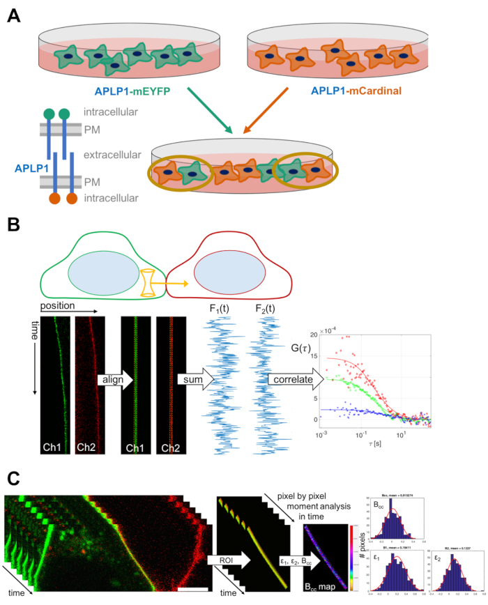
This video demonstrates the use of fluorescence fluctuation spectroscopy to detect the interaction among cell surface proteins at cell-cell contacts. By expressing the transmembrane adhesion receptor of interest labeled with a fluorescent protein and mixing two different cell populations harboring two spectrally separated fluorescent labels, the trans-interaction between the receptors of two neighboring cells with different-colored fluorescence is assessed via cross-correlation in the fluctuations of fluorescence intensity.
Protocol

1. Sample Preparation: Cell-Cell Mixing Assay

NOTE: The following protocol describes the mixing procedure for adherent cells. It may be modified for cells cultured in suspension.

  1. Seed an appropriate number of cells on a 6-well plate, e.g., 800,000 HEK 293T cells (counted with a Neubauer counting chamber), a day before transfection. The number can be modified depending on the time between seeding and transfection and adjusted for other cell types. To perform a basic experiment (i.e., proteins of interest and negative control), prepare at least 4 wells. Culture cells at 37 °C, 5% CO2

Access restricted. Please log in or start a trial to view this content.

Results

Fluorescence correlation spectroscopy diagram shows APLP1 protein interactions and pixel analysis data.
Figure 1. Experimental workflow and schematic representation of scanning fluorescence cross-correlation spectroscopy and cross-correlation number and brightness analysis at cell-cell contacts. (A) Scheme of sample preparation: Two ce.......

Access restricted. Please log in or start a trial to view this content.

Disclosures

No conflicts of interest declared.

Materials

List of materials used in this article
NameCompanyCatalog NumberComments
DMEM growth mediumPAN-BiotechP04-01548
DPBS w/o: Ca<sup>2+</sup> and Mg<sup>2+</sup>PAN-BiotechP04-36500
DPBS w: Ca<sup>2+</sup> and Mg<sup>2+</sup>PAN-BiotechP04-35500
Trypsin EDTAPAN-BiotechP10-023100
TurboFect Transfection ReagentThermo Fisher ScientificR0531
HEK 293T cellsDSMZACC 635
Alexa Fluor 488 NHS EsterThermo Fisher ScientificA20000
Rhodamine BSigma-Aldrich83689-1G
Plasmid DNAAddgeneNASee Dunsing et. al., MBoC 2017, for a detailed description of all plasmids
6-well plateStarlabCC7672-7506
35-mm glass bottom dishesCellVisD35-14-1.5-N
Zeiss LSM780 confocalCarl ZeissNA
Neubauer cell counting chamberMarienfeld640110

Explore More Articles

Protein-Protein InteractionsCell-Cell ContactsConfocal MicroscopyFluorescence Intensity FluctuationsCross-Correlation AnalysisFluorescent Protein LabelingTransmembrane Adhesion ReceptorsLine Scan AcquisitionPhoton Counting Mode