A subscription to JoVE is required to view this content. Sign in or start your free trial.

Method Article

Real-Time Analysis of Resident T-cell Migration in Different Tumor Regions Using Confocal Microscopy

865 views

⸱

July 8th, 2025

In This Article

Abstract

Source: Peranzoni, Elisa, et al. Ex Vivo Imaging of Resident CD8 T Lymphocytes in Human Lung Tumor Slices Using Confocal Microscopy. J. Vis. Exp. (2017).

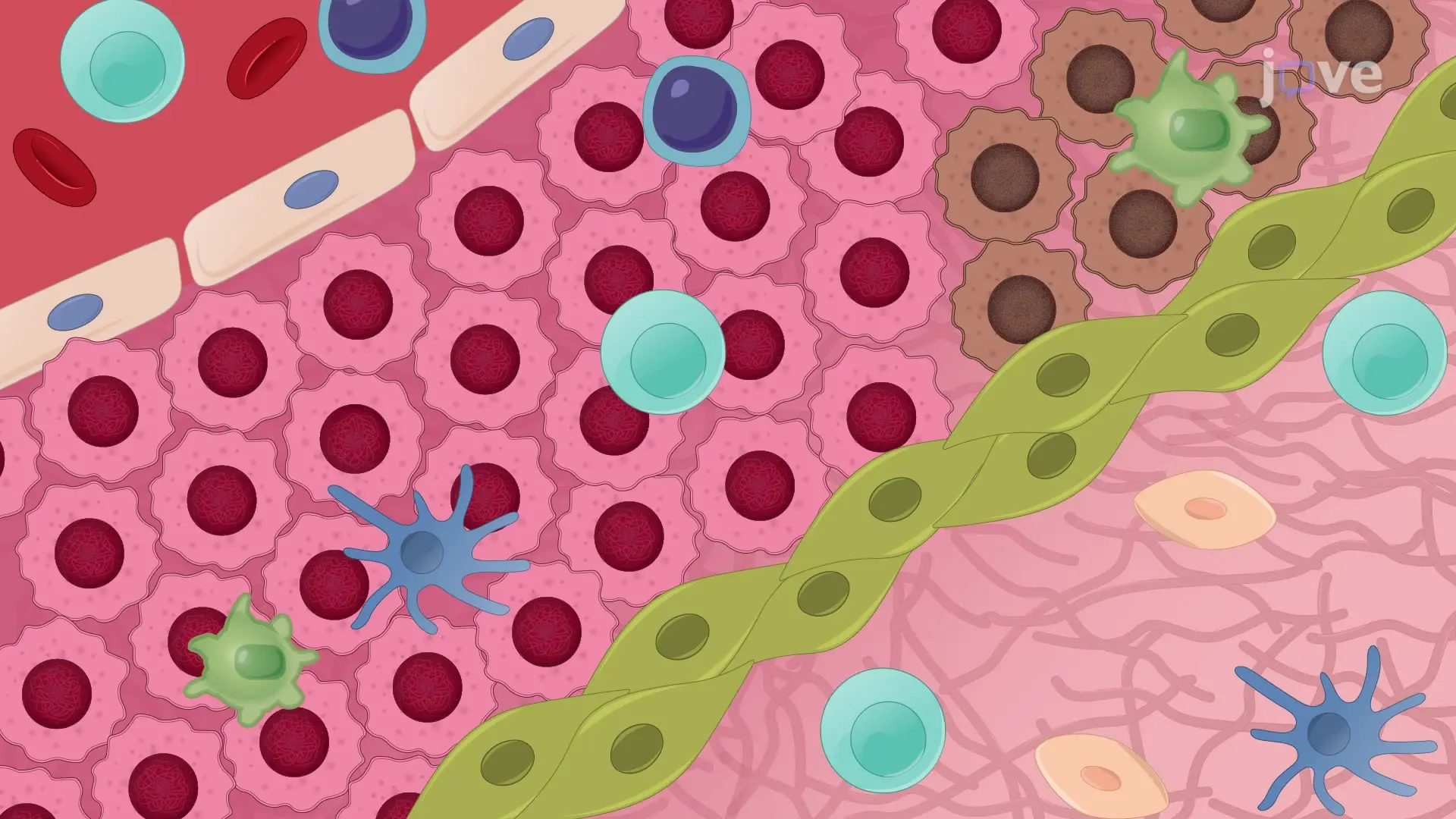
This video describes the imaging of resident tumor-infiltrating CD8 T cells labeled with fluorescently coupled antibodies within human lung tumor slices. This technique permits real-time analyses of CD8 T cell migration using confocal microscopy.
Protocol

1. Immunostaining of resident CD8 T cells and tumor cells

  1. Dilute the antibodies (final concentration 10 µg/mL) and the nuclear dye (final concentration 1 µg/mL) in RPMI-1640 media without phenol red.
    NOTE: Use fluorescently-coupled anti-CD8 (murine IgG, clone SK1) and anti-EpCAM (epithelial cell adhesion molecule) (murine IgG HEA-125) antibodies to label CD8 T lymphocytes and tumor epithelial cells, respectively. Use fluorescently-coupled anti-CD90/Thy1 (murine IgG, clone 5E10) antibody to label fibroblasts and activated endothelial cells. Use 4',6-diamidino-2-phenylindole (DAPI) to label the nucleus of ce....

Access restricted. Please log in or start a trial to view this content.

Results

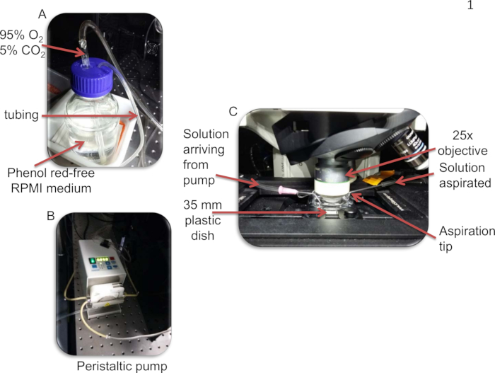
Cell culture setup: RPMI medium, peristaltic pump, 35mm dish, microscope observation, solution flow.
Figure 1: Photographs of the perfusion system. A) The phenol red-free RPMI solution is enriched in 5% CO2, 95% O2. B) The solution is then perfused by a peristaltic pump. C) The RPMI solution enters the culture dish via the inlet. .......

Access restricted. Please log in or start a trial to view this content.

Disclosures

No conflicts of interest declared.

Materials

List of materials used in this article
NameCompanyCatalog NumberComments
Upright confocal microscopeLeicaThe use of a spinning disk confocal is recommended
Imaris softwareBitplaneThis sofware is used for visualization of images and tracking of T cell migration
Fine ForcepsWorld Precision Instruments14142
30 mm Culture insertsMilliporePICM0RG50
Roswell Park Memorial Institute (RPMI) medium, GlutaMAX SupplementThermoFisher61870010Complete RPMI-medium is made by adding 10 % heat-inactivated fetal calf serum and Penicillin/ streptomycin
Phosphate-buffered saline (PBS)ThermoFisher20012019
Hanks' Balanced Salt Solution (HBSS)Invitrogen14170088
Low gelling temperature Agarose, type VII-ASigma-AldrichA0701
Non-toxic butyl Cyanoacrylate Glue, Vetbond3M1469
Slice anchors, 19.7 diameter, 2 mm spacing threadsWarner Instruments64-1415
Stainless steel washers, 5 mm of inner diameterAmazonB004K1FDGQ
BV605-conjugated anti-CD8 (clone SK1)BD Biosciences565289
FITC-conjugated anti-EpCAM (clone HEA-125)Miltenyi Biotec130-098-113
BV510-conjugated anti-CD90 (clone 5E10)Biolegend328125
4',6-diamidino-2-phenylindole (DAPIThermoFisherD1306

Explore More Articles

Tumor MicroenvironmentFluorescent LabelingZ-Stack ImagingCD8 T CellsTumor SlicesPerfusion SystemTracking Software