Canalostomy for Local Drug Delivery into the Inner Ear of an Adult Mouse

Published: May 31, 2024

Abstract

Source: Guo, J. et al., Canalostomy as a Surgical Approach to Local Drug Delivery into the Inner Ears of Adult and Neonatal Mice. J. Vis. Exp. (2018)

This video showcases canalostomy, a procedure that allows local drug delivery into the inner ear of a mouse through the semicircular canal with minimal damage to hearing and vestibular function. This method enables the inoculation of viral vectors, pharmaceuticals, and small molecules into the mouse's inner ear.

Protocol

All procedures involving animal models have been reviewed by the local institutional animal care committee and the JoVE veterinary review board.

1. Device Preparations

  1. To make the injection cannula (Figure 1A), connect polyimide tubing (inner diameter 114.3 µm, outer diameter 139.7 µm, length ~3 cm) to polyethylene tubing (inner diameter 280 µm, outer diameter 640 µm, length ~40 cm). Using superglue, seal the connection with at least three applications. Sterilize the injection cannula with ethylene oxide.       
    NOTE: When sealing the tubing, prevent the superglue from entering the polyimide tubing, which can result in a blockage of the cannula. Seal the tubing at least three times, because gas sterilization may cause leakage at the connection.
  2. Using a 30 G needle, connect the end of the polyethylene tubing to a 1 cc syringe containing normal saline. Fill the cannula with normal saline by injection with the 1-cc syringe to check for any leakage or blockage at the connection between the polyimide tubing and polyethylene tubing.       
    NOTE: If there is a leakage or a blockage of the connection, the cannula cannot be used in the following procedures.
  3. Evacuate a 10 µL micro-syringe with normal saline and connect it to the aforementioned 30 G needle with the injection cannula. Install the micro-syringe on a microinjection pump (Figure 1B). Set the injection speed to 0.5 µL/min and the volume to 1 µL for fast-green dye and adeno-associated virus tagged with green fluorescent protein (AAV8-GFP), or 2 µL for streptomycin.
    NOTE: The recommended range of injection speed is 0.1−0.5 µL/min, and the recommended range of injection volume is 0.5−2 µL.
  4. Pull back the micro-syringe to 1 µL and extract the injection reagent. An air gap will form between the normal saline and the injected reagent (Figure 1C).
  5. Optionally label the position of the gas-fluid border on the polyethylene tubing using a marker pen. This can be used to monitor reagent flow during injection.

2. Canalostomy in Adult Mice

  1. Anesthetize an adult mouse (female, FVB/N, 5 to 6-week old) by intraperitoneal injection of ketamine hydrochloride (ketamine HCl) (120 mg/kg) and xylazine HCl (7 mg/kg). Wait for 5−10 min until the animal exhibits no response to painful stimuli (the toe-pinch reflex). Place the animal on a preheated electric pad after anesthesia. An analgesic, Meloxicam (1 mg/kg), was applied subcutaneously before surgery.
  2. Cover the animal's eyes with eye ointment. Shave the left post-auricular region with an electric animal clipper and disinfect the skin three times with 75% ethanol.
  3. Place the animal on a preheated electric pad. Set the temperature of the electric pad to ~37°C. Place the animal in the right lateral position to facilitate surgery on the left ear.
  4. Make a 1-1.5-cm post-auricular incision ~3 mm from the left retroauricular groove (Figure 2A).
  5. Positioning the posterior semicircular canal (PSC) and lateral semicircular canal (LSC): When the root of the pinna (bold dot in Figure 2B) is defined as the origin and the plane parallel to the calvarium as 3 to 9 o'clock, the PSC and the LSC are typically located ~3 mm from the root of the pinna between 2 and 3 o'clock (Figure 2B).
  6. Bluntly dissect the muscle covering the temporal bone with micro-forceps to expose the PSC and LSC, whose margins are clearly visible as dark stripes in the temporal bone (Figure 2C). The LSC is at an approximately 30° angle from the plane parallel to the calvarium, and the PSC is vertical to the LSC (Figure 2C). Collect a small piece of muscle with micro-forceps and let it dry. In the following step, this muscle will be used to seal the hole.   
    NOTE: Avoid excessive tearing of the tissues to minimize injury to muscles. Avoid damaging the adjacent vessel when exposing the PSC (Figure 2D).
  7. Make a small hole in the middle portion of the PSC using a 26 G needle (Figure 2D). Fluid leakage through the hole indicates successful penetration of the bony wall of the PSC. Enlarge the hole to an appropriate size, slightly larger than the diameter of the polyimide tubing.       
    NOTE: Drill and enlarge the hole gently and gradually to avoid fracture of the PSC.
  8. Clean the effusion surrounding the hole of the PSC using a cotton pellet.
  9. Insert the tip of the polyimide tubing gently into the PSC toward the crus commune to a depth of 1−2 mm (Figure 2E). Start the injection by pressing the 'run' button on the pump.
  10. After the injection, wait ~2 min to allow the reagent to spread. Trim the small piece of muscle collected in step 2.6 with micro-scissors. Next, remove the injection cannula and immediately place the muscle into the hole in the PSC.       
    NOTE: Check for any liquid leaking from the hole after plugging to ensure that the hole is completely sealed.
  11. Return the separated muscles and subcutaneous tissues. Suture the incision using a 5-0 suture. Disinfect the incision region with povidone iodine.       
    NOTE: All the above surgical procedures take about 25 min.
  12. Position the animal on an electric pad preheated to ~37 °C. Place the animal in the right lateral position for recovery.

Representative Results

Figure 1
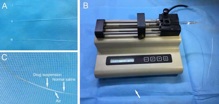
Figure 1: Device preparation. (A) The polyimide tubing (i) and the polyethylene tubing (ii) are sealed to make an injection cannula (iii). (B) The cannula is connected to a 10-µL micro-syringe with a 30 G needle and then installed on a pump. Arrow indicates the cannula tip. (C) Injection reagent and normal saline are separated by an air gap.

Figure 2
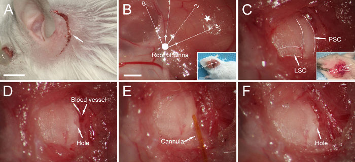
Figure 2: Canalostomy in an adult mouse. (A) A post-auricular incision (arrow). (B) Muscles covering the temporal bone are exposed. If we define the root of the pinna (bold dot) as the origin and the plane parallel to the calvarium as 3/9 o'clock, the posterior semicircular canal (PSC) and lateral semicircular canal (LSC) are generally located in the region between 2 and 3 o'clock (pentagram), ~3 mm from the origin. Inset: lower-magnification image of the orientation. The dotted line indicates the calvarium plane. (C) The PSC and LSC are exposed (dotted lines). Inset: The LSC is at an approximately 30° angle from the plane parallel to the calvarium, and the PSC is vertical to the LSC. (D) A small hole is made in the PSC. (E) The tip of the cannula is inserted into the PSC, and the reagent is injected. (F) The hole is sealed with a small piece of muscle. Scale bar in A is 5 mm, that in B is 1 mm (B for B-F), that in the inset to B is 1 cm, and that in the inset to C is 5 mm.

Disclosures

The authors have nothing to disclose.

Materials

Polymide Tubing A-M Systems 823400
Polyethylene Tubing Scientific Commodities Inc. BB31695-PE/1
10μl Microsyringe Hamilton Company 80001
Xylazine HCL Sigma-Aldrich Co. Llc. X-1251
Operating Miroscope Carl Zeiss Optical LLC. Pico
Micro Forceps Dumont Dumostar 10576
Fast-green Dye Sigma-Aldrich Co. Llc. F7252
AAV8-GFP BioMiao Biological Technology Co. Ltd (Beijing, China) 20161101 Titer: 2×10e12 vg/mL
Streptomycin Sulfate Sigma-Aldrich Co. Llc. S9137
Microinjection Pump Stoelting Co. 789100S
Electric Pad Pet Fun 11072931136
1 cc Syringe Mishawa Medical Industries Ltd. (Shanghai, China) 2011-3151258
Ketamine HCL Gutian Pharmaceutical Co., Ltd. (Fujian, China) H35020148
Electric Animal Clipper Codos Electrical Appliances Co., Ltd. (Guangdong, China) CP-8000
Cotton Pellet Yatai Healthcare Ltd. (Henan, China) Yu-2008-1640081
Suture Shanghai Pudong Jinhuan Medical Products Co., Ltd. (Shanghai, China) Hu-2013-2650207
Eye Ointment Beijing Shuangji Pharmaceutical Ltd. (Beijng China) H11021270

Tags

This article has been published
Video Coming Soon
Keep me updated:

.

Cite This Article
Canalostomy for Local Drug Delivery into the Inner Ear of an Adult Mouse. J. Vis. Exp. (Pending Publication), e22275, doi: (2024).

View Video