This protocol provides a combination strategy of two herbs to treat injured PC12 cells. The protocol provides a reference for optimizing the best application mode of traditional Chinese medicine (TCM).
In view of the advantages of the combination of traditional Chinese medicine (TCM) in the treatment of cerebral ischemia, we studied the differences in the efficacy and mechanism between the preparation combination and the component combination in order to explore the two herb combination strategy to treat injured PC12 cells. Cobalt chloride (CoCl2) combined with a glucose-free medium was employed to induce oxidative damage of PC12 cells. Then, the optimal combination of Astragalus mongholicus (Ast) and Erigeron breviscapus (Eri) injection was selected and combined following uniform design methods after screening their safe and effective concentration on PC12 cells. Further, the component combination screened comprises 10 µM astragaloside A, 40 µM scutellarin, and 75 µM chlorogenic acid in two herbs. Then, MTT, Annexin V-FITC/PI, immunofluorescence, and Western blot analysis were used to evaluate the efficacy and mechanism of the preparation combination and the component combination on injured PC12 cells. The results showed that the optimal preparation combination for cell pro-survival was Ast injection and Eri capsule with a concentration of 6:1.8 (µM). The component combination (10 µM astragaloside A, 40 µM scutellarin, and 75 µM chlorogenic acid) was more effective than the preparation combination. Both combinations remarkably reduced apoptotic rate, the fluorescence intensity of caspase-3, and intracellular reactive oxygen species (ROS) level; meanwhile, they upregulated the expression levels of p-Akt/Akt, Bcl-2/Bax, and Nrf2. These effects were more evident in the component combination. In conclusion, both combinations can inhibit the injury induced by CoCl2 combined with a glucose-free medium on PC12 cells, thus promoting cell survival. However, the efficiency of the component combination over the preparation combination may be due to its stronger regulation of the PI3K/Akt/Nrf2 signaling pathway related to oxidative stress and apoptosis.
Chronic cerebral ischemia, caused by cerebral hypoperfusion, is a common disease in middle-aged and elderly people1. As a long-term occult ischemia disease, it can lead to progressive or persistent neurological dysfunction. The main pathological mechanisms include cell apoptosis and oxidative damage, leading to progressive or persistent neurological dysfunction; this is the pathological basis of Alzheimer’s disease, vascular dementia, and other diseases, and seriously affects the quality of life of patients. However, there is still a lack of ideal drugs in modern medicine to treat chronic cerebral ischemia2. At the same time, the combination of Astragalus mongholicus (Ast) and Erigeron breviscapus (Eri) has been widely used in the clinical practice of traditional Chinese medicine (TCM)4. The combination strategy has remarkably improved in promoting the recovery of nerve function after cerebral ischemia, which is better than that of a single drug; however, there are large differences in the dosage ratio in the combination of the two drugs4. The effective components and mechanism of action are not well defined, which is the key issue restricting its clinical application.
Previous studies have demonstrated the synergistic effect of the preparation combination of Ast injection and Eri injection in treating cerebral ischemia in rats. It can upregulate the expression of p-Akt protein and downregulate the expression of Bcl-2 associated death promoter (BAD) protein5,6, which is one of the B-lymphoblastoma 2 (Bcl-2) family proteins and has the effect of promoting apoptosis. However, the material basis and mechanism of its synergistic effect are not clear. Further, the injury model of PC12 cells was established with CoCl2 combined glucose-free medium, and the optimal component combination in Ast and Eri has been screened and defined7.
In this study, the effective doses of Ast and Eri are screened by using the injured PC12 cell model. The optimal combination of the two drugs is screened using this model combined with the homogeneous design method. The cell model is used to further evaluate the difference in the effects and mechanisms of the preparation combination and the component combination in injured PC12 cells. This strategy aims to explore the protective effect and regulatory mechanism of the two types of combinations on injured PC12 cells through the PI3K/Akt/Nrf2 signal pathway to determine the best combination mode. This study provides a reference for optimizing the best application mode of TCM.
1. Preparation of reagent
2. Cell viability assay
3. Annexin V-FITC/PI assay for apoptosis rate
4. Immunofluorescence detection of caspase-3 generation
5. Immunofluorescence assay of ROS level
6. Western blot detection of protein expression of Nrf2, p-Akt, Akt, Bcl-2, and Bax11,12
The screening of the optimal combination of Ast injection and Eri capsule is shown in Figure 1. The cell survival rate of Ast injection and Eri capsule on the normal PC12 cells is shown in Figure 1A. The cell viability was lower than 95% with Ast injection at concentrations greater than 12 µM (Figure 1A) and Eri capsule at concentrations greater than 5 µM (Figure 1A), indicating that the maximum nontoxic concentration was 12 µM and 5 µM, respectively. Their cytotoxicity was greater than that of astragaloside A and scutellarin used alone. The viability of Ast injection and Eri capsule on the injured PC12 cells induced by CoCl2 is shown in Figure 1B,C. Compared with the model group, Ast injection could improve the survival rate of the injured PC12 cells in the concentration range of 6-12 µM (p < 0.05 or p < 0.01), and Eri capsules at the concentration of 2-5 µM could improve the survival rate (p < 0.05 or p < 0.01). Ast injection and Eri capsule at the ratios of 10:1, 8:4.2, and 6:1.8 µM can significantly increase the viability of the injury PC12 cells (p < 0.01) (Figure 1D). The ratio of 6:1.8 µM exhibited the highest cell viability, indicating that it is the optimal preparation combination and pharmacological activity compared with the best component combination.
The evaluation of the protective effects of two kinds of combination on the injured PC12 cells is shown in Figure 2. Compared with the model group, the cell viability of the two combinations was significantly promoted (p < 0.001), and the component combination was superior to the preparation combination (Figure 2A). This suggests that the component combination can promote cell survival better than the preparation combination. The apoptosis rate was tested by flow cytometry (Figure 2B,C). Compared with the normal group, the percentages of early, late, and total apoptotic cells were significantly higher in the model group (p < 0.01 or p < 0.001). Compared with the model group, the percentages of apoptotic cells at each stage were significantly lower in the treatment groups (p < 0.05 or p < 0.01, or p < 0.001). The fluorescence intensity of caspase-3 protein expression in each group is shown in Figure 2D,E. Compared with the normal group, the fluorescence intensity of caspase-3 was significantly higher in the model group. Compared with the model group, the fluorescence intensity of caspase-3 was significantly lower in each treatment group (p < 0.001 or p < 0.01) and was relatively lower in the component combination group than the preparation combination group. These results suggest that the cell model can induce apoptosis, and the anti-apoptosis effect of the component combination is better than that of the preparation combination.
Western blot detected the p-Akt, Akt, Bcl-2, and Bax protein expression (Figure 3). Compared with the normal group, the expression levels of p-Akt/Akt and Bcl-2/Bax were significantly lower in the model group (p < 0.01 or p < 0.001). Compared with the model group, the expression of p-Akt/Akt and Bcl-2/Bax were significantly higher in the two combinations (p < 0.05 or p < 0.01, or p < 0.001), specifically higher in the component combination. The results suggest that the component combination is superior to the preparation combination in promoting cell survival, which is related to the stronger anti-apoptosis effect produced by upregulating the Akt/Bcl-2/Bax signal pathway.
The fluorescence of DCFH can be measured by a fluorescence microscope to determine the level of ROS in the cells (Figure 4A,B). As shown in Figure 4A, the fluorescence was almost invisible in the normal cells, and the fluorescence in the model group was significantly enhanced. The fluorescence in both combinations was significantly reduced compared to the model group. As shown in Figure 4B, the relative fluorescence intensity was significantly weaker in each treatment group compared with the model group (p < 0.001). Fluorescence had a tendency to decrease in the component combination group compared with the preparation combination group, indicating that the cell model can induce oxidative damage. The anti-oxidative damage effect of the component combination is better than that of the preparation combination.
Western blot detected the expression of the Nrf2 protein (Figure 4C). Compared with the normal group, the expression of Nrf2 was significantly reduced in the model group (p < 0.05). Compared with the model group, the expression of Nrf2 protein was significantly higher in the treatment groups (p < 0.01 or p < 0.05), specifically higher in the component combination group (Figure 4D). This suggests that the component combination is better than the preparation combination in promoting cell survival, which is related to the stronger anti-oxidative damage produced by upregulating the Nrf2 signal pathway.
In conclusion, the component combination (10 µM astragaloside A, 40 µM scutellarin, and 75 µM chlorogenic acid) could promote the survival of injured cells better than the preparation combination (6 µM Ast injection and 1.8 µM Eri capsule) through stronger regulation of signal pathways related to apoptosis and oxidative damage.
SPSS statistical software 26.0 was used for statistical analysis, and all data are expressed as the means ± standard deviation (SD). For comparisons between groups, the data were evaluated by a one-way ANOVA followed by Tukey's test. p < 0.05 was considered to indicate a statistically significant difference.
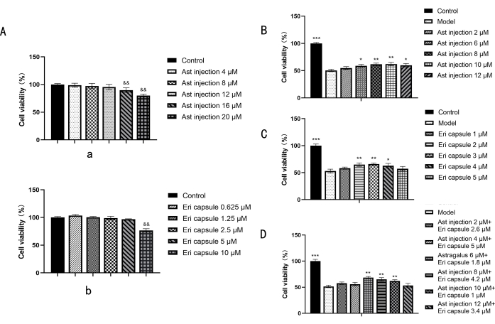
Figure 1: Effects of drugs on the viability of normal and injured PC12 cells. (A) Effects of various concentrations of Ast injection and various concentrations of Eri capsule on normal PC12 cells. (B) Screening of the effective concentration of Ast injection on injured PC12 cells. (C) Screening of the effective concentration of Eri capsule on injured PC12 cells. (D) Effects of the combinations of two drugs in different proportions on the survival rate of injured PC12 cells. Statistical values are expressed as the mean ± SD from six independent experiments. &&p < 0.01 compared with the control group. *p < 0.05 and **p < 0.01 compared with the model group. Please click here to view a larger version of this figure.
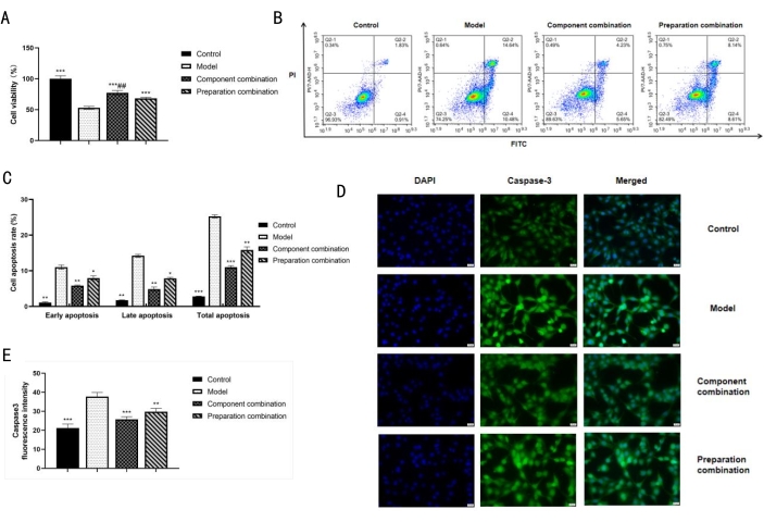
Figure 2: Effects of two combinations on the survival and apoptosis of injured PC12 cells. (A) Effect of the component combination and the preparation combination on the survival rate of injured PC12 cells. (B) Graph of apoptosis rate detected by Annexin V-PI. (C) Statistical histogram of apoptosis rate. (D) The relative fluorescence intensity of caspase-3 protein expression (400x). Scale bars: 20 µm. (E) Statistical histogram of fluorescence intensity of caspase-3 protein expression. Statistical values are expressed as the mean ± SD from three independent experiments, except for cell viability for six independent experiments. *p < 0.05, **p < 0.01, and ***p < 0.001 compared with the model group. ##p < 0.01 compared with the preparation combination group. Please click here to view a larger version of this figure.
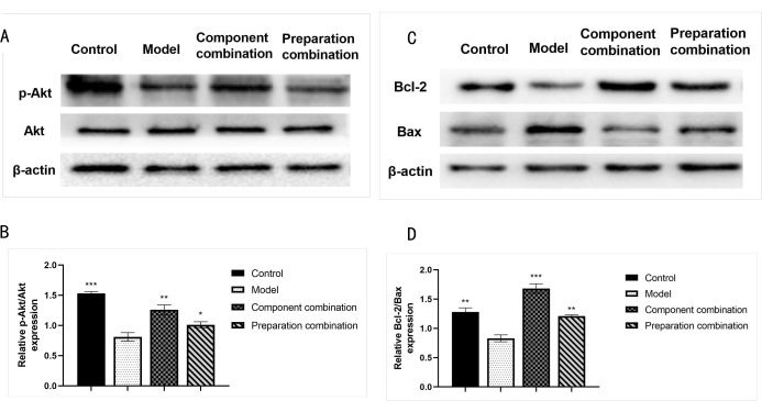
Figure 3: Effects of the two combinations on the expression levels of p-Akt, Akt, Bcl-2, and Bax. (A) Protein expression of p-Akt and Akt determined by Western blot analysis. (B) Statistical histogram of p-Akt/Akt ratio statistics in each group. (C) Protein expression of Bcl-2 and Bax determined by Western blot analysis. (D) Statistical histogram of Bcl-2/Bax ratio in each group. Statistical values are expressed as the mean ± SD from three independent experiments. *p < 0.05, **p < 0.01, and ***p < 0.001 compared with the model group. Please click here to view a larger version of this figure.
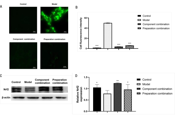
Figure 4: Effect of two combinations on the Nrf2 protein expression and ROS levels. (A) Levels of ROS were detected by fluorescence microscopy (400x). Scale bars: 20 µm. (B) Statistical histogram of ROS fluorescence intensity in each group. (C) Nrf2 protein expression determined by Western blot analysis. (D) Statistical histogram of Nrf2 protein expression. Statistical values are expressed as the mean ± SD from three independent experiments. *p < 0.05, **p < 0.01, and ***p < 0.001 compared with the model group. Please click here to view a larger version of this figure.
There is still a lack of ideal drugs for the treatment of cerebral ischemia in modern clinical practice2. Under the guidance of supplementing qi and activating the blood circulation method, Ast, Eri, and other preparations have been used in combination in the clinical practice of TCM and have achieved good comprehensive advantages13,14,15. A large number of studies have shown that Ast can improve the permeability of the blood-brain barrier, increase cerebral blood flow, improve the ability of nerve cells to tolerate hypoxia and resist oxygen free radical damage, and can significantly improve neurological dysfunction16,17. It is particularly suitable for senile ischemic cerebrovascular disease16,17. Eri can dilate blood vessels, increase blood supply to the brain, remove ROS, and reduce lipid peroxidation18,19. However, the synergistic effect, pharmacodynamic components, and the mechanism of the combination of the two drugs are still unclear, which is a common problem restricting the clinical application of TCM.
The ischemic and hypoxic environment of chronic cerebral ischemia induces oxidative stress damage and, at the same time, activates the brain cell apoptosis signaling pathway to promote neuronal apoptosis20,21. The PI3K/Akt pathway is a classic anti-apoptotic and pro-survival signal transduction pathway22,23, among which Bcl-2 family proteins and the caspase family are the downstream executive proteins of this signal pathway. Phosphorylated Akt can directly or indirectly regulate the Bcl-2 family and inhibit activation of the downstream pathway caspase-3, thereby exerting an anti-apoptotic effect11. In addition, excess ROS generated by hypoxia can induce oxidative stress injury, while phosphorylated Akt can activate Nrf2 to eliminate excess ROS to combat oxidative stress damage12,24. Therefore, activation of the PI3K/Akt/Nrf2 pathway can effectively prevent and control oxidative stress and apoptosis, thereby reducing hypoxic-ischemic brain injury25,26.
In the study, the preparation combination was screened using the injured PC12 cell model, and the effect and mechanism in this model were compared with the component combination screened previously7. In addition, the maximum nontoxic concentration of Ast injection and Eri capsule is 12 µM (calculated by astragaloside A) and 5 µM (calculated by scutellarin), respectively, while the maximum nontoxic concentration of astragaloside A and scutellarin is 20 µM and 50 µM, respectively7. It shows that the component combination is safer than the preparation combination, with more controllable quality and comprehensive advantages of high efficiency and low toxicity.
The current cell models that can be used to simulate cerebral ischemia mainly include physical hypoxia (induced by OGD) and chemical hypoxia (induced by Na2S2O4 or CoCl2)27. Among them, both OGD and Na2S2O4 injuries have a short hypoxia time and are not suitable for simulating chronic ischemia27. CoCl2-induced hypoxia is one of the most commonly used hypoxia mimics compared to OGD and the use of other hypoxia mimics. It can lead to persistent and stable oxidative damage by ROS and produce typical apoptotic changes in different cell lines27. Therefore, in this study, CoCl2 was used for 24 h to simulate the neuronal hypoxia28,29. Its modeling concentration (0.1, 0.2, 0.4, and 0.8 mM) and the validity period (1 and 7 days) were screened. In addition, given the inevitable lack of glucose and oxygen after cerebral ischemia, the glucose-free and hypoxic environment can better simulate cerebral ischemic injury. This study showed that 0.4 mM CoCl2 combined with a glucose-free medium for 24 h could establish a stable and controllable chronic hypoxic cell model. In the follow-up study, the animal model of chronic cerebral ischemia will be used to further validate the therapeutic advantages of the component combination in vivo.
This cell model can increase the apoptosis rate, caspase-3 fluorescence intensity, and intracellular ROS level (Figure 2 and Figure 4), which can better simulate the pathological changes of chronic cerebral ischemia, such as apoptosis, oxidative stress, etc. Based on this model, it was found that the two types of combinations can inhibit apoptosis and oxidative stress damage. The effect of the component combination on promoting cell survival was significantly better than that of the preparation combination (Figure 1); both combinations could upregulate the expression levels of P-Akt/Akt, Bcl-2/Bax, and Nrf2 in different degrees (Figure 3 and Figure 4). In particular, the component combination had a stronger effect on the up-regulation of Akt/Bcl-2/Bax and Nrf2 signaling pathway. These results indicate that the component combination can better resist cell damage than the preparation combination, which is related to stronger anti-apoptosis and anti-oxidative stress.
In conclusion, these results provide a combination strategy of two herbs to treat injured PC12 cells and provides a new idea for evaluating and optimizing the combined application mode of two herbs. Overall, this study provides a reference for selecting the active component combination of TCM.
The authors have nothing to disclose.
This research was funded by Key R & D projects of the Sichuan Provincial Department of science and technology (2020YFS0325).
1300 series class II biosafety cabinet | Thermo Fisher Scientific Instruments Company | 1374 | |
3-(4,5-Dimethylthiazol-2-yl)-2,5-diphenyltetrazolium bromide | Guangzhou saiguo biotech Company | 1334GR001 | MTT |
ACEA NovoExpress 1.4.0 | ACEA Biosciences, Inc | – | |
Akt | Wuhan Three Eagles Proteintech Group, Inc | 10176-2-AP | |
Analytical flow cytometry | Thermo Fisher Scientific Instruments Company | 62-2-1810-1027-0 | |
Annexin V-FITC apoptosis detection kit | Beyotime Biotechnology Company | C1062L | |
Astragalus injection | Heilongjiang Zhenbao Island Pharmaceutical Company | A03190612144 | Ast injection |
Astragaloside A | Chongqing Gao Ren Biotechnology Company | 84687-43-4 | |
Bax | Wuhan Three Eagles Proteintech Group, Inc | 60267-1-Ig | |
BCA protein quantification kit | Beyotime Biotechnology Company | P0012 | |
Bcl-2 | Wuhan Three Eagles Proteintech Group, Inc | 26593-1-AP | |
Carbon dioxide incubators | Thermo Fisher Scientific Instruments Company | 0816-2014 | |
Caspase-3 | Wuhan Three Eagles Proteintech Group, Inc | 19677-1-AP | |
Chlorogenic acid | Chongqing Gao Ren Biotechnology Company | 327-97-9 | |
Cobalt chloride hexahydrate | Merck Biotechnology, Inc. | 7791-13-1 | CoCl2 |
Dimethyl sulfoxide | Guangzhou saiguo biotech Company | 2020112701 | DMSO |
Disodium hydrogen phosphate dodecahydrate | Chengdu Kolon Chemical Company | 2020090101 | |
DMEM high sugar medium | Thermo scientific Hyclone | 2110050 | |
DMEM sugar free medium | Beijing Solarbio life sciences Company | 2029548 | |
ECL luminous fluid | Lianshuo Biological Company | WBKLS0500 | |
Electronic balance | Haozhuang Hengping Scientific Instrument Co., Ltd., Shanghai, China | FA1204 | |
Electrophoresis instrument | Bio-Rad Laboratories (Shanghai) Co., Ltd | 1658026 | |
Erigeron breviscapus capsule | Yunnan Biogu Pharmaceutical Company | Z53021671 | Eri capsule |
Fetal bovine serum | Four Seasons Institute of Biological Engineering | 20210402 | FBS |
Fluorescent microscope | Olympms Corporation | IX71-F32PH | |
Gel imager | Bio-Rad Laboratories (Shanghai) Co., Ltd | 1708265 | |
Goat anti-mouse secondary antibody | Wuhan Three Eagles Proteintech Group, Inc | SA00001-1 | |
Goat anti-rabbit secondary antibody | Wuhan Three Eagles Proteintech Group, Inc | SA00001- 2 | |
IBM SPSS Statistics version 26.0 | International Business Machines Corporation, USA | – | |
ImageJ 1.8.0 | National Institutes of Health, USA | – | imaging software |
Immunol fluorescence staining kit | Beyotime Biotechnology Company | P0196 | |
KCl | Chengdu Kolon Chemical Company | 2021070901 | |
Marker | Thermo Fisher Scientific Instruments Company | 26616 | |
Microplate reader | Perkin Elmer Corporate Management (Shanghai) Co. | HH35L2018296 | |
Motic Inverted microscope | Nanda Scientific Instruments Co. | AE2000LED | |
NaCl | Chengdu Kolon Chemical Company | 2014081301 | |
Nonfat milk | Beyotime Biotechnology Company | P0216 | |
Nrf2 | Wuhan Three Eagles Proteintech Group, Inc | 16396-1-AP | |
P-Akt | Wuhan Three Eagles Proteintech Group, Inc | 66444-1-Ig | |
PC12 cells | Chinese Academy of Sciences | CBP60430 | |
Penicillin-Streptomycin solution | Thermo scientific Hyclone | SV30010 | |
Phosphatase protease inhibitor mixture | Beyotime Biotechnology Company | P1045 | |
Potassium dihydrogen phosphate | Chengdu Kolon Chemical Company | 2015082901 | |
Reactive oxygen species assay kit | Beyotime Biotechnology Company | S0033S | |
RI-PA lysis solution | Beyotime Biotechnology Company | P0013B | |
Scutellarin | Chongqing Gao Ren Biotechnology Company | 27740-01-8 | |
SDS-PAGE sample loading buffer, 5x | Beyotime Biotechnology Company | P0015L | |
Sterile filter tips (0.22 µm) | Merck Biotechnology, Inc. | SLGP033RB | |
Tris-base | Guangzhou saiguo biotech Company | 1115GR500 | TBS |
Trypsin | Thermo scientific Hyclone | J190013 | |
Tween-20 | Chengdu Kolon Chemical Company | 2021051301 | |
Vortex oscillator | OHAUS International Co., Ltd., Shanghai, China | VXMNAL | |
β-actin | Wuhan Three Eagles Proteintech Group, Inc | 66009-1-Ig |