Electromyographic Endotracheal Intubation in Pig: A Technique to Insert Electromyographic Tube Inside the Pig Trachea

Published: April 30, 2023

Abstract

Source: Wu, C. W., et al. Intra-Operative Neural Monitoring of Thyroid Surgery in a Porcine Model. J. Vis. Exp. (144) (2019).

This video demonstrates a procedure to insert an electromyography tube inside the pig's trachea. It helps to keep the pig ventilated and monitor the electrical activity of laryngeal muscles in real-time.

Protocol

All procedures involving animal models have been reviewed by the local institutional animal care committee and the JoVE veterinary review board.

1. Tracheal intubation (Figure 1B)

  1. Prepare the equipment and materials required for electromyography (EMG) tube intubation: a size #6 EMG endotracheal tube, a face mask for assisted ventilation, two slings to hold the mouth open, one gauze strip to pull the tongue, a blunt tip suction catheter, a veterinary laryngoscope with 20cm straight blades, an elastic bougie, a 20-mL syringe, a stethoscope, and adhesive tape.
  2. Position the anesthetized piglet in a prone position on the operating table. Align the head and body to ensure clear visualization of the upper airway.
  3. Direct the assistant to apply traction of the upper and lower jaw to maintain an adequate mouth opening and to avoid rotation or overextension of the head. Cover the tongue with gauze and pull the tongue out to optimize the visual field.
  4. Hold the laryngoscope and place it directly in the oral cavity to depress the tongue.
  5. Directly visualize the epiglottis and use the laryngoscope to press the epiglottis downward toward the tongue base.
  6. When the vocal cords are clearly identified, gently advance the elastic bougie into the trachea. Slight rotation of the elastic bougie may be required to overcome resistance. Next, advance the EMG tube at the mouth angle to a depth of 24 cm.
  7. Inflate the EMG tube cuff to a volume no larger than 3 mL. If ventilation by manual bagging reveals no obvious air leakage, in situ deflation of the EMG tube is feasible.
  8. When the EMG tube is placed at the proper depth, confirm the free passage of fresh gas by manual bagging. Further confirm the proper tracheal intubation by end-tidal carbon dioxide (etCO2) monitoring (capnography) and chest auscultation for early identification of inadvertent esophageal or endobronchial intubation.
    NOTE: Capnography showed both the etCO2 waveform and the digital value in mmHg. When esophageal intubation occurred, etCO2 was absent or near zero after 6 breaths. When the EMG tube was in the correct place, the typical etCO2 waveform and adequate value (usually >30 mmHg) was noted. Furthermore, the breathing sound of a bilateral lung filled is clear and symmetric as determined by chest auscultation.
  9. Use medical tape to fix the EMG tube at the mouth angle. Since the tube usually requires adjustment during intraoperative neural monitoring (IONM) experiments, do not fasten the tube to the snout.
  10. Connect the EMG tube to the ventilator. Continuous capnography is mandatory for monitoring the etCO2 value and curve throughout the experiment.

2. Anesthesia maintenance (Figure 1C)

  1. After the EMG tube is fixed, position the piglet on its back with the neck extended (Figure 1C). Maintain general anesthesia with 1-3% sevoflurane in oxygen at 2 L/min.
  2. Ventilate the lungs in volume-control mode at a tidal volume of 8-12 mL/kg, and set the respiratory rate to 12-14 breaths/min.
  3. Begin physiologic monitoring, including capnography, electrocardiography (ECG)and monitoring of oxygenation (SaO2).

Representative Results

Figure 1
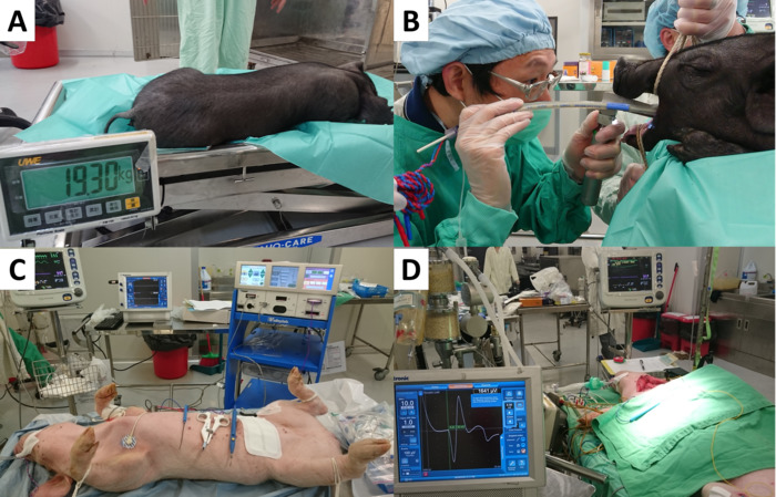
Figure 1. Preparation and anesthesia of KHAPS Black/Duroc-Landrace Pigs for IONM research. (A) Net weight of each piglet was measured before anesthesia. (B) An assistant maintained an adequate mouth opening while traction was applied to the upper and lower jaw. A laryngoscope was then used to press the epiglottis downward toward the base of the tongue. When the vocal cords were clearly identified, the elastic bougie was gently advanced into the trachea. The EMG tube was then inserted to a depth of 24 cm at the appropriate mouth angle. (C) The piglet was placed on its back with the neck extended. The channel leads from the recording electrodes were connected to the monitoring system. Physiologic monitoring was performed during the study. (D) The neck and the larynx were exposed for experiments.

Offenlegungen

The authors have nothing to disclose.

Materials

Criticare systems nGenuity 8100E physiologic monitoring, including capnography, electrocardiography (ECG) and monitoring of oxygenation (SaO2)
Intraoperative NIM nerve monitoring systems Medtronic NIM-Response 3.0 monitor EMG activity from multiple muscles. If there is a change in nerve function, the NIM system may provide audible and visual warnings to help reduce the risk of nerve damage.
NIM TriVantage EMG Tube Medtronic 8229706 6 mm ID, 8.2 mm OD. The NIM TriVantage EMG Tube is a standard size, non-reinforced, DEHP-free PVC tube that features smooth, conductive silver ink electrodes and a cross-band to guide placement. It has reduced sensitivity to rotation and movement while offering increased EMG responses that facilitate improved nerve dissection.
NIM Contact Reinforced EMG Endotracheal Tube Medtronic 8229506 6 mm ID, 9 mm OD. The NIM Contact EMG Tube continuously monitors electromyography (EMG) activity during surgery. An innovative design allows the tube to maintain contact, even upon rotation. Vocal cords are more easily visible against the white band. Recording electrode leads are twisted pair. Packaged sterile with one green and one white subdermal needle. Single use.
NIM Standard Reinforced EMG Endotracheal Tube Medtronic 8229306 6 mm ID, 8.8 mm OD. The NIM Standard EMG Tube continuously monitors electromyography (EMG) activity during surgery. Recording electrode leads are twisted pair. Packaged sterile with one green and one white subdermal needle. Single use.
NIM Flex EMG Endotracheal Tube Medtronic 8229960 6 mm. The NIM Flex EMG Tube monitors vocal cord and recurrent laryngeal nerve EMG activity during surgery. An updated, dual-channel design allows the tube to maintain contact with the vocal cords, even upon rotation. Recording electrode leads are twisted pair. Packaged sterile with one green and one white subdermal needle. Single use.
APS (Automatic Periodic Stimulation) Electrode* Medtronic 8228052 / 8228053 2 mm/ 3mm. The APS Electrode offers continuous, real-time monitoring. The electrode is placed on the nerve and can provide early warning of a change in nerve function.
Neotrode ECG Electrodes ConMed 1741C-003 The electrode is made of a clear tape material, which allows for continuous observation of the patient's skin during monitoring.

Tags

check_url/de/20931?article_type=t

Play Video

Diesen Artikel zitieren
Electromyographic Endotracheal Intubation in Pig: A Technique to Insert Electromyographic Tube Inside the Pig Trachea. J. Vis. Exp. (Pending Publication), e20931, doi: (2023).

View Video