A Dissection Method to Harvest Murine Popliteal Draining Lymph Nodes for Ex Vivo Imaging

Published: August 31, 2023

Abstract

Source: Rezende, R. M., et al. Visualizing Lymph Node Structure and Cellular Localization using Ex-Vivo Confocal Microscopy. J. Vis. Exp. (2019)

This video demonstrates a dissection technique to harvest popliteal draining lymph nodes from the mouse hind legs. Lymph nodes play a critical role in bridging innate and adaptive immunity, and harvesting intact lymph nodes — with a preserved tissue structure and cellular viability — can help study local and systemic immune responses.

Protocol

All procedures involving animal models have been reviewed by the local institutional animal care committee and the JoVE veterinary review board.

1. Surgery procedure to remove the popliteal draining lymph node

  1. Euthanize mice using CO2 asphyxiation followed by cervical dislocation.
  2. Immobilize mice at a prone position on the acrylic stage with adhesive tape and apply mineral oil with a cotton swab in the calf and knee (Figure 1A-D).
  3. Perform a midline incision in the calf from the heel to the knee (Figure 1E, F).
  4. Dissociate the calf musculature from the skin (Figure 1F, G).
  5. Expose the popliteal fossa (Figure 1G). The popliteal lymph node will appear as a translucid sphere in the popliteal fossa.
  6. Remove the popliteal lymph node using microsurgery curved forceps (Figure 1G).
  7. Alternatively, turn the mouse over on the supine position, and approach the popliteal fossa between biceps femoris and semitendinosus for popliteal LN removal by performing a midline incision in the calf from the heel to the knee followed by dissociation of the calf musculature from the skin.
    NOTE: Popliteal fossa is a shallow depression located at the back of the knee joint. Open carefully to see the popliteal lymph node.

Representative Results

Figure 1
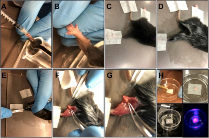
Figure 1: Popliteal lymph node preparation. (A, B) Subcutaneous injection of FACS antibody master mix into the paw pad. (C, D) 3 h after the injection, euthanize the mouse, immobilize the mouse on an acrylic plate with adhesive tape (C), and apply mineral oil to the abdominal skin to prevent fur deposition around the incision (D). (E) Perform a midline incision in the calf from the heel to the knee. (F) Expose the popliteal fossa. (G) Remove the popliteal lymph node using microsurgery-curved forceps. (H) Place the organ in a petri dish and remove the fat that surrounds the organ, centralize the organ in the middle of the Petri dish, cover the organ with a piece of delicate task wipers, and keep soaked with warm saline 0.9% or 1x PBS. Position the Petri dish in the microscope slot and scan the organ.

Offenlegungen

The authors have nothing to disclose.

Materials

Cellview culture dish Greiner-Bio 627861 35×10 mm with glass bottom
Kimwipes Kimtech Science Brand 7557 size 21 x 20 cm / 100 sheets per box
Microsurgery curved forceps WEP Surgical Instruments Custom-made 12.5 cm
Microsurgery curved scissors WEP Surgical Instruments Custom-made 11.5 cm

Tags

check_url/de/21578?article_type=t

Play Video

Diesen Artikel zitieren
A Dissection Method to Harvest Murine Popliteal Draining Lymph Nodes for Ex Vivo Imaging. J. Vis. Exp. (Pending Publication), e21578, doi: (2023).

View Video