A Method to Induce Ocular Surface Inflammation in a Murine Model
Abstract
Source: Singh, J., et al. Induction of Ocular Surface Inflammation and Collection of Involved Tissues. J. Vis. Exp. (2022)
This video demonstrates a method to induce ocular surface inflammation and meibomian gland dysfunction in a murine model. Stimulation of the mouse's immune system using an immunogen and later restimulation with the same immunogen promotes ocular surface inflammation, which causes plugging of meibomian gland secretion through neutrophil-produced aggNETs and leads to dry eye disease.
Protocol
All procedures involving animal models have been reviewed by the local institutional animal care committee and the JoVE veterinary review board.
1. Induction of murine ocular surface inflammation
- Perform immunogen preparation for immunization.
- Prepare the immunogen freshly on the day of immunization, mixing ovalbumin (OVA, 50 mg/mL) and pertussis toxin (100 µg/mL) in saline and the adjuvant aluminum hydroxide (40 mg/mL) (see Table of Materials) in a 1:1 proportion.
CAUTION: Perform the opening, reconstitution, and immunogen preparation of pertussis toxin in a laminar safety cabinet. Wear protective clothing and avoid any contact with the skin. - Incubate the immunogen and adjuvant mixture at room temperature for 30 min and load 100 µL into a 1 mL syringe.
- Perform intraperitoneal injection of the immunogen solution in a non-anesthetized mouse.
- Hold the mouse softly by the tail while grasping the cage grid. Hold firmly the skin of the back and the neck region between the thumb and index finger and fix the tail and lower limbs between the ring and little finger against the palm of the hand.
- Keep the fixed mouse with its head downward.
- Inject 100 µL of the prepared immunogen solution in the right or left quadrant of the lower abdominal cavity.
- Prepare the immunogen freshly on the day of immunization, mixing ovalbumin (OVA, 50 mg/mL) and pertussis toxin (100 µg/mL) in saline and the adjuvant aluminum hydroxide (40 mg/mL) (see Table of Materials) in a 1:1 proportion.
- Perform ocular surface challenge 2 weeks after immunization.
- Anesthetize the mouse with isoflurane (2.5%).
- Apply 5 µL of OVA (50 mg/mL) or saline (0.9 % NaCl) per eye on both eyes and wait until the drop gets absorbed by the eye. This takes ~5 min.
- Repeat the procedure 1x daily for 7 days.
NOTE: The topical administration of medications can be done in the same way.
2. Collection of ocular exudates
- Recover the ocular exudates formed during the challenge phase by applying 50 µL of sterile saline to the eye immediately after the challenge.
3. Excision of ocular surface tissues
- Dissect the eyelids and eye globe following the steps below.
- Euthanize the mouse by CO2 asphyxiation and cervical dislocation.
- Place the mouse on an even surface.
- Disinfect the orbital area around the eye with a swab impregnated with 70% ethanol.
- Make an incision between the ear and the retro-orbital sinus and along the surface above the squamous bone vertically, extending the incision horizontally below the lower eyelid along the maxillary bone and above the upper eyelid along the frontal bone. This forms an incision around the eye (Figure 1).
- Carefully hold the dissected tissue around the eye using curved forceps and pull the tissue and eyeball out.
- Place the excised organs in sterile PBS.
- Trim excess facial muscle tissue around the excised upper eyelids using a scalpel and under the stereomicroscope.
- Collect the conjunctiva following the steps below.
- Place the upper eyelid on a dry Petri dish under the stereomicroscope.
- Using fine tweezers and a scalpel, peel the whitish-mucous layer very gently out of the inner surface of the eyelid.
- Next, dissect the cornea following the steps below.
- Place the eye globe on a new dry Petri dish on top of dry ice for 3 min.
- Take the Petri dish onto the bench surface in a stable position.
- Make a small incision at the border of the cornea next to the limbus using fine sharp scissors.
- Prolong the incision with the scalpel around the eye globe, separating the sclera from the cornea.
- Remove remnants of the iris and the lens from the backside of the cornea by generously flushing with saline solution.
Representative Results
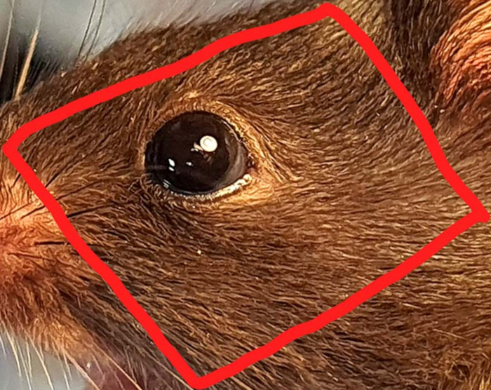
Figure 1: Excision of the facial tissue surrounding the orbital region. The incision trail is indicated in red. This allows for excising the ocular organs and investigating the influence and effects of various topically administered therapeutics on accessory ocular organs such as the conjunctiva and cornea.
Offenlegungen
The authors have nothing to disclose.
Materials
| 1x PBS | Gibco | ||
| Aluminium Hydroxide | Imject alum Adjuvant | 77161 | 40 mg/ mL Final Concentration: in vivo: 1 mg/ 100 µL |
| C57Bl/6 mice, aged 7–9 weeks | Charles River Laboratories | ||
| Curved forceps | FST by Dumont SWITZERLAND | 5/45 11251-35 | |
| Fine sharp scissor | FST Stainless steel, Germany | 15001-08 | |
| Laminar safety cabinet | Herasafe | ||
| NaCl 0.9% (Saline) | B.Braun | ||
| Ovalbumin (OVA) | Endofit, Invivogen | 9006-59-1 | 10 mg/200 µL in saline |
| Pertussis toxin | ThermoFisher Scientific | PHZ1174 | 50 µg/ 500 µL in saline Final Concentration: in vivo: 100 µg/ 100 µL |
| Petridish | Greiner bio-one | 628160 | |
| Scalpel | Feather disposable scalpel | No. 21 | Final Concentration: in vivo: 300 ng/ 100 µL |
| Stereomicroscope | Zaiss | Stemi508 | |
| Syringe (corneal/iris washing) | BD Microlane | 27 G x 3/4 – Nr.20 0,4 x 19 mm | |
| Syringe (i.p immunization) | BD Microlane | 24 G1"-Nr 17, 055* 25 mm |
