Generating a Multivalent-Displaying Outer Membrane Vesicle Vaccine
Abstract
Source: Feng, R., et al. A "Plug-And-Display" Nanoparticle Vaccine Platform Based on Outer Membrane Vesicles Displaying SARS-CoV-2 Receptor-Binding Domain. J. Vis. Exp. (2022).
This video demonstrates a method to generate multivalent-displaying outer membrane vesicle (OMV) vaccines. Mammalian cells are transfected to express and secrete viral antigens fused to a SpyTag and a polyhistidine tag. These antigens are then purified using a nickel-nitrilotriacetic acid (Ni-NTA) affinity column. The purified antigen is mixed with outer membrane vesicles expressing surface-bound SpyCatcher (SC) protein. Multiple antigens covalently attach to SpyCatchers via their SpyTags (ST), creating OMVs that display the antigen, serving as a multivalent vaccine.
Protocol
1. Plasmid construction
- Insert DNA encoding SpyCatcher sequence (Figure 1) into an ampicillin-resistant pThioHisA-Cytolysin A (ClyA) plasmid (see Table of Materials) between the BamH I and Sal I sites to construct the plasmid pThioHisA ClyA-SC following a previously published report.
- Ligate the synthesized SpyTag-SARS-CoV-2 receptor-binding domain (RBD)-polyhistidine tag (Histag) fusion gene (Figure 1) into a pcDNA3.1 plasmid (see Table of Materials) between the BamH I and EcoR I sites to construct the plasmid pcDNA3.1 RBD-ST following a previously published report.
2. OMV-SC preparation
- Perform ClyA-SC transformation following the steps below.
- Add 5 µL of pThioHisA ClyA-SC plasmid solution (50 ng/µL) to 50 µL of BL21 competent strain, gently blow, and let cool on ice for 30 min.
- Place the solution for 90 s in the water bath at 42 °C, and then immediately put the mixed solution on ice for 3 min.
- Add 500 µL of Luria-Bertani (LB) medium to the bacterial suspension and, after mixing, culture at 220 rpm at 37 °C for 1 h.
- Plate all the transformation onto an LB agar plate containing ampicillin (100 µg/mL, see Table of Materials) and culture overnight at 37 °C.
NOTE: In subsequent experiments, the ampicillin was kept at the same concentration in the medium. If not, this may cause loss of plasmids in the bacteria.
- For OMV-SC production, perform the steps below.
- Isolate a single colony from the plate (step 2.1.4.) to 20 mL of LB (ampicillin-resistant) medium and culture overnight at 220 rpm at 37 °C.
- Inoculate the bacterial solution (from step 2.2.1.) into 2 L medium, culture at 220 rpm, 37 °C for 5 h until the logarithmic growth stage (the OD600nm is between 0.6 to 0.8).
- When the OD at 600 nm of the bacterial solution reaches 0.6-0.8, add isopropyl beta-D-thiogalactopyranoside (IPTG, see Table of Materials) to make the final concentration of the bacterial solution to be 0.5 mM, and then culture overnight at 220 rpm at 25 °C.
- Perform OMV-SC purification.
- Centrifuge the bacterial solution at 7,000 x g at 4 °C for 30 min.
- Filter the supernatant with a 0.22 µm membrane filter, then concentrate it using a 100 kD ultrafiltration membrane or hollow fiber column (see Table of Materials).
- Filter the concentrate through a 0.22 µm membrane filter, then centrifuge it at 150,000 x g at 4 °C for 2 h using an ultracentrifuge (see Table of Materials), and discard the supernatant with a pipette.
- Resuspend the precipitation with PBS and store it at −80 °C. The solution can maintain long-term stability at −80 °C.
3. RBD-ST preparation
- Perform RBD-ST transfection following the steps below.
- Select an appropriate eukaryotic expression system (e.g., HEK293F) and culture the cells overnight at 130 rpm at 37 °C after recovery.
- Add 20 µL of HEK293F cell solution into the automated cell counter (see Table of Materials), record the number of cells, adjust the concentration to 1 x 10⁶ cells/mL, and then culture the cells at 130 rpm at 37 °C for 4 h.
- Filter RBD-ST plasmid through a 0.22 µm membrane filter, and add 300 µg of plasmids into the cell culture medium (see Table of Materials) until the final volume is 10 mL; shake for 10 s.
- Heat polyethylenimine (PEI, 1 mg/mL, see Table of Materials) to 65 °C in the water bath, mix 0.7 mL of PEI with 9.3 mL of cell culture medium, and shake intermittently for 10 s.
NOTE: Do not shake the solution vigorously. Otherwise, the resulting bubbles may affect the transfection efficiency. - Add plasmid solution to the PEI solution, shake the mixture intermittently for 10 s, and incubate it at 37 °C for 15 min.
- Add the mixture to 280 mL of cell culture medium and culture at 130 rpm at 37 °C for 5 days.
- Perform RBD-ST purification.
- Centrifuge the cells at 6,000 x g at 25 °C for 20 min and use a pipette to collect the supernatant.
- Fill the column with 2 mL of Ni-NTA agarose (see Table of Materials) and wash it 3x with 3x PBS.
- Add imidazole (see Table of Materials) into the cell supernatant to make the final concentration of 20 mM, and load the cell supernatant 2x.
- Add 3 column volumes (CV) of PBS containing 20 mM of imidazole for washing, and collect the washing fraction.
- Gradient elute with 3 CV of PBS containing low to high concentrations (e.g., 0.3 M, 0.4 M, 0.5 M) of imidazole; elute 2x for each concentration.
- Use SDS-PAGE to identify the RBD-ST in different concentration gradients.
4. OMV-RBD bioconjugation and purification
- Determine the protein concentration by the bicinchoninic acid assay (BCA) method (see Table of Materials).
- Serially dilute the standard bovine serum albumin (BSA) protein solution from 2 mg/mL to 0.0625 mg/mL, dilute the purified OMV-SC and RBD-ST 10x, then mix BCA working solutions A and B (provided in the assay kit) at a ratio of 50:1 (v/v).
- Add diluted protein solution (25 µL/well) and mix with BCA working solution (200 µL/well); incubate at 37 °C for 30 min.
- Measure the absorbance (OD) at 562 nm of each well and calculate the protein concentration from the standard curve.
- Perform bioconjugation of OMV-SC and RBD-ST following the steps below.
- Mix OMV-SC and RBD-ST in PBS at a 40:1 (w/w) ratio.
- Vertically rotate to blend the mixture overnight at 15 rpm at 4 °C.
NOTE: Different antigens may react in different proportions. One could try different reaction ratios based on the characteristics of the antigen.
Representative Results
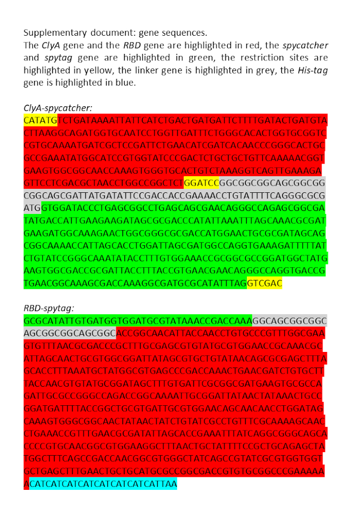
Figure 1: Plasmid sequences used in the present study.
Offenlegungen
The authors have nothing to disclose.
Materials
| Ampicillin sodium | Sangon Biotech | A610028 | |
| Automated cell counter | Countstar | BioTech | |
| BCA protein quantification Kit | cwbio | cw0014s | |
| Electrophoresis apparatus | Cavoy | Power BV | |
| High speed freezing centrifuge | Bioridge | H2500R | |
| His-Tag mouse mAb | Cell signaling technology | 2366s | |
| Imidazole | Sangon Biotech | A600277 | |
| Isopropyl beta-D-thiogalactopyranoside | Sangon Biotech | A600118 | |
| Ni-NTA His-Bind Superflow | Qiagen | 70691 | |
| OPM-293 cell culture medium | Opm biosciences | 81075-001 | |
| pcDNA3.1 RBD-ST plasmid | Wuhan genecreat biological techenology | ||
| Phosphate buffer saline | ZSGB-bio | ZLI-9061 | |
| Polyethylenimine Linear | Polysciences | 23966-1 | |
| Prestained protein ladder | Thermo | 26616 | |
| pThioHisA ClyA-SC plasmid | Wuhan genecreat biological techenology | ||
| Quixstand benchtop systems (100 kD hollow fiber column) | GE healthcare | ||
| SDS-PAGE loading buffer (5x) | Beyotime | P0015 | |
| Sodium chloride | Sangon Biotech | A100241 | |
| Suspension instrument | Life Technology | Hula Mixer | |
| Tryptone | Oxoid | LP0042B | |
| Ultracentrifuge | Beckman coulter | XPN-100 | |
| Ultraviolet spectrophotometer | Hitachi | U-3900 | |
| Yeast extract | Sangon Biotech | A610961 |
Tags

.