SIVQ-LCM Protocol for the ArcturusXT Instrument
Summary
SIVQ-LCM is an innovative approach that harnesses a computer algorithm, Spatially Invariant Vector Quantization (SIVQ), to drive the laser capture microdissection (LCM) process. The SIVQ-LCM workflow greatly improves the speed and accuracy of microdissection, with applications in both the research and clinical settings.
Abstract
SIVQ-LCM is a new methodology that automates and streamlines the more traditional, user-dependent laser dissection process. It aims to create an advanced, rapidly customizable laser dissection platform technology. In this report, we describe the integration of the image analysis software Spatially Invariant Vector Quantization (SIVQ) onto the ArcturusXT instrument. The ArcturusXT system contains both an infrared (IR) and ultraviolet (UV) laser, allowing for specific cell or large area dissections. The principal goal is to improve the speed, accuracy, and reproducibility of the laser dissection to increase sample throughput. This novel approach facilitates microdissection of both animal and human tissues in research and clinical workflows.
Introduction
Originally developed in the mid-1990s, laser capture microdissection (LCM) enables the user to precisely capture specific cells or cellular regions from a histological tissue section via microscopic visualization1,2. Many studies comparing molecular analysis of LCM versus tissue scrapes illustrate the value of the method3-12. In addition, there are three video protocol publications on the technology that are available for viewing13,14. However, despite its proven value, LCM can be tedious and laborious when the target of interest is a dispersed cell population in a heterogeneous tissue section, or when large numbers of cells are required for specific downstream applications such as proteomics. The burden placed on the human operator led us to develop a semi-automated dissection approach for LCM by combining a powerful image analysis algorithm to guide the LCM process15.
In collaboration with the University of Michigan, our laboratory at the NIH extended the previously developed and reported Spatially invariant vector quantization (SIVQ) algorithm in a manner to allow it to semi-automate the tissue selection process intrinsic to guided microdissection, thus making available a tool with the pathologist or life scientist in mind. Spatially invariant vector quantization (SIVQ) is an algorithm that allows the user to simply “click” on a histological feature of interest to create a ring vector (predicate image feature) that can be used to search the entire histological image, adjusting the statistical threshold as needed16-21. The resultant heat map displays the quality of matches to the initial predicate image feature and is subsequently converted into a single color (red) annotation map that can be imported into the LCM instrument. The automated selection software, AutoScanXT, is then used to draw a map based on SIVQ's annotation guiding the capture of the target cells from the tissue sample. The detailed protocol below describes the implementation of SIVQ into the microdissection workflow.
Protocol
The described protocol was employed in accordance with NIH rules on the use of human tissue samples.
1. Tissue Preparation
- Prior to beginning, obtain human tissue specimens according to Institutional Review Board (IRB) protocols.
- Choose the type of tissue/cell block and corresponding processing method [formalin-fixed paraffin-embedded (FFPE), frozen, or ethanol-fixed paraffin-embedded (EFPE)]. Formalin fixation provides optimal histology, followed by ethanol fixation and flash frozen. However, fixation and tissue processing methods can affect DNA, RNA, and protein quantity and quality for downstream molecular analysis and should be considered.
- Cut the tissue/cell block sections onto the slide type (glass, membrane glass, or metal frame membrane) of choice. SIVQ analysis works equally well on all three slide types. Please note that the pseudo-coverslip method (described below) with xylenes and ethanol cannot be performed on the metal frame membrane slides since the slide must be inverted on the stage.
- Select a chemical- or IHC-based tissue stain to identify cells of interest from the background. Note that staining methods can also affect DNA, RNA, and protein quality and quantity of the tissue. Test the tissue after staining to assess baseline quality of biomolecules before proceeding with the protocol. SIVQ-LCM has been performed on tissue/cytology slides stained with: immunohistochemistry (IHC) with DAB15, immunofluorescence, fast red, de novo red, toluidine blue, and hematoxylin and eosin (H&E) (unpublished data).
2. Specimen Imaging
- Load slides onto the motorized stage of the microdissection instrument and launch the related software. Select the check boxes to designate the positions of the loaded slides and ensure the captured image files are to be saved in jpeg format.
- Optimize the image quality by adjusting the brightness and focus of the image on the screen, using either the manual focus wheel or through the dissection instrument’s software.
- Using the Image Toolbox within the dissection instrument’s software, set the lamp brightness and camera gain appropriately. Example values are brightness = 60 and gain = 220, with the diffuser.
- Focus either manually or with the autofocus feature in the software.
- Capture a thumbnail overview image of the slide to provide a roadmap for the dissection process.
- For optimal image quality of an uncoverslipped slide, utilize the diffuser below the condenser on the instrument, or, add a small amount (~30 μl) of either ethanol or xylenes to improve the refractory index (pseudo-coverslip). When using xylenes, be aware they are toxic and proper safety measures must be used, including the use of a fume hood and protective lab coat, eye goggles, and gloves.
- Do NOT place the LCM cap on the slide until the ethanol or xylenes solution has completely cleared or the polymer on the cap will be distorted.
- Navigate the slide and capture images of the areas to be dissected at 10X, 20X, or 40X magnification. If necessary, enhance the image with software such as AutoCorrect (in Microsoft Office Picture Manager) as previously described22. Images must be captured in jpeg format to allow them to be re-imported into the automated selection software.
3. Algorithm Analysis of the Image
- Transfer captured images from the microdissection instrument to the SIVQ folder. Install and open the ArcturusXT, AutoScan and SIVQ software packages on the computer attached to the dissection instrument. For access to the SIVQ software, please contact Dr. Ulysses Balis (ulysses@med.umich.edu).
- Open SIVQ and load the captured image (jpeg) of interest.
- Navigate to the area of interest and/or adjust the size of the display windows (Viewport 5 & 6). In the SIVQ software, Viewport 5 shows the pre-processing image and Viewport 6 shows the post-processing image16.
- Choose the size of the ring vector and number of rings to be used.
- Select the predicate image feature to be captured by right clicking on it in Viewport 6.
- Click “scan” to analyze the image.
- Adjust the statistical probability of image matching using the two sliding bars. The upper bar adjusts the overall vector specificity, and is used to exclude area from the initial scan (with sensitivity as defined by the selected “Stat” variable) that may represent excessive included area. Conversely, the lower slider is utilized to increase sensitivity, after a scan is performed, with the intent of increasing area that is classified as being a match. Both of these slider controls utilize the “Stat” variable as the initial baseline sensitivity threshold.
- To save the image, click “save as jpeg” (image is now saved in c:/vq_test folder/pics).
- Analyze the image with the algorithm. The output of the algorithm analysis needs to result in an annotated image for it to be used in SIVQ-LCM. At present, the current version of the SIVQ core engine is in beta version testing with the expectation that the full production version (available Q1 2014) will incorporate a full software development kit (SDK) and application programming interface (API) for simplified integration of user-generated spatial filters and downstream workflow data processing steps with the core ring matching engine. This SDK will be distributed with a complete set of documentation.
- Ensure that the SIVQ heatmap is changed to a uniform red color.
- Export the image. It is essential to re-embed the positional coordinates in the post-analysis jpeg image by using a HEX editor to paste in the file header from the original dissection instrument image. The appropriate data may be found between “Start of Image” marker (0xFF, 0xD8) and the first “Define Quantization Table” marker (0xFF, 0xDB).
4. Microdissection
- Place the LCM cap at the center of the region of interest, where the images were captured for the SIVQ analysis.
- Calibrate and Quality Control (QC) the UV/IR lasers and optimize the parameters, including power, duration, laser locations, and UV cutting speed (as recommended by the manufacturer). Perform these calibrations before re-importing the analyzed image.
- Open AutoScanXT (automated selection software) and import the analyzed image from c:/vq_test folder/pics.
- Train the automated selection software to recognize the SIVQ annotation and create the dissection map.
- To create a training file, select four regions of interest (marked by “blue circles”) on the “red paint” of the SIVQ analyzed image.
- Select the background areas (marked by “red squares”) that are not to be dissected.
- Click on the “Analyze” button to generate the training file, which can be saved for subsequent uses.
- Perform microdissection using the appropriate infrared (IR) and/or ultraviolet (UV) lasers.
- Copy the selected areas onto the “live” image.
- In the “microdissect” toolbox, select the appropriate IR capture or UV cutting buttons.
- After the dissection is complete, move the LCM cap to the QC station and capture an image of the dissected tissue/cells. Another approach is to place the cap on a blank area of the slide prior to moving it to the QC station, this allows the user to take pictures at various magnifications.
- Capture an image of the tissue area of interest after microdissection to further assess lifting efficiency.
- If the desired cells have been dissected successfully, click on the "present stage" button in the ArcturusXT software and remove the LCM cap to initiate the molecular extraction procedure for downstream analysis.
Representative Results
A FFPE human breast tissue section was immunostained for cytokeratin AE1/AE3 using a standard IHC protocol23. After staining, the tissue slide was placed on the ArcturusXT stage and the SIVQ-LCM protocol was initiated as described above. Since the tissue cannot be coverslipped for microdissection, the IHC+ stained cells can be difficult to discern visually (Figure 1A). Thus, to provide better index matching and an improved image, xylenes were added to the tissue section to create a pseudo-coverslip15 (Figure 1B). A jpeg image was then captured of the pseudo-coverslipped area and imported into SIVQ for the algorithm analysis. A predicate image feature (dark brown DAB stain) selected by the user initiated the SIVQ algorithm to analyze the image (Figure 1C). The SIVQ heatmap was then converted to a “red paint” that is recognized by the automated selection software (Figure 1D). The xylenes were allowed to evaporate and the SIVQ heatmap was imported into the automated selection software (Figure 1E) and the highlighted cells were dissected with the IR laser. The LCM cap was then moved to the QC station of the ArcturusXT instrument and visually inspected to assess dissection efficiency (Figure 1F). The remaining tissue was also inspected (Figure 1G) and the SIVQ heatmap was re-imported to further assess the microdissection efficiency (Figure 1H).
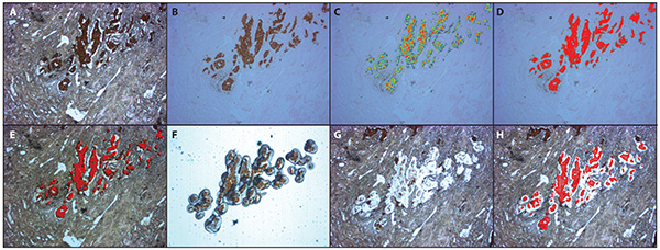
Figure 1. SIVQ-LCM of IHC stained FFPE breast tissue. A) Uncoverslipped image of cytokeratin AE1/AE3 stained FFPE breast tissue. B) Xylenes coated (pseudo-coverslipped) image of panel A. C) SIVQ generated heatmap using a ring vector (predicate image) captured from the brown chromagen. D) The heatmap was converted to a single color (red). In the ArcturusXT, the AutoScanXT software was trained to recognize the red annotation. E) The microdissection map generated from the AutoScanXT algorithm. F) Image of the microdissected breast epithelial cells on the LCM cap. G) Image of the tissue on the slide after microdissection. H) Overlap of the microdissection map (panel E) on the tissue area that was dissected. Please click here to view a larger version of this figure.
Discussion
We present a protocol for the application of SIVQ-LCM to microdissect immunostained epithelial cells from FFPE human breast tissue. The use of an image analysis algorithm, such as SIVQ, reduces the amount of hands-on time required for the microdissection process. This is a potentially important advance for the field since operator time and effort is typically the rate-limiting step for the precise dissection of cells of interest. In the present protocol, we specifically adapted our procedure to the ArcturusXT instrument, although it likely will be possible to adapt SIVQ and other algorithms to other commercially available microdissection instruments as well. In addition to improving efficiency, this new approach may allow users who are non-histopathology experts to perform microdissection. Finally, the use of an algorithm to drive the microdissection process may enable greater reproducibility from site-to-site by removing user subjectivity in identifying cells of interest.
While SIVQ exhibits broad adaptability to multiple classes of image subject matter, including histology, it is important to recognize that it is not intended to serve as a universal image processing/segmentation tool, but rather, as a high-efficiency first-pass foreground selection tool. Used in this capacity, it has high utility for predicting whether or not a particular class of imagery subject matter is suitable for numerical segmentation approaches. When SIVQ is successful, there is high predictive power that either: a) a more refined and targeted image analysis/segmentation approach will be highly successful and/or b) an optimized, machine-selected SIVQ vector will be effective in rendering a clinical-workflow-ready solution. In either circumstance, the use of SIVQ serves as an effective triaging tool to identify imagery subject matter that poses computationally tractable solutions in terms of foreground segmentability.
While we previously described the use of the SIVQ-LCM protocol only for mRNA expression microarray experiments15, we believe the protocol is applicable to most downstream molecular assays, and may be particularly useful for molecular assays that require large numbers of cells, such as proteomics. For these studies, large amounts of cells need to be procured due to the inability to amplify proteins. In addition, the advent of newer technologies, such as Next-Gen Sequencing (NGS), emphasizes the need to begin with as pure of a sample as possible. The ability for SIVQ-LCM to isolate cells via both morphological features and/or staining qualities could enable the ability to more easily isolate pure cell populations.
It is critical to have properly prepared tissue for reproducible results for both the SIVQ analysis and microdissection. For optimal conditions for LCM it is best to perform dissects in a low humidity room and maintain properly dehydrated tissue. In addition, ensure that the staining of the tissue is reproducible. Finally, familiarity with the dissection instrument is essential to successful SIVQ-LCM.
The SIVQ-LCM protocol presented here is the first steps towards semi-automated microdissection, which may have applications in future clinical testing since current dissection methods are not easily capable of handling large workloads. In summary, we believe that SIVQ-LCM is a new foray into the field of automated microdissection and provides a starting point from which investigators and algorithm developers can further improve the technology.
Offenlegungen
The authors have nothing to disclose.
Acknowledgements
The study was supported in part by the Intramural Research Program of the National Institutes of Health, National Cancer Institute, Center for Cancer Research.
Materials
| Positive Charged Glass Slides | Thermo Scientific | 4951Plus-001 | |
| Xylenes, ACS reagent, ≥98.5% xylenes + ethylbenzene basis | Sigma Aldrich | 247642 | CAUTION: PLEASE USE PROPER SAFETY PROCEDURES. |
| Ethyl Alcohol, U.S.P. 200 Proof, Anhydrous | The Warner-Graham Company | 6.505E+12 | CAUTION: PLEASE USE PROPER SAFETY PROCEDURES. |
| Arcturus CapSure Macro LCM Caps | Life Technologies | LCM0211 | |
| ArcturusXT Laser Microdissection Instrument | Life Technologies | ARCTURUSXT | |
| AutoScanXT Software | Life Technologies | An optional image analysis program for the ArcturusXT Laser Microdissection Device. This is software is required for SIVQ-LCM. | |
| Spatially Invariant Vector Quantization (SIVQ) | University of Michigan | This tool suite is publicly available for academic collaborations. For access to the SIVQ algorithm, please contact Dr. Ulysses Balis [Ulysses@med.umich.edu] |
Referenzen
- Bonner, R. F., et al. Laser capture microdissection: molecular analysis of tissue. Science. 278, 1481-1483 (1997).
- Emmert-Buck, M. R., et al. Laser capture microdissection. Science. 274, 998-1001 (1996).
- Edwards, R. A. Laser capture microdissection of mammalian tissue. J Vis Exp. (8), (2007).
- El-Serag, H. B., et al. Gene expression in Barrett’s esophagus: laser capture versus whole tissue. Scandinavian journal of gastroenterology. 44, 787-795 (2009).
- Espina, V., et al. Laser-capture microdissection. Nature. 1, 586-603 (2006).
- Harrell, J. C., Dye, W. W., Harvell, D. M., Sartorius, C. A., Horwitz, K. B. Contaminating cells alter gene signatures in whole organ versus laser capture microdissected tumors: a comparison of experimental breast cancers and their lymph node metastases. Clinical & experimental metastasis. 25, 81-88 (2008).
- Rodriguez-Canales, J., et al. Optimal molecular profiling of tissue and tissue components: defining the best processing and microdissection methods for biomedical applications. Methods in molecular biology. 980, 61-120 (2013).
- Silvestri, A., et al. Protein pathway biomarker analysis of human cancer reveals requirement for upfront cellular-enrichment processing. Laboratory investigation; a journal of technical methods and pathology. 90, 787-796 (2010).
- Eberle, F. C., et al. Immunoguided laser assisted microdissection techniques for DNA methylation analysis of archival tissue specimens. The Journal of molecular diagnostics : JMD. 12, 394-401 (2010).
- Kim, H. K., et al. Distinctions in gastric cancer gene expression signatures derived from laser capture microdissection versus histologic macrodissection. BMC medical genomics. 4, 48 (2011).
- Klee, E. W., et al. Impact of sample acquisition and linear amplification on gene expression profiling of lung adenocarcinoma: laser capture micro-dissection cell-sampling versus bulk tissue-sampling. BMC medical genomics. 2, 13 (2009).
- Zheng, J., Garg, S., Wang, J., Loose, D. S., Hauer-Jensen, M. Laser capture microdissected mucosa versus whole tissue specimens for assessment of radiation-induced dynamic molecular and pathway changes in the small intestine. PloS one. 8, e53711 (2013).
- Boone, D. R., Sell, S. L., Hellmich, H. L. Laser capture microdissection of enriched populations of neurons or single neurons for gene expression analysis after traumatic brain injury. J Vis Exp. (74), (2013).
- Iyer, E. P., Cox, D. N. Laser capture microdissection of Drosophila peripheral neurons. J Vis Exp. (39), (2010).
- Hipp, J., et al. SIVQ-aided laser capture microdissection: A tool for high-throughput expression profiling. Journal of pathology informatics. 2, 19 (2011).
- Hipp, J. D., Cheng, J. Y., Toner, M., Tompkins, R. G., Balis, U. J. Spatially Invariant Vector Quantization: A pattern matching algorithm for multiple classes of image subject matter including pathology. J Pathol Inform. 2, 13 (2011).
- Hipp, J., et al. Optimization of complex cancer morphology detection using the SIVQ pattern recognition algorithm. Anal Cell Pathol (Amst). , (2011).
- Hipp, J., et al. Integration of architectural and cytologic drive n image algorithms for prostate adenocarcinoma identification. Analytical cellular pathology. 35, 251-265 (2012).
- Hipp, J., et al. Automated area calculation of histopathologic features using SIVQ. Anal Cell Pathol (Amst. 34, (2011).
- Cheng, J., et al. Automated vector selection of SIVQ and parallel computing integration MATLAB: Innovations supporting large-scale and high-throughput image analysis studies. Journal of pathology. 2, 37 (2011).
- Roy Chowdhuri, S., et al. Semiautomated laser capture microdissection of lung adenocarcinoma cytology samples. Acta Cytol. 56, 622-631 (2012).
- Hipp, J., et al. Image Microarrays (IMA): Digital Pathology’s Missing Tool. Journal of pathology. 2, (2011).
- Hanson, J. C., et al. Expression microdissection adapted to commercial laser dissection instruments. Nature. 6, 457-467 (2011).
