1. Focusing the Nanosecond Laser and Measuring Fluence
- Assemble the ablation apparatus by placing a magnetic stir bar and a porous ablation stage inside a 50 mL glass beaker.
NOTE: The ablation stage consists of a 3.81 cm diameter, 1.6 mm thick stainless steel platform with ten 0.65 cm diameter holes and six 0.50 cm diameter holes drilled in the pattern illustrated in Figure 1. The purpose of these holes is to allow the liquid to move across the target so that particles do not accumulate immediately above the target. Insufficient mixing leads to deleterious interactions between the laser and the already-formed particles. Additionally, three #29 (8-32) tapped holes are located near the perimeter of the platform to accept set screws which serve as legs to raise the platform and provide space for a magnetic stir bar (Figure 1A).- Place the beaker on a magnetic stir plate and set the stir plate upon an xy-translation stage to enable movement of the target during ablation (Figure 1A).
- Set the Nd:YAG laser to operate at the fundamental wavelength of 1,064 nm, with a pulse duration of 5 ns, and a pulse repetition rate of 10 Hz. Measure the energy per pulse with a laser power and energy meter. The energy required is 240-250 mJ.
- Focus the beam beneath the target on the ablation stage using a 250 mm focal length converging lens (NA = 0.05).
NOTE: The incoming beam has a radius of 4.025 mm and a lens height of 161 mm is required to attain the desired spot size. The optimal spot size is determined empirically. A larger spot size is utilized to reduce the effect of shielding by NPs present in solution. This is balanced with the fact that increasing spot size requires higher energy to maintain adequate fluence. - Determine the spot size by placing a metal target (see section 2) on the stage and ablating with several laser pulses. View the ablated target together with a micrometer slide on a CCD camera-equipped light microscope (4X objective) to measure the spot size (Figure 1A).
NOTE: For the apparatus here, the ablation system yields a spot size with a mean area of 5.51 mm2. The spot size remains in this range for each ablation. - Calculate fluence by dividing the pulse energy by the spot area. For the apparatus here, the fluence is 4.80 J/cm2.
2. Synthesis of Silver Nanoparticles by Pulsed Laser-ablation in Liquid
- Weigh a flat silver target using a microbalance to obtain the pre-ablation mass.
- Adhere the silver target to the porous stage using double-sided carbon tape. Add 40 mL of ablation liquid to the beaker (Figure 1A). The liquid height above the target is 11 mm.
NOTE: Typical ablation liquids are aqueous solutions containing either 60 mM SDS or 2 mM PVP to enhance monodispersity. - Under constant stirring, move the computer-controlled motorized xy-stage in an elliptical pattern (dimensions: major axis = 2.09 cm, minor axis = 0.956 cm, area = 1.57 cm2) at a speed of 0.42 cm/s and ablate the target for 20-40 min.
NOTE: The concentration of NPs increases with longer ablation times. Ensure that the stirring is sufficiently vigorous to keep the NP concentration uniform throughout the solution to minimize shielding effects7.
3. Characterizing Metal Nanoparticles
- Collect the nanoparticle solution from the beaker by decanting. Confirm the presence of nanoparticles by measuring their UV-visible light spectra (200-1,100 nm).
NOTE: The NPs have a peak-absorption at the surface plasmon resonance (SPR) wavelength. For silver, the SPR is centered at 400 nm. Highly concentrated NP solutions require dilution prior to measuring the UV-VIS spectrum to ensure that the absorbance readings remain within the linear range of the spectrophotometer. - Measure the hydrodynamic diameter of the NPs by dynamic light scattering (DLS) utilizing a number distribution analysis method29.
- Dilute the NP solution 1:40 in the ablation solution and pipette 1 mL into a 1 cm plastic cuvette. Utilizing a measurement angle of 180°, measure the light scattering at a wavelength of 633 nm to determine the NP diameter according to the Stokes-Einstein equation:

where d is the hydrodynamic radius, k is Boltzmann's constant, T is absolute temperature, η is viscosity, and D translational diffusion coefficient or velocity of Brownian motion.
- Dilute the NP solution 1:40 in the ablation solution and pipette 1 mL into a 1 cm plastic cuvette. Utilizing a measurement angle of 180°, measure the light scattering at a wavelength of 633 nm to determine the NP diameter according to the Stokes-Einstein equation:
- Confirm NP size and shape using transmission electron microscopy (TEM)30.
NOTE: The hydrodynamic diameter measured using DLS is larger than the size measured using TEM due to the solvent layer surrounding the NPs.- Dilute the NP solution 1:40 in double-distilled water to remove any excess additives (e.g. SDS or PVP) that may interfere with imaging. Drop 2 µL of the solution onto a copper TEM grid pre-coated with lacey/thin carbon film (commercially available; see the Materials List) and dry overnight at room temperature under vacuum in a desiccator.
- Image the NPs to assess size and shape as described in reference30.
- To calculate the NP concentration, dislodge any loosely attached NPs from the ablated metal target (step 2.3) by placing the target in a sonicating water bath containing distilled water for 1 min.
- Dry the target under a stream of compressed air for 1 min. Measure the mass of the target on a microbalance. Quantify the mass of NPs in solution as the difference in weight before and after ablation, which is assumed to be the result of ejection of metal nanoparticles into the solution.
4. Post-irradiation
- Dilute the NPs to a maximum concentration of 100 µg/mL in the same ablation solution used in 2.2. This concentration limit is critical to ensure uniform irradiation.
- Transfer 15-17 mL of the diluted NPs to a quartz cuvette containing a stir bar (Figure 1B). Place the cuvette on a magnetic stir-plate aligned parallel with the incoming laser.
- Use a Nd:YAG laser system to produce 25 ps 532 nm laser pulses and a 75 mm focal-length lens to focus the laser on the center of the solution. Irradiate the solution for 30 min to multiple hours, depending on the desired size.
NOTE: The total energy delivered depends on the concentration of the solution and time of irradiation and can range from 0.5 mJ to 3.5 mJ. For the apparatus here, 30 min of post-irradiation of a transparent, low concentration sample (<50 µg/mL) yields silver NPs with a diameter of 10 nm.
5. Measuring the Antibacterial Properties of the Nanoparticles
NOTE: The toxicity of silver NPs against both Gram-positive (Bacillus subtilis) and Gram-negative (Escherichia coli) was tested31. The method is easily adapted to any species; however the efficacious dose of nanoparticles may vary considerably and must be determined empirically. Here, E. coli is used as the model system for the description of the method.
- Grow E. coli cultures (strain MG1655) overnight at 37 °C in Luria Broth (LB) containing 10 g/L Bacto tryptone, 5 g/L yeast extract, and 10 g/L sodium chloride. Dilute the overnight cultures to an optical density (λ = 600 nm) of 0.01 in fresh LB.
- If the NPs were synthesized in ablation media containing additives (e.g. SDS or PVP), add the respective chemical to the LB such that the concentration remains constant upon adding the NPs.
NOTE: For example, in a typical experiment a silver target is ablated into a 60 mM SDS solution to yield a 100 µg/mL solution of NPs. If the final concentration of NPs in the culture media is 10 µg/mL, prepare LB containing 6 mM SDS (i.e. 1/10 the concentration of SDS in the ablation liquid). There is no negative effect on the bacteria's growth when using these concentrations. This is shown in the -AgNP control in Figure 3. - Add the NPs to the diluted cultures at concentrations ranging from 0-50 µg/mL and grow the cultures with shaking at 37 °C for an additional 2 h. As a positive control for toxicity, treat E. coli with an antibiotic (e.g. 30 µg/mL kanamycin).
- After the 2 h incubation, serially dilute the culture samples 1:10 into fresh LB and spot 10 µL drops of each dilution onto LB agar plates. Typically, 104-108 fold dilutions are sufficient to see individual colonies.
- Once the droplets have been absorbed, incubate the plates overnight at 37 °C and count colony forming units (cfu) the following morning.
Using silver targets, the laser parameters described above, and 60 mM SDS in the ablation liquid, silver NPs are generated with the characteristic UV-VIS absorbance at the SPR (Figure 2A). TEM and DLS measurements reveal a mean NP diameter of approximately 25 nm before post-irradiation (Figure 2B). Ablation of the silver target for 30 min typically yields an NP concentration of 200 µg/mL. In assessing the antimicrobial toxicity of silver NPs, 15 µg/mL strongly inhibits E. coli growth (Figure 3).
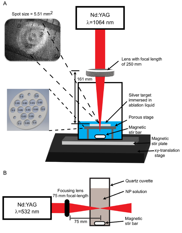
Figure 1: Apparatus configurations. (A) For the PLAL process, the Nd:YAG laser operating at a wavelength of 1,064 nm is focused through a 250 mm focal length lens to produce a spot size of 5.51 mm2 on the target stage. The spot size image is captured using a CCD camera coupled with an optical microscope. The ablation target is set on a porous stage with ten 0.65 cm diameter holes and six 0.50 cm diameter holes. An additional 3 holes are tapped for set screws which function as legs to support the stage above the stir bar. (B) For post-irradiation, the Nd:YAG laser output is set to 532 nm and focused through a 75 mm focal-length lens onto the center of a quartz cuvette containing NPs. Please click here to view a larger version of this figure.
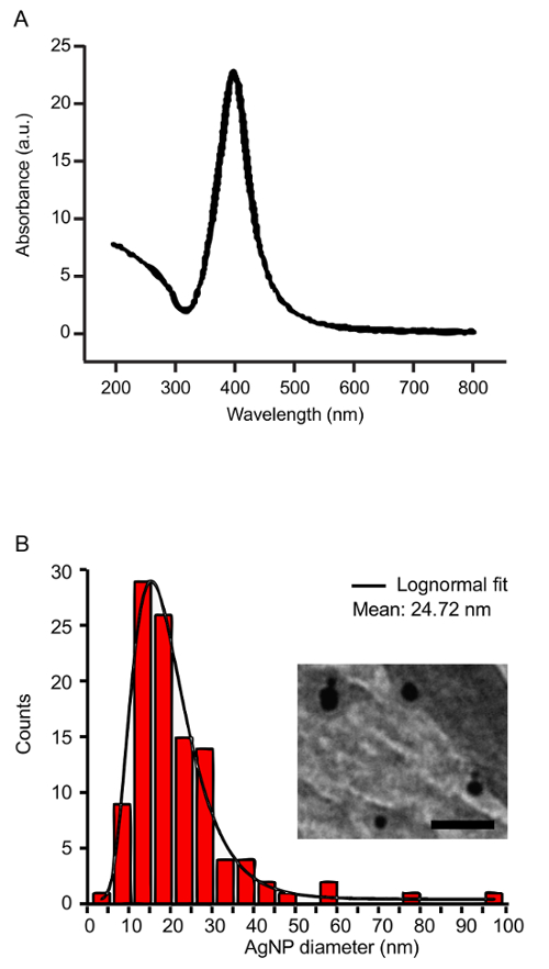
Figure 2: Characterization of silver nanoparticles. (A) The UV-VIS spectrum of silver NPs shows a characteristic peak at the SPR wavelength (400 nm). (B) The size distribution of the silver NPs before post-irradiation was measured by TEM. The inset shows a representative TEM image of AgNPs (85,000X magnification, Scale bar = 100 µm). Please click here to view a larger version of this figure.
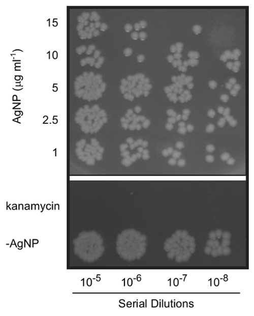
Figure 3. Antimicrobial effects of silver nanoparticles. E. coli cells were treated for 2 h with varying concentrations of silver NPs. Serial dilutions of cultures were plated on LB-agar to determine bacterial viability. Cells were treated with 30 µg/mL kanamycin as a positive control. Note that the cells not receiving AgNPs (-AgNP sample) were grown in the presence of 6 mM SDS to ensure that the surfactant did not independently result in toxicity. The figure is a composite of colonies on two plates from the same experiment and is a representative result (n = 5). Please click here to view a larger version of this figure.
