This study assesses the fracture toughness of bovine cortical bone at the sub-meso levels using microscopic scratch tests. This is an original, objective, rigorous, and reproducible method proposed to probe fracture toughness below the macroscopic scale. Potential applications are studying changes in bone fragility due to diseases like osteoporosis.
Bone is a complex hierarchical material with five distinct levels of organization. Factors like aging and diseases like osteoporosis increase the fragility of bone, making it fracture-prone. Owing to the large socio-economic impact of bone fracture in our society, there is a need for novel ways to assess the mechanical performance of each hierarchical level of bone. Although stiffness and strength can be probed at all scales – nano-, micro-, meso-, and macroscopic – fracture assessment has so far been confined to macroscopic testing. This limitation restricts our understanding of bone fracture and constrains the scope of laboratory and clinical studies. In this research, we investigate the fracture resistance of bone from the microscopic to the mesoscopic length scales using micro scratch tests combined with nonlinear fracture mechanics. The tests are performed in the short longitudinal orientation on bovine cortical bone specimens. A meticulous experimental protocol is developed and a large number (102) of tests are conducted to assess the fracture toughness of cortical bone specimens while accounting for the heterogeneity associated with bone microstructure.
In this study, we measure the fracture toughness of bovine compact bone from the mesoscale (osteons) to the microscale (lamellar level) using a novel micro scratch technique1,2,3,4,5. Fracture processes including crack initiation and crack propagation in bone are directly influenced by length scales owing to the different structural constituents and organization at different levels of hierarchy. Therefore, assessing bone fracture at smaller length scales is essential to yielding a fundamental understanding of bone fragility. On the one hand, conventional tests such as three-point bending, compact tension, and flexure tests are commonly conducted on bovine femur and tibia for fracture characterization at the macroscopic scale6,7,8. On the other hand, to measure the fracture toughness at the microscopic scale, Vicker's indentation fracture was proposed9. Micro indentation was performed using the Vicker's indenter to generate radial cracks. Furthermore, the Oliver Pharr nanoindentation fracture toughness method was performed using a sharp cube corner indenter10.
In the above nanoindentation based fracture toughness studies, the lengths of the cracks thus generated were measured by the observer and a semi-empirical model was used to calculate the fracture toughness. However, these methods are irreproducible, subjective, and the results are highly dependent on the observer's skill due to the need to measure the crack lengths using optical microscopy or scanning electron microscopy. Moreover, scratch tests were conducted at the nano-scale, but the underlying mathematical model is not physics-based as it does not account for the reduction in strength due to cracks and defects11. Thus, a gap of knowledge exists: a method for fracture assessment at the microscopic level based on a physics-based mechanistic model. This gap of knowledge motivated the application of micro scratch tests to compact bone by focusing first on porcine specimens5. The study has now been further extended to understand bovine cortical bone.
Two different orientations of the specimens are possible: longitudinal transverse and short longitudinal. Longitudinal transverse corresponds to fracture properties perpendicular to the longitudinal axis of the femur. Whereas, short longitudinal corresponds to the fracture properties along the longitudinal axis of femur5. In this study, we apply scratch testing to bovine cortical bones to characterize the bone's fracture resistance in the short longitudinal direction.
NOTE: The protocol described here, follows the animal care guidelines of the Illinois Institutional Animal Care and Use Committee.
1. Specimen Procurement
2. Cutting, Cleaning, and Embedding the Specimens
3. Grinding and Polishing Protocols
NOTE: A pre-requisite to high-precision testing at small-length scales is a smooth and levelled surface of specimens. Previous polishing protocols13,17 result in a large surface roughness, leading to substantial inaccuracy in measurement. The challenge lies in achieving low average surface roughness, less than 100 nm, over a large area 3 x 8 mm2 surface.
4. Micro Scratch Test
NOTE: Micro scratch tests are performed on the polished bovine cortical bone specimens using a micro scratch tester (Figure 3). A diamond Rockwell indenter with a tip radius of 200 µm and apex angle of 120° is used for the study. The instrument allows the application of a linear progressive load up to 30 N. Furthermore, the instrument is equipped with high-accuracy sensors to measure the horizontal load, penetration depth, and acoustic emissions generated due to scratching. The instrument can capture the panoramas of scratch grooves.
Atomic force microscopy was used to measure the roughness of the polished surface. As a rule of thumb, the specimen qualifies as a well-polished one if the surface roughness is an order of magnitude smaller than the surface features of interest. In this case, the measured surface roughness of 60 nm over a 40 µm x 40 µm area clearly falls within this criterion.
Figure 4 shows the force versus penetration depth graphs of representative scratch tests performed on the short longitudinal bovine cortical bone specimen. While the vertical force is the prescribed incremental load, the horizontal force is the measured resistance experienced by the probe. Figure 5 shows the scanning electron microscopy images of the fractured short longitudinal bovine cortical bone surface. This image shows chipping and flaking of the surface and occurrence of intrinsic toughening mechanisms such as micro cracking, crack deflection, and crack bridging. The micro scratch test data is analyzed using MATLAB scripts based on non-linear fracture mechanics modelling2. Prior to the occurrence of the fracture process, there would be plastic dissipation18. As the penetration depth increases, fracture processes are activated.
Based on microscopic observation, we consider a single crack propagating as shown in Figure 3b. We build a nonlinear fracture mechanics model1,2 to predict the scaling of the scratch force. A homogeneous transverse isotropic microstructure is considered for the cortical bone at the tissue level. Figure 6 shows the force scaling of the fracture toughness of the short longitudinal cortical bone specimens. A ductile-to-brittle transition is introduced by varying the penetration depth. In the brittle and fracture-driven regime, the scratch force is proportional to the quantity , where is the probe shape function1,2,3,4,5. Therefore, the fracture toughness, 1,2,3,4,5 converges toward a constant. Furthermore, a Kc value which corresponds to a brittle fracture is reported on the force scaling plot for a single test as shown in Figure 6. 102 micro scratch tests were conducted on the short longitudinal bovine cortical bone specimens as shown in Figure 7. Outlier tests correspond to the specimens which were tested after one week of preparation and storage in the saline solution. Storing the specimen for a very long duration altered the surface due to precipitate formation from the saline solution leading to different fracture toughness values. The overall fracture toughness value obtained is 4.05±0.63 MPa. The literature reported fracture toughness values in the range of 2.5 to 5.5 MPa 6,8. These results show that the fracture toughness values reported from the micro scratch tests are in accordance with literature.
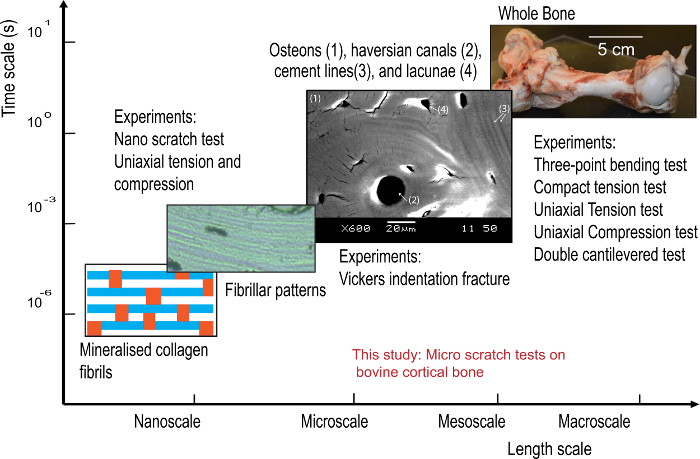
Figure 1: A graph showing the different hierarchical levels of bone specimens and the experimental investigations conducted at each level. The horizontal axis corresponds to the length scale ranging from macroscale to nanoscale and the vertical axis corresponds to time scale at which the experiments corresponding to each level are conducted. (Image Credit: Kavya Mendu). Please click here to view a larger version of this figure.
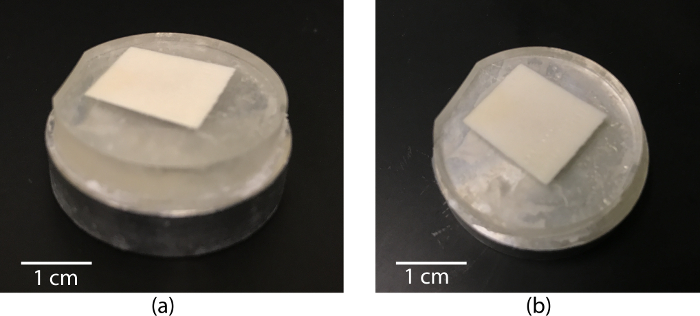
Figure 2: Digital photographs of (a) aluminum discs used as a base for the specimens and (B) well-polished short longitudinal bone specimen. Please click here to view a larger version of this figure.
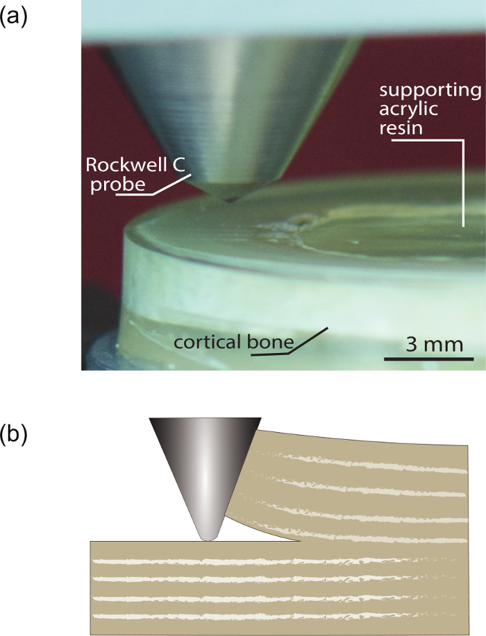
Figure 3: Micro scratch test. Digital photograph of the micro scratch test on the bovine cortical bone specimen (A). A Rockwell probe having an apex angle of 120o probing the cortical bone specimen embedded in Polymethyl Methacrylate. (B) Schematic of a scratch probe ploughing the bone material showing the advent of a mixed mode of fracture in a short longitudinal specimen. (Credits: Ange-Therese Akono, Amrita Kataruka, and Kavya Mendu). Please click here to view a larger version of this figure.
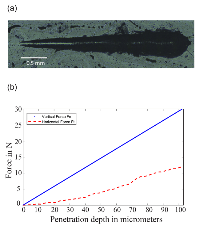
Figure 4: Scratch groove. Optical microscopy image of the panorama of the scratch groove (A). (B) Corresponding plot of the force versus depth along the length of the scratch groove. Horizontal force corresponds to the resistive frictional force detected by the sensors attached to the micro scratch tester stage and the vertical force corresponds to the progressive linear force applied onto the cortical bone specimen. Please click here to view a larger version of this figure.
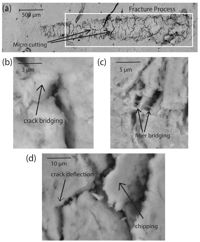
Figure 5: Scanning electron microscopy (SEM) images. SEM images of the scratch groove showing micro mechanisms such as crack deflection, crack bridging, fiber bridging, and chipping at different magnification levels (A) 40X (B) 10,000X (C) 2,400X (D) 5,000X. Captured using the low vacuum Scanning Electron Microscope at the Frederick Seitz Material Science Laboratory and Beckman Institute, University of Illinois at Urbana Champaign. Please click here to view a larger version of this figure.
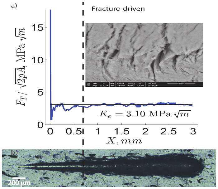
Figure 6: Scratch force and micro scratch image. (A) Scaling of the scratch force along the length of the scratch shows the convergence of fracture toughness. is the horizontal force and is the probe shape function that depends on the geometry and penetration depth. (B) Panoramic optical microscopy image of a micro scratch on bovine bone in the short longitudinal direction. Please click here to view a larger version of this figure.
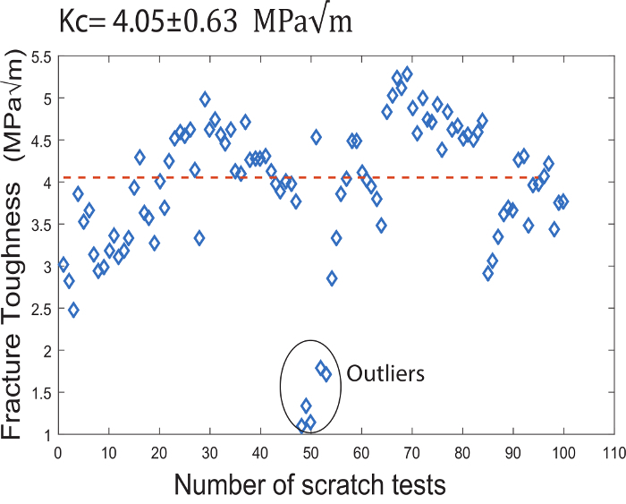
Figure 7: Fracture toughness. Plot showing the fracture toughness values of the 102 micro scratch tests conducted on the short longitudinal bovine cortical bone specimens. Please click here to view a larger version of this figure.
Micro scratch tests induce a mixed-mode fracture3. Furthermore, in the short longitudinal bovine cortical bone specimens, fracture processes are activated as the probe digs deeper. For a 3-mm long scratch, the prismatic volume probed is around 3,600 µm long, 600 µm wide, and 480 µm deep. This large volume helped in predicting a homogenized response. A non-linear fracture mechanics model enabled us to extract the fracture resistance based on the J-integral calculation1,2,4.
Bovine cortical bone specimens provide a larger area for testing when compared to the porcine specimens which were used for the earlier publication5. However, there is a corresponding difference in the size of microstructure features from porcine to bovine cortical bone specimens. This led to the development of a new polishing protocol for the bovine specimens. Furthermore, during the development of the method, it was observed that the prepared bovine cortical bone specimens need to be tested within one week after preparation. This is to avoid residue formation on the bovine specimens due to saline solution, which might drastically affect the test results.
In addition, the tests conducted on the short longitudinal bovine cortical bone specimens had controlled environmental conditions and standardized specimen preparation protocols. This led to a reduction in the variability of the test results from the previously reported 23% for short longitudinal porcine cortical bone specimens5 to 15% for the short longitudinal bovine cortical bone specimens in this study. However, in Figure 7, outlier test results can be attributed to various reasons like duration of storage in saline water or location of the scratch itself. Nevertheless, given that bone is heterogeneous at the meso- and microscopic length-scales, a certain amount of variability is expected.
Scanning electron microscopy shows the incidence of fracture processes during these scratch tests. Toughening mechanisms such as micro cracking at the meso scale, crack deflection, and crack bridging at the microscale and fiber bridging at the sub-micron scale were observed (see Figure 5). This is in accordance with the toughening mechanisms reported earlier in the literature19. Thus, micro scratch tests determine the fracture properties of bovine cortical bone specimens from the meso scale to micro scale.
The method that we propose here requires a small number of specimens and enables testing of the specimens at smaller length scales. For instance, ductile to brittle transition is introduced at the macroscopic scale by working with specimens of different sizes while having a constant aspect ratio. According to the size effect fracture assessment technique, at least 5 different sized specimens are required to estimate a fracture toughness value20,21. Thus, to estimate 102 fracture toughness values, macroscopic testing needs around 510 specimens which involves a lot of time and resources. Thus, this method we propose estimates the fracture toughness at a faster rate and is more economical. Furthermore, understanding the fracture characteristics at different hierarchical levels enables us to comprehend the mechanics of bone more efficiently. In addition, testing is efficient, reproducible, and can easily be carried out under a wide range of environmental controls. For instance, testing specimens submerged in a saline solution in an environmental chamber may be carried out to simulate in vitro conditions. In addition, the method will also be applied to test bone fracture toughness in the longitudinal transverse direction to capture anisotropy in bone. Thus, our method is a novel means for the fracture assessment of biological tissues.
The authors have nothing to disclose.
This work was supported by the Department of Civil and Environmental Engineering and the College of Engineering at University of Illinois at Urbana Champaign. We acknowledge the Ravindra Kinra and Kavita Kinra Fellowship for supporting the graduate studies of Kavya Mendu. Scanning Electron Microscopy investigation was carried out at the facilities of the Frederick Seitz Material Research Laboratory and Beckman Institute at the University of Illinois at Urbana Champaign.
Table Top Diamond Band Saw | McMaster Carr, Elmhurst, IL | Model C-40 | Blade speed of 40 mph; Blade dimensions: 37 inch in diameter, 0.02 inch wide and 0.14 inch deep |
Buehler Isomet 5000 Precision Cutter | Buehler,41 Waukegan Rd, Lake Bluff, IL 60044 | 112780 | Blade speed in the range of 200-5000 rpm in 50 rpm incrments; 8 inch diamond wafering blade |
Branson 5800 Ultrasonic Cleanser | (Through) Grainger, Peoria, Illinois | 39J365 | Bransonic CPXH ultrasonic bath has a tank capacity of 2.5 gal |
Buehler Ecomet 250 Grinder – Polisher | Buehler,41 Waukegan Rd, Lake Bluff, IL 60044 | 497250 | 8 inch base plate with a speed range from 10-500 rpm |
Anton Paar, CSM Instruments Micro scratch tester | Anton Paar Switzerland AG | 163251 | Compact Platform, Acoutstic Emission Sensor |
JEOL 6060LV general purpose scanning electron microscope | JEOL USA, Inc., Peabody, MA | Environmental scanning electron microscope which enables imaging at low vacuum levels. | |
Philips XL30 ESEM FEG | FEI Company | Wet mode working of the instrument enables imaging of non conductive samples without altering them | |
Name | Company | Catalog Number | Comments |
Consumables | |||
Bovine Femur | L&M Slaughter house, Georgetown, IL | Corn fed, 24-30 month old mature bovine specimens. | |
Alconox Powdered Precision Cleaner | Alconox, Inc., 30 Glenn St., Ste. 309, White Plains, NY, 10603 | 1104-1 | Biodegradable, Non caustic, Interfering-residue free |
Acrylic Plastic Casting | Electron Microscopy Sciences | 24210-02 | Polymethyl Methacrylate |
CarbiMet SiC Abrasive Paper 400 grit, 8 inch, PSA backed | Buehler,41 Waukegan Rd, Lake Bluff, IL 60044 | 36080400 | Grinding – Abrasive Papers |
CarbiMet SiC Abrasive Paper 600 grit, 8 inch, PSA backed | Buehler,41 Waukegan Rd, Lake Bluff, IL 60044 | 36080600 | Grinding – Abrasive Papers |
MicroCut Discs 800 grit, 8 inch, PSA backed | Buehler,41 Waukegan Rd, Lake Bluff, IL 60044 | 36080800 | Grinding – Abrasive Papers |
MicroCut Discs 800 grit, 8 inch, PSA backed | Buehler,41 Waukegan Rd, Lake Bluff, IL 60044 | 16081200 | Grinding – Abrasive Papers |
Texmet P For 8'' Wheel PSA | Buehler,41 Waukegan Rd, Lake Bluff, IL 60044 | 407638 | Polishing Cloth |
8'' Microcloth PSA | Buehler,41 Waukegan Rd, Lake Bluff, IL 60044 | 407518 | Polishing Cloth |
Meta Di Supreme Polycrystalline Diamond Suspension, 3 µm | Buehler,41 Waukegan Rd, Lake Bluff, IL 60044 | 406631 | Polishing suspension |
Meta Di Supreme Polycrystalline Diamond Suspension, 1 µm | Buehler,41 Waukegan Rd, Lake Bluff, IL 60044 | 406630 | Polishing suspension |
Meta Di Supreme Polycrystalline Diamond Suspension, 0.25 µm | Buehler,41 Waukegan Rd, Lake Bluff, IL 60044 | 406629 | Polishing suspension |
MasterPrep Polishing Suspension, 0.05µm | Buehler,41 Waukegan Rd, Lake Bluff, IL 60044 | 40-6377-032 | Polishing suspension |
HBSS, calcium, magnesium, no phenol red | Thermo Fisher Scientific | 14025126 | Buffer Solution |