Patient-derived GBM cell lines (GS122 and GS304) were provided by Professor David Nathanson (our collaborator), who developed these lines under a protocol approved by the UCLA Institutional Review Board (IRB# 10-000655). Cells were provided de-identified so that the cell lines could not be linked back to the individual patients.
1. Preparation of hydrogel solution
- Prepare HEPES-buffered solution by dissolving HEPES powder at 20 mM in Hank's balanced salt solution (HBSS). Adjust pH to 7 following full solvation.
- In the HEPES-buffered solution, dissolve thiolated HA (700 kDa nominal molecular weight, see Table of Materials), prepared following the previous report31, so that 6%-8% of carboxylic acid residues on each glucuronic acid are modified with a thiol, at a concentration of 10 mg/mL in buffer solution.
NOTE: An amber vial is recommended to prevent thiol oxidation by ambient light.- Stir using a magnetic stir plate (<1,000 rpm) at room temperature until fully dissolved, typically around 45 min.
- While HA is dissolving, prepare separate solutions of (1) 100 mg/mL of 8-arm-PEG-Norbornene (20 kDa), (2) 100 mg/mL of 4-arm-PEG-Thiol (20 kDa), (3) 4 mM of cysteine or cysteine-containing peptide (e.g., GCGYGRGDSPG), and (4) 4 mg/mL of LAP in microcentrifuge tubes (see Table of Materials).
- Prepare each of these four solutions in the HEPES-buffered solution prepared in step 1.1. Vortex the solutions to ensure full dissolution of each reagent prior to performing step 4.
NOTE: If testing multiple different peptides, each must contain a cysteine or other source of thiol moiety for this conjugation chemistry. - Prepare solutions (4 mM available thiol) of all peptides to be tethered within a single hydrogel at this point.
NOTE: Peptide sequences and ECM proteins from which they were derived and used in this study are listed in Table 1. N-acetyl cysteine (see Table of Materials), to which cells do not bind, can be substituted for a bioactive, thiol-containing peptide to titrate the concentration of an adhesive peptide or act as a negative control31.
- Prepare each of these four solutions in the HEPES-buffered solution prepared in step 1.1. Vortex the solutions to ensure full dissolution of each reagent prior to performing step 4.
- Mix the individual solutions of HA, PEG-Norbornene, PEG-thiol, and cysteine/thiol-containing peptides (see Table of Materials) to achieve the final concentrations for the final hydrogel matrices listed in Table 2. Stir (<1,000 rpm) on a magnetic stir plate for at least 30 min to mix fully.
NOTE: HA solutions are highly viscous and best handled using a positive displacement pipette (see Table of Materials). If a positive displacement pipette is unavailable, viscous solutions can also be dispensed with a standard micropipette by slowly pipetting using wide-orifice tips.
2. Illumination and photocrosslinking of hydrogels via an LED array
CAUTION: Wear UV protective eyewear and cover the illumination field with UV-absorbing material.
NOTE: The LED array described in this protocol consists of six sets of eight LEDs placed in series, as illustrated by the provided circuit diagram (Figure 1A). Each set of LEDs can be independently powered, which allows for up to six different irradiances per run. Supplementary File 1 contains screenshots corresponding to the following directions for further guidance.
- Download the Illumination Device.zip file from the Supplementary Coding Files. This directory contains the following files: Arduino.zip (Supplementary Coding File 1), Drivers.zip (Supplementary Coding File 2), GUI.zip (Supplementary Coding File 3), and Holder.zip (Supplementary Coding File 4).
NOTE: 3D Print the top and bottom portions for holding the circuit board in place (see Supplementary Coding Files for details). - Download and install the microcontroller software (see Table of Materials).
- Download and install the GUI software (see Table of Materials). Refer to Supplementary File 1 for software operating instructions.
- Open Processing and install the controlIP5 library via clicking on Sketch > Import Library > Add Library. Then, search for controlIP5 in libraries and click on Install. Perform this for the very first time.
- Power the illumination device (see Table of Materials) using the 36 Volt power supply and connect it to a PC using a micro-USB cable.
NOTE: Some devices will not install drivers automatically for various Arduino nano boards. One set of drivers is provided in the device zip file. - Open the Arduino.ino file, located in the Adruino.zip folder, using Arduino IDE.
- Compile the Arduino.ino file by clicking on the Checkmark button. Upload the compiled code by clicking on the Arrow button.
- Open the GUI.pde file, located in the GUI.zip folder, using Processing.
- Click on Run in the processing program to launch the graphical user interface for controlling the illumination device.
- In the graphical user interface window, click on Intensity for the column containing hydrogel precursor solution to be crosslinked and input the desired intensity. Click on the Time box and input desired time. For the solution provided in Table 2, this will be 15 s.
NOTE: End-users need to calibrate digital intensity values to irradiance using a radiometer. Examples of typical intensities are provided in Figure 2A. - Align the samples with the illumination device (Figure 2B) with every other LED in a single column of the silicone molds (see Table of Materials) or 384-well plate. Click on Finish to begin illumination. Repeat this process as necessary for illumination of multiple slides or other wells of a 384-well plate.
NOTE: The holder is designed such that the 384-well plate sits flush with one corner of the inner chamber during illumination.- Following illumination, when placed in one corner, move the well plate to the next corner and repeat. To illuminate wells on the other half of the plate, lift the plate out of the holder and rotate 180°.
- Generate hydrogels with varying mechanics for mechanical characterization following the steps below.
- Clean the glass slides and silicone molds using tape to remove debris. Adhere the silicone molds to the glass slide, press down to ensure a good seal, and displace any air bubbles.
- Pipette 80 µL of hydrogel precursor solution, as prepared in step 1.4, into each silicone mold on the glass slide.
- Place the glass slide onto the illumination device aligned with every other LED in a single column. Expose the hydrogel precursors to UV light for 15 s, as described in step 2, to photocrosslink.
- Once illumination has stopped, retrieve the slides, and loosen the gels from the molds by tracing the inner circumference of the mold with a fine tip (10 µL pipette tip, 30 G needle, etc.). Remove silicone molds with tweezers/forceps.
- Move crosslinked hydrogels into individual wells of a 12-well plate by wetting a spatula and gently pushing them off the glass slide. Fill each well with 2 mL of DPBS (see Table of Materials) prior to adding the hydrogel. Swell the gels in DPBS solution for at least 12 h (typically overnight) at room temperature (for the next day's mechanical characterization).
3. Atomic Force Microscopy (AFM) measurements
- Turn on the atomic force microscope (AFM) according to the manufacturer's instructions (see Table of Materials). This protocol provides brief instructions for using the instrument and the related software.
- Install the AFM probe (see Table of Materials).
NOTE: For the present study, a triangular silicon nitride cantilever with a nominal spring constant of 0.01 N/m was modified with a spherical 2.5 µm silicon dioxide particle. - Following installation, align the laser to the apex of the triangular probe, and then adjust mirror and laser deflection to maximize signal sum (typically between 1.5-2.2 Volts).
- Immerse the probe in DPBS and wait for up to 15 min to obtain thermal equilibrium. Click on the Calibration button and select Contact-Dependent calibration. Click on the Collect Thermal Tuning button, and following data collection, select the peak around 3 kHz for calibration.
NOTE: Slight adjustment of the mirror and laser deflectors may be necessary following immersion into a liquid due to refractive index changes. - Approach the surface of a Petri dish (plastic) by setting the Approach Parameters auf Constant Velocity, a target height of 7.5 µm, and an approach speed of 15 µm/s. Enable Baseline Measurement Per Run for Approach so that the approach runs continuously and does not stop early due to drift in the deflector.
- Upon approach, set acquisition parameters for force mapping to 4 nN turnarounds, 2 µm indentation distance, 1 µm/s velocity, and 0 s contact time. Press the Start button to begin collecting a force curve on the plastic surface (e.g., a well plate).
- Return to the calibration window and select the portion of the force curve corresponding to contact and indentation of the plastic. Accept the calculated sensitivity and stiffness values for the probe to complete calibration.
- Following calibration, raise the AFM probe and place the hydrogel sample for interrogation. Approach hydrogel following the settings provided in step 5.
NOTE: During the approach procedure toward the hydrogel surface, the unit may mistakenly trigger the approached state. To verify the actual approach, obtain a force curve as in step 4.6. Repeat the approach procedure if the resulting curve does not show contact and resulting indentation. - When the surface approach is successful, switch to the Force Mapping mode and set acquisition parameters to a map of 8 x 8 size with 40 µm length per axis. Obtain force maps in various regions to assess the uniformity of stiffness measurements.
- Interpret force curves using the software program JPK SPM Data Processing through a Hertz/Sneddon model fit (Equations 1 and 2, see Table 3 for the definition of all the variables) with the spherical geometry selected32,33,34.
 Equation 132
 Equation 232
- Interpret force curves using the software program JPK SPM Data Processing through a Hertz/Sneddon model fit (Equations 1 and 2, see Table 3 for the definition of all the variables) with the spherical geometry selected32,33,34.
4. Setting up and drug treatment of 3D, matrix-embedded cultures
- Prepare desired cells as a single cell solution.
NOTE: Different cell types may require different passaging methods. A typical protocol for passaging a suspension culture of GBM spheroids from a T-75 flask is reported in reference31. - Collect GBM spheroids (roughly 150 µm in diameter) from a T-75 flask suspension culture into a 15 mL conical tube. Rinse the culture flask with 5 mL of DPBS to remove any residual cells and media and add this volume to the conical tube.
- Centrifuge the conical tube containing cells at 200 x g for 5 min at room temperature. Following centrifugation, remove the supernatant with a 5 mL serological pipette, taking care not to disturb the cell pellet, and resuspend in 5 mL of DPBS.
- Centrifuge at 200 x g for 5 min at room temperature to wash cells. Aspirate the supernatant with a 5 mL serological pipette, taking care not to disturb the cell pellet, and then resuspend cells in 2 mL of cell dissociation reagent (see Table of Materials).
- Incubate at room temperature for 10-15 min. Add 3 mL of complete medium (see Table of Materials) and gently pipette 3-5 times to break down the spheroids to a single cell suspension31.
- Centrifuge the single-cell suspension at 400 x g (single-cell suspensions may be spun faster for pellet formation) for 5 min to pellet cells at room temperature. Aspirate the supernatant with a 5 mL serological pipette, taking care not to disturb the cell pellet. Resuspend cells in 1 mL of complete medium.
NOTE: If the cells remain in clumps, rather than as single cells in suspension, following passaging, cells can be passed through a 40 µm cell strainer to achieve a single cell suspension. - Remove a portion of the cells for counting using a hemocytometer. Dilute this portion two-fold with trypan blue, which permeates cells with compromised viability. Count only the live, colorless cells. Typically, a T-75 seeded at 800,000 cells per flask yields 2-3 million cells after a week in culture.
- Determine the number of cells necessary for encapsulation. Transfer a volume of media containing the total number of cells needed into a sterile 1.7 mL microcentrifuge tube. Spin down at 400 x g for 5 min at room temperature.
NOTE: For example, a minimum of 2.5 million cells resuspended in 1 mL of gel volume is needed to encapsulate cells at 2.5 million cells/mL. A gel volume of 1 mL allows users to dispense 100 gel drops, where each gel drop is of 10 µL volume. Preparing an extra ~20% volume of cells suspended in hydrogel solution is recommended to account for loss during pipette transfer. Thus, one would prepare 3 million cells and 1.2 mL of hydrogel precursor solution in this example. A minimum density of 500 thousand cells/mL is recommended. - Aspirate the supernatant with a micropipette, taking care not to disturb the cell pellet. Resuspend the cell pellet in the hydrogel precursor solution, as prepared in step 1.4, mixing well by pipetting up and down with a 1,000 µL micropipette 4-5 times.
- Load the cells into a repeat pipettor (see Table of Materials) set to dispense 10 µL. To avoid bubbles and uneven dispensing, prime the repeat pipettor by dispensing an additional 1-2 times into a waste container.
- In each well of a 384-well plate, dispense 10 µL of cells suspended in hydrogel solution from the repeat pipettor. Using the LED array, illuminate each well containing cells (step 2) for 15 s with intensities (example results in Figure 2A utilized intensities of 1.14, 1.55, 2.15, 2.74 mW/cm2) to achieve the desired mechanical properties.
NOTE: It is suggested to start with five replicates per experimental condition and scale up or down depending on the desired throughput and variance of the endpoint assay. - Add 40 µL of complete media to each well containing the cells. Add 50 µL of DPBS to non-experimental, dry wells surrounding the gels to minimize losses due to evaporation.
- For GBM cells, add 40 µL of the media-containing drug (e.g., TMZ, see Table of Materials) to achieve the final desired concentration (10 µM-100 µM in dimethylsulfoxide (DMSO) or vehicle (DMSO), accordingly, starting 3 days after encapsulation.
5. CCK8 proliferation assay
- Add 10 µL of CCK8 reagent (see Table of Materials) to each well containing the cells.
NOTE: If performing this assay for the first time, include negative control wells such as media only or cell-free hydrogel in media. - Incubate for 1-4 h according to the manufacturer's instructions.
NOTE: This time may vary as a function of cell type and density, and thus incubation times need to be tested for each application so that absorbance values fall within a linear range, a requirement for applying Beer's Law35. - Read absorbances at 450 nm for all wells following incubation.
- Calculate the average absorbance at 450 nm obtained in step 3 for the vehicle condition for each group. Divide each drug-treated well by the average of the vehicle control per group.
- Calculate confidence intervals by generating bootstrap distributions (N = 10,000) through the percentile method36.
NOTE: Generally, one may utilize 95% confidence intervals and interpret conditions whose confidence intervals do not cross over 1 to be significant and warrant further investigation. Setting confidence intervals to 95% is congruent with setting a significance cutoff of p = 0.05. For the data shown in the results, there is utility in distinguishing conditions that either promote or inhibit matrix-mediated drug resistance, requiring a two-side analysis.
AFM measurements confirmed precise control of hydrogel mechanics as a function of UV irradiance (mW/cm2) during photo-crosslinking using a custom-built, Arduino-controlled LED array (Figure 2A). The hydrogel formulation used in this protocol can be found in Table 2. The spacing of the LEDs on the provided template matches the spacing for every other well of a 384-well plate, allowing for the formation of gels inside the plate (Figure 2B). AFM interrogation of micron-scale regions at the surfaces of single hydrogels showed that hydrogels with softer average Young's moduli also had smaller ranges of moduli than stiffer hydrogels (Figure 2C–E).
Cell seeding densities that maximize viability should be determined empirically for each cell type. This study demonstrates that 3D cultures of GS122 cells seeded at densities of 2,500,000 cells/mL exhibited substantially higher viabilities when assessed after 7 days in culture compared to those seeded at densities of 500,000 cells/mL (Figure 3A). Furthermore, GS122 and GS304 cells were used as models for culturing patient-derived GBM cells to investigate the dependence of chemotherapy response on the stiffness and biochemical composition of the matrix microenvironment (Figure 3B–D). Cell viability was assessed through the CCK8 assay after treatment with TMZ for 4 days leading to a total culture time of 7 days by scaling OD450 measurement by a corresponding vehicle control and generating 95% confidence intervals by bootstrapping (N = 10,000) with the percentile method36. With these distributions, conditions in which confidence intervals did not overlap with a value of 1 (dashed line) were considered significant. Compared to more commonly used statistical methods such as t-tests or ANOVA, estimation of confidence intervals, using bootstrapping to estimate distributions that would be present for larger sample sizes, is preferred for screening assays whose goal is to identify a smaller subset of conditions for further investigation. One additional benefit of this method is that conditions with a smaller spread in a confidence interval can be prioritized over other conditions with a similar mean value but a higher spread in the confidence interval. This study indicated that GS122 cells gained survival benefits from microenvironmental interactions (Figure 3B). This survival benefit was significant in the 0.8, 1, and 4 kPa conditions but not in the 8 kPa condition for GS122 cells. GS304 cells were insensitive to both stiffness and TMZ treatment.
The effect of the biochemical composition of the matrix microenvironment was then examined by varying the inclusion of ECM-derived, integrin-binding peptides (Table 1) known to be upregulated in the GBM tumor microenvironment at two stiffnesses, 0.8 kPa, and 8 kPa. Again, GS304 cells received no significant survival benefit from matrix inclusion and were insensitive to TMZ. However, GS122 cells showed survival gains in the 8 kPa condition when osteopontin-derived peptides were included in the matrix, while the incorporation of integrin-binding sialoprotein (IBSP)- or tenascin-C-derived peptides provided minimal survival benefits, such as culture in matrices with the general RGD peptide (Figure 3C). In contrast, no peptides conferred survival gains in the 0.8 kPa culture condition (Figure 3D). Together, the results suggest intrinsic differences in both matrix and drug responses between the two patient-derived cell lines evaluated.
3D hydrogel cultures can be visualized using standard light microscopy to assess how cell morphology and invasive behaviors are affected by culture conditions in a cell-line-dependent manner (Figure 4). Both GS122 and GS304 cells spread when cultured in soft or stiff hydrogel matrices including RGD-containing peptides (Figure 4A). While peptides affected cell spreading, the ability of a cell to spread did not necessarily predict the ability of the culture to acquire TMZ resistance. For example, GS122 cells show a similar lack of spreading in both 0.8 and 8 kPa with osteopontin; however, GS122 only showed enhanced resistance to TMZ in the 8 kPa condition (Figure 4B). Finally, this miniaturized, 3D culture platform can be used to culture human cells from other tumor types, including viable organoids of terminally differentiated, neuroendocrine prostate cancer cells (Figure 4C).
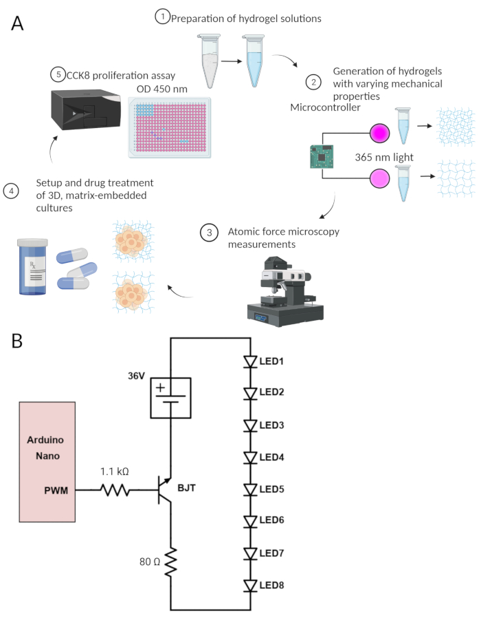
Figure 1: Cartoon depiction of the protocol. (A) Cartoon depiction of the process for 3D culture generation and monitoring. (1) HA-based hydrogel solutions are prepared. (2) Hydrogel solutions are then crosslinked with variable intensity through LEDs controlled by an Arduino microcontroller. (3) Resulting hydrogel mechanics are assessed by AFM to verify the difference in gel mechanics. (4) Solutions matching the formulation from Step 1 are then used to encapsulate patient-derived GBM cells and treated with the drug. (5) Following 7 days, cell viability is read out via CCK8 colorimetric assay. (B) Circuit diagram for custom LED illumination array used in this protocol. The individual components are listed in the Table of Materials. Please click here to view a larger version of this figure.
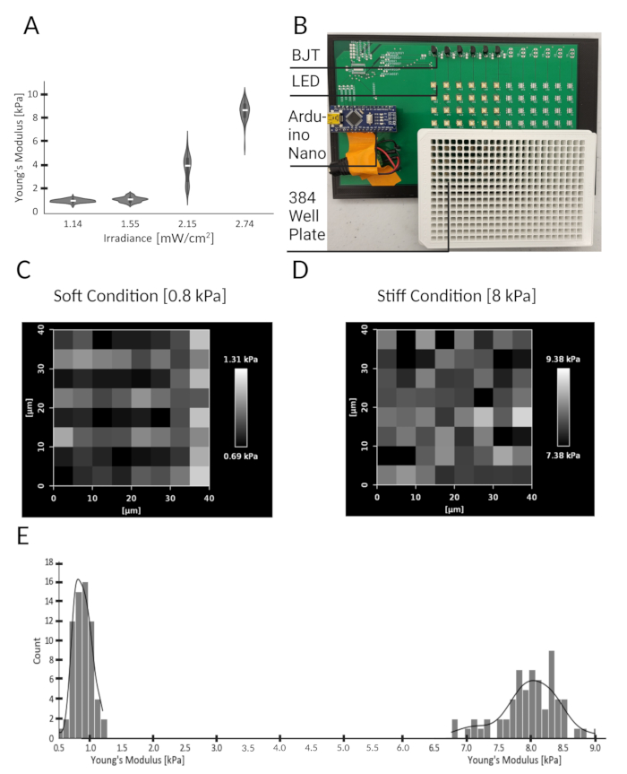
Figure 2: Hydrogels fabricated with varying stiffness using tunable LEDs to modify irradiance. (A) Violin plots show the calculated Young's Modulus from force curves generated by AFM across three surface regions, spanning 40 µm x 40 µm, of individual hydrogels. Young's modulus of each hydrogel is shown as a function of UV irradiance during photocrosslinking. Horizontal white lines indicate the median for each experimental group. (B) LED array with spacing matching the pitch of multi-well plates (384 wells). (C) Heat map showing the regional variation of Young's modulus (mean = 0.8 kPa) for a typical gel crosslinked by exposure to 1.55 mW/cm2 for 15 s. (D) Heat map showing the regional variation of Young's modulus (mean = 8 kPa) for a typical gel crosslinked upon exposure to 2.74 mW/cm2 for 15 s. (E) Histogram illustrating the range of Young's modulus measurements across the surface of hydrogels shown in C and D. Please click here to view a larger version of this figure.
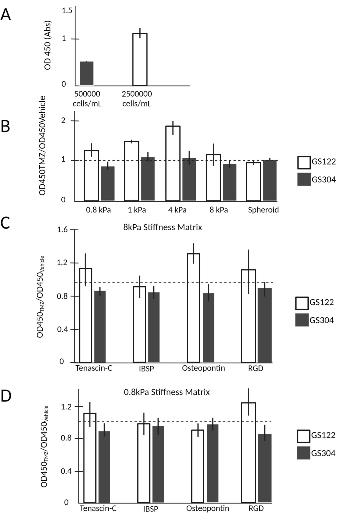
Figure 3: Orthogonal presentation of stiffness and integrin-binding peptide reveals intrinsic biological differences between GBM cell lines. (A) Typical absorbance values for GS122 cells encapsulated in a 10 µL hydrogel at a density of 500,000 or 2,500,00 cells per mL. (B) Drug response data for GS122 and GS304 cell lines are visualized by normalizing the OD450 value of the drug-treated wells (N = 5) by the average of the vehicle-treated wells (N = 5). Viability in the context of drug treatment was observed to vary nonlinearly for hydrogel stiffness, demonstrating variation between cell lines. (C,D) Drug response data for GS122 and GS304 cell lines when the type of integrin-binding peptide was included and matrix stiffness was varied orthogonally. All error bars represent the 95% confidence intervals obtained from bootstrapping each condition by N = 10,000. The dashed line (y-axis = 1) corresponds to the case where OD450 for the treatment conditions equals the vehicle. All experimental values were obtained after 7 days in culture; TMZ was added 3 days after initial encapsulation for drug studies. Please click here to view a larger version of this figure.
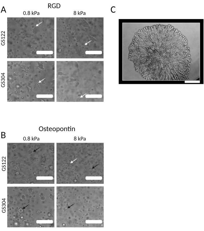
Figure 4: Morphological differences between cells encapsulated in different HA-based hydrogel environments. (A) Phase-contrast images of GS122 and GS304, when cultured in a hydrogel (0.8 and 8 kPa), displayed an RGD motif. White arrows indicate cells with spread morphologies. (B) Phase images of GS122 and GS304, when cultured in a hydrogel (0.8 and 8kPa), displayed a peptide derived from osteopontin. White arrows indicate cells with spread morphologies. Black arrows indicate cells with rounded morphologies. After 7 days in culture, images were taken, and TMZ was added 3 days after initial encapsulation. (C) Phase image of a terminally differentiated neuroendocrine prostate organoid. Scale bars = 200 µm (for A,B); 100 µm (for C). Please click here to view a larger version of this figure.
| Protein | Peptide Sequence |
| RGD | GCGYGRGDSPG |
| Tenascin-C | GCGYGRSTDLPGLKAATHYTITIRGV |
| Integrin-binding sialoprotein (IBSP) | GCGYGGGGNGEPRGDTYRAY |
| Osteopontin | GCGYGTVDVPDGRGDSLAYG |
Table 1: ECM proteins and derived peptide sequences.
| Reagent | Initial Concentration | Volume (µL) | Final Concentration |
| HA-SH Solution | 10 mg/mL | 2300 | 5 mg/mL |
| PEG-SH | 100 mg/mL | 503 | Varies per experiment |
| PEG-Norbornene | 100 mg/mL | 443 | Varies per experiment |
| Peptide | 4 µM | 288 | 0.250 µM |
| LAP | 4 mg/mL | 288 | 0.25 mg/mL |
| HEPES-HBSS | N/A | 798 |
Table 2: Typical final formulation components for hydrogel.
| Variable | Parameter |
| F | Force |
| E | Young’s Modulus |
| ν | Poissons’s Ratio |
| δ | Indentation (vertical tip position) |
| a | Radius of contact circle |
| RS | Radius of Sphere |
Table 3: Variables and corresponding parameters for AFM calculations.
Supplementary File 1: Guidance regarding the use of the LED Array for Illumination and photocrosslinking of hydrogels. Please click here to download this File.
Supplementary Coding File 1: Arduino file for LED array microcontroller (Arduino.zip). Please click here to download this File.
Supplementary Coding File 2: Third-party drivers for Arduino nano (Drivers.zip). Please click here to download this File.
Supplementary Coding File 3: Processing file for GUI control of LED array microcontroller (GUI.zip). Please click here to download this File.
Supplementary Coding File 4: File for 3D printing external holder for LED array microcontroller (Holder.zip). Please click here to download this File.
