The present protocol describes the concepts and technical application of the tensometric myograph technique using a multi-chamber myograph system in the experimental ex vivo assessment of mouse aortic endothelial function.
Small volume chamber tensometric myography is a commonly used technique to evaluate the vascular contractility of small and large blood vessels in laboratory animals and small arteries isolated from human tissue. The technique allows researchers to maintain isolated blood vessels in a tightly controlled and standardized (near-physiological) setting, with the option of adjusting to various environmental factors, while challenging the isolated vessels with different pharmacological agents that can induce vasoconstriction or vasodilation. The myograph chamber also provides a platform to measure vascular reactivity in response to various hormones, inhibitors, and agonists that may impact the function of smooth muscle and endothelial layers separately or simultaneously. The blood vessel wall is a complex structure consisting of three different layers: the intima (endothelial layer), media (smooth muscle and elastin fibers), and adventitia (collagen and other connective tissue). To gain a clear understanding of the functional properties of each layer, it is critical to have access to an experimental platform and system that would allow for a combinational approach to study all three layers simultaneously. Such an approach demands access to a semi-physiological condition that would mimic the in vivo environment in an ex vivo setting. Small volume chamber tensometric myography has provided an ideal environment to evaluate the impact of environmental cues, experimental variables, or pharmacological agonists and antagonists on vascular properties. For many years, scientists have used the tensometric myograph technique to measure endothelial function and smooth muscle contractility in response to different agents. In this report, a small volume chamber tensometric myograph system is used to measure endothelial function in the isolated mouse aorta. This report focuses on how small volume chamber tensometric myography can be used to evaluate the functional integrity of the endothelium in small segments of a large artery such as the thoracic aorta.
For the last few decades, the small chamber myography system has been used to measure the reactivity of different layers of blood vessel walls in response to various pharmacological agents and neurotransmitters in an ex vivo, real-time setting. Vascular reactivity is a major component of a healthy functional blood vessel and is critical for the regulation of blood flow and perfusion in peripheral and cerebral vasculature1. Within the blood vessel wall, the interaction between endothelial and smooth muscle layers is a major determinant of vascular tone, which is also constantly impacted by structural changes in the connective tissue layer surrounding the blood vessel wall (adventitia).
The endothelial layer controls vasomotion by releasing a few vasodilatory factors, including nitric oxide (NO), prostacyclin (PGI2), and endothelium-derived hyperpolarizing factor (EDHF), or by producing vasoconstrictive agents such as endothelin-1 (ET-1) and thromboxane (TXA2)2,3,4. Among these factors, NO has been extensively studied, and its important regulatory roles in other critical cellular functions such as inflammation, migration, survival, and proliferation have been highly cited in scientific literature2,5.
In the field of vascular biology, chamber myography has provided vascular physiologists and pharmacologists with a valuable and reliable tool to measure endothelial function in a tightly controlled semi-physiologic system1. Currently, there are two different myograph systems available to scientists: wire (or pin) tensometric (isometric) myography and pressure myography. In a wire myography system, the blood vessel is stretched between two wires or pins, allowing for the isometric measurement of force or tension development in the wall of the blood vessel, while pressure myography is a preferable platform for measurements of vascular reactivity in small resistance arteries, where changes in blood pressure are considered the main stimulus for changes in vascular tone and vasomotion. There is a general agreement that, for small resistance arteries such as mesenteric and cerebral arteries, pressure myography creates a condition that is closer to the physiological conditions in the human body. The small chamber myograph can be utilized for vessels with very small diameters (200-500 µm) to much larger vessels such as the aorta.
While the wire myograph is a powerful system for recording blood vessel tension under isometric conditions, the pressure myograph is a more appropriate system for measuring changes in vessel diameter in response to changes in isobaric conditions. The diameter changes in the vessel in response to changes in pressure or flow are much larger in a small muscular artery (arteriole) compared to large elastic arteries such as the aorta. For these reasons, the pressure myograph is considered a better tool for small blood vessels with substantial vasoreactivity1. One of the other practical strengths of multi-chamber small volume chamber tensometric myography is that one can discern the contribution of different mechanisms to vascular reactivity by studying multiple (up to four) segments of the same artery and from the same animal to reduce variability and produce robust and conclusive data. It is also relatively easy to set up and maintain technically. Vessels of almost any size can be studied with a wire myograph. It is a more cost-effective solution for assessing vascular function and is a good alternative to pressure myography in experiments where the length of the dissected vessel is too short for the pressure myograph protocol.
This report provides a detailed protocol for the assessment of endothelial function in the isolated mouse thoracic aortic ring using mounting pins in the small volume chamber tensometric myography technique using the DMT-620 multi-chamber myograph system (DMT-USA). This protocol utilizes a 6-month-old male C57BL6 mouse with an average weight between 25-35 g. Fortunately, this protocol can be applied to various animal types and weights, considering the broad range of vessel types and diameters that this protocol can be used for.
All surgical procedures and animal care were approved by the Institutional Animal Care and Use and Care Committee (IACUC) of Midwestern University (IACUC# AZ-3006, AZ-2936).
1. Buffer preparation
NOTE: Although the HEPES physiological salt solution (HEPES-PSS) buffer is stable at 4 °C for 7 days, it is recommended that all buffers are freshly made on the day of each experiment. All other reagents and agonists must be prepared freshly for each experiment. The HEPES-PSS buffer used in this protocol is a well-established buffer for ex vivo vascular studies that has been shown to be cytoprotective for more than 12 h while preserving the vessel's vasodilatory responses-the main focus of this experimental protocol6,7.
2. Myograph unit preparation
3. Mouse aorta isolation
4. Mounting of the aortic segments onto the myograph chambers
5. Normalization
NOTE: A normalization procedure is necessary to ensure that the experimental conditions are properly standardized and the collected data is reliable and reproducible. The "IC1/IC100", or "Normalization Factor", is defined as the ratio of the internal circumference of the artery at which it is possible to record the maximum response to a vasoconstrictor (e.g., 60 mM KCl) divided by the internal circumference at which a transmural wall pressure of 100 mm Hg (i.e., IC100) is recorded. Therefore, by multiplying the IC100 by this ratio, we can determine the internal circumference of the artery at which an optimal response (i.e., IC1) can be established.
6. Measurement of endothelium-dependent vasorelaxation in aortic rings
7. Effects of general inhibitors of NO production on endothelium-mediated vasorelaxation
8. Contribution of the endothelial layer to aortic vasorelaxation
The tensometric small chamber myography protocol explained here is the standard method for measuring vascular reactivity in small and large arteries and allows for simultaneous measurements of vascular reactivity in up to four blood vessel segments from the same experimental small laboratory animal. In this report, we specifically use the system to measure endothelial function in the isolated mouse aorta (Figure 1). In this protocol, isolated aortic segments are mounted onto a small organ chamber (Figure 2) between two small stainless steel pins (Figure 3). The myograph chamber can hold up to 8 mL of buffer solution and provide a semi-physiologic environment for the isolated vessels for the duration of the experiments. It is very important that, prior to each experiment, the viability of each isolated segment is tested and verified. The standard protocol to establish the integrity and viability of each isolated vessel segment is to challenge the tissue with a high concentration of potassium chloride to induce smooth muscle membrane depolarization. In the scenario that the isolated vessel is healthy and responsive, we would be able to record the contractile force generation on the display (Figure 4). The peak of the recorded force is later used to normalize the force generation for the same segment in response to the agonists used during the protocol (e.g., phenylephrine). In order to measure endothelium-mediated vasorelaxation, it is necessary to pre-contract the aortic tissue with the sub-maximum concentration (10 µM) of phenylephrine, which causes smooth muscle-mediated contraction and force generation (Figure 5). When the phenylephrine-induced contraction reaches a plateau (Figure 6), increasing doses of acetylcholine are applied in multiple steps to achieve the maximum vasorelaxation in the isolated segment (Figure 6). The level of vessel relaxation is an indirect measurement of endothelium-mediated nitric oxide production. To further confirm that acetylcholine-induced vasorelaxation in aortic rings is due to the production of nitric oxide, aortic segments are pre-treated with a general inhibitor of nitric oxide production (200 µM of L-NAME) for 30 min prior to phenylephrine application. As shown in Figure 7, L-NAME is able to completely block acetylcholine-induced vasorelaxation in the pre-contracted aorta, highlighting the fact that acetylcholine induces aortic vasorelaxation through increasing nitric oxide production. On the other hand, the removal of the endothelial layer from the aortic segments also blocks acetylcholine-induced vasorelaxation, underscoring the role that the endothelium plays in blood vessel relaxation (Figure 8).
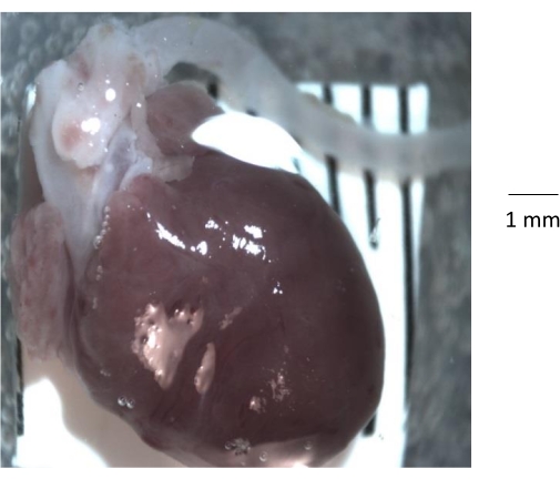
Figure 1: Gross anatomic view of the heart, aortic root, and descending aorta isolated from a 6-month-old control mouse. After removing the rib cage from the mouse, the heart and aorta are isolated from the rib cage and transferred to a clean silicone elastomer-coated Petri dish. Prior to isolating the aorta, it is important to remove all the fat and connective tissue and any blood clot from the lumen of the aorta. Please click here to view a larger version of this figure.
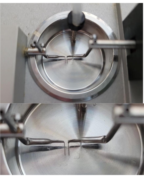
Figure 2: A representative image of a chamber of the myograph unit showing the 200 µm mounting pins. As shown, the two pins inside the myograph chamber are barely touching. Prior to using the chamber, it is critical to make sure that the pins are properly aligned. Please click here to view a larger version of this figure.
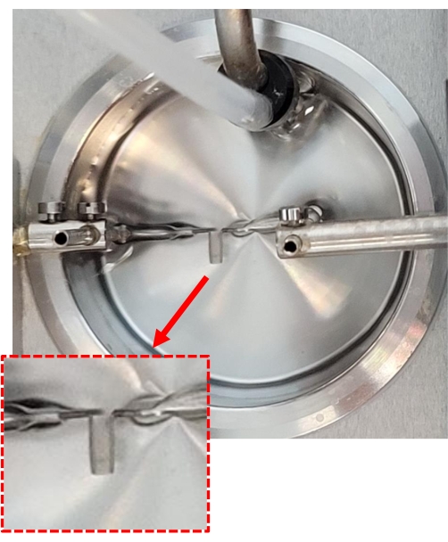
Figure 3: Anchoring the aortic segments onto the myograph chamber. A 2 mm mouse aortic segment isolated from a 6-month-old C57BL/6 mouse is held by two pins inside a myograph chamber. This is achieved by gently sliding the aorta onto the two mounting pins using forceps. The red dotted box shows the zoomed-in image of the 2 mm aortic segment that is mounted between two pins inside the myograph chamber. Please click here to view a larger version of this figure.
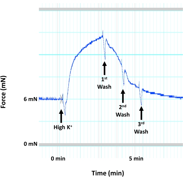
Figure 4: Aortic contraction due to smooth muscle membrane depolarization. Representative image showing the trace for mouse aortic contraction (force generation) in response to a high concentration of K+ (60 mM KCl) that would induce smooth muscle membrane depolarization and contraction within the medial layer of the aorta. The application of high K+ solution is followed immediately by three consecutive washes using warm, aerated HEPES-PSS solution. Please click here to view a larger version of this figure.
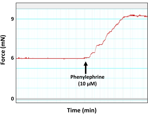
Figure 5: Aortic contraction in response to the vasoconstricting agent phenylephrine. Representative myograph trace showing force generation (contraction) by the aortic ring in response to the sub-maximum concentration of phenylephrine (10 µM). As shown, the peak of phenylephrine-induced contraction eventually reaches a plateau. Please click here to view a larger version of this figure.
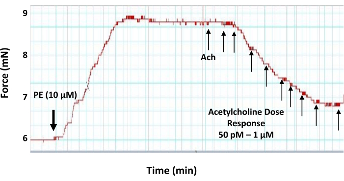
Figure 6: Dose-response effects of acetylcholine on the pre-contracted aortic ring. Representative myograph trace showing the dose-response (50 pM-1 μM) vasodilatory effect of the vasodilator neurotransmitter acetylcholine on a 2 mm pre-contracted aortic ring. The aortic ring is pre-contracted with 10 µM of phenylephrine prior to the application of acetylcholine. The first dose of acetylcholine is added when the phenylephrine-induced tension reaches a plateau. Please click here to view a larger version of this figure.
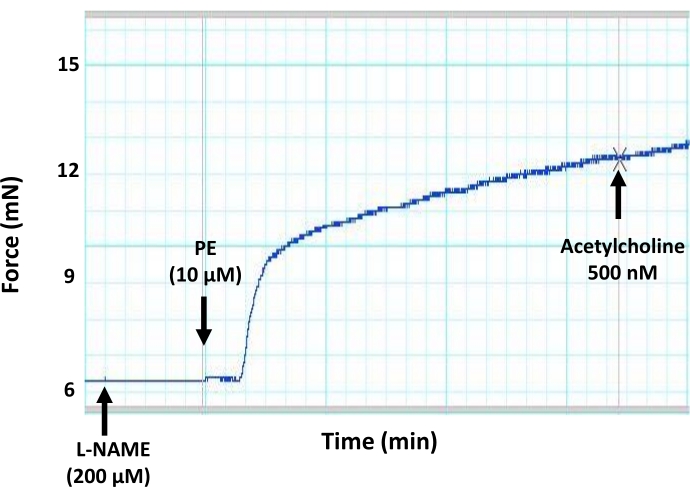
Figure 7: Effects of a general inhibitor of NO production (L-NAME) on endothelium-mediated vasorelaxation in mouse aorta. Representative myograph trace showing that the preincubation of aortic segments with a general inhibitor of NO production (L-NAME, 200 µM final concentration) blocks acetylcholine-induced vasodilation in a pre-contracted aortic ring. This is due to the inhibition of NO production by the endothelium due to the inhibitory action of L-NAME on eNOS. Acetylcholine was added to the pre-contracted aortic segment at the sub-maximum concentration of 500 nM. Please click here to view a larger version of this figure.
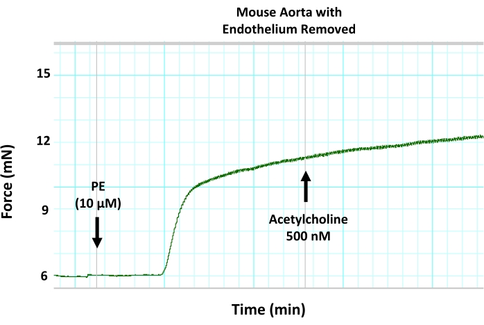
Figure 8: Effects of mechanical endothelium removal on endothelium-mediated vasorelaxation in mouse aorta. Representative myograph trace showing that removing the endothelium from aortic segments using wire denudation blocks acetylcholine-induced vasodilation in a pre-contracted aortic ring. This is due to the inhibition of endothelium-mediated vasorelaxation. Acetylcholine was added to the pre-contracted aortic segment at the sub-maximum concentration of 500 nM. Please click here to view a larger version of this figure.
The field of vascular biology heavily relies on tools that help researchers to assess the functional and structural integrity of the blood vessel wall. It also demands special attention on the direct and indirect interactions between the three layers of blood vessels: the intima, media, and adventitia. Among those three layers, the intima is formed by a monolayer of endothelial cells and has a very important function in regulating vascular health and hemostasis.
It is well established that any damage to the endothelial layer can negatively affect its ability to release NO and other vasodilatory factors, leading to dysregulation of vascular function, which is observed in various vascular disorders such as atherosclerosis, aneurysm, and vasculitis10,11,12. In order to understand the underlying mechanisms that control normal endothelial function and assess the vasodilatory function and integrity of the endothelium within the vascular wall, it is imperative to utilize a standard experimental system that mimics the in vivo physiological conditions.
For large arteries, such as the aorta, the small chamber tensometric (isometric) myography is vastly recognized as a reliable tool that creates the best available, near-physiologic conditions for the blood vessel in an ex vivo setting. The system also allows for maintaining the viability of tissue for a considerably long period of time (up to 6-8 h) in the laboratory setting, making the technique a valuable and versatile tool. One other advantage is that the myograph chamber allows the blood vessel rings to be kept and reused for back-to-back different experiments, thus making it a cost-effective approach while reducing the need for high numbers of experimental mice. Up to four blood vessel segments can be tested simultaneously using a four-chamber myograph system, increasing consistency while reducing variations across experiments.
Various pharmacological and mechanical tools can be used to study the function of the endothelial layer in blood vessels. The major marker of a functional endothelium is the normal production of NO, which is known as the most important vasodilatory agent produced and released by the endothelial layer. Endothelial dysfunction is mainly associated with a significant drop in NO production and has been shown to be involved in the progression of different vascular disorders such as hypertension, thrombosis, and atherosclerosis.
Within the vascular bed, NO production is mainly controlled by changes in blood flow and pressure, or by other intracellular events that may lead to changes in cytoplasmic calcium concentration or the activation of signaling pathways in response to hormones and growth factors13,14. Changes in NO production are considered one of the early and reliable markers of endothelial dysfunction, and they are usually detectible early during the progression of cardiovascular disorders. Regardless of the disease model, vascular biologists are very much interested in tools and assays that allow for measuring endothelial function. It is especially important that one can differentiate between the contribution of various layers of the blood vessel using a platform that mimics the physiological conditions.
In a small chamber myograph, researchers can utilize pharmacological and mechanical tools to measure endothelial function in a tightly controlled environment. Inside the myograph chamber, an artificial environment is created that can support the normal function of the blood vessel. In such an artificial environment, since the isolated blood vessel segments are not supported by the surrounding connective tissue and other organs, it is important to determine the optimal passive tension at which the isolated segments can generate the maximum possible contraction in response to vasopressors. At the optimal tension, one can measure the normal maximum contractile response to vasoconstricting agents such as phenylephrine or norepinephrine to test the structural and functional integrity of the smooth muscle layer of the blood vessel wall. In the laboratory, it was determined that the passive tension of 6 mN is a proper tension for 2 mm mouse aortic segments15. However, the optimal passive tension must be determined for different types of arteries in different species16.
In addition, before performing any experiments with isolated blood vessels, it is imperative to test the viability of isolated rings to make sure they meet the inclusion and exclusion criteria for viable and usable tissue. This is usually achieved by subjecting the isolated rings to high concentration K+ solution (60 mM KCl). This results in depolarization of the smooth muscle membrane due to the opening of voltage-gated calcium channels (VGCC), leading to smooth muscle contraction and aortic vasoconstriction. This method is used to validate the viability of the aortic segments before using those segments for further experiments.
On the other hand, vasodilating agents such as acetylcholine can be used to test the functional properties of the endothelial layer. If the endothelium layer is intact and functional, then a sub-maximum concentration of acetylcholine can induce relaxation in a pre-contracted blood vessel segment17. The magnitude of acetylcholine-induced relaxation is an indication of the level of NO release from the endothelial layer. Any damage to the endothelial layer (mechanical or functional) will have an impact on NO production and the vessel vasodilatory response. In this protocol, the provided data shows that acetylcholine can induce relaxation in the pre-contracted mouse aorta in a dose-dependent fashion, with sub-maximum relaxation achieved at the final concentration of 500 nM (Figure 6).
In some experiments, researchers are interested in measuring the smooth muscle direct response to the vasodilator NO. Since the focus of such experiments is on smooth muscle function only, there is a protocol in place that would allow researchers to bypass the endothelium contribution by removing (denuding) the endothelium layer from the blood vessel segment. The endothelium can be removed through various methods, including air, rolling between fingers, or removal by wire. In such experimental settings, the denuded blood vessel (with no endothelial layer) is subjected to NO donors such as nitroglycerin and sodium nitroprusside. This allows for determining whether the smooth muscle cyclic GMP-protein kinase G signaling pathway that responds to NO is intact and functional7,18. This manuscript explained how the removal of the endothelial layer in the isolated aortic segment completely blocks acetylcholine vasodilatory effects, highlighting the importance of NO release in blood vessel relaxation and vasodilation (Figure 8).
While the wire and tensometric myograph techniques have wide utility in vascular biology experiments, it is worth noting the potential limitations. Specifically, the tensometric system has tissue size limitations (i.e., smaller vasculature). In addition, the ex vivo nature of this technique does not allow for manipulation of the intraluminal pressure and flow for more precise mimicry of in vivo vascular function and hemodynamic parameters. Pressure myograph setups would, in fact, account for these variables and simulate real-time vascular dynamics while also allowing for the use of smaller resistance arteries. In addition, wire myograph experiments cannot fully replicate physiological conditions or accurately model the interactions between circulating blood, vessel walls, and surrounding tissue, which also play important roles in regulating vascular function in the body.
Regardless of the experimental protocol used in the myography experiments or the selected vasoconstrictor/vasodilator in the experiment, the small chamber myography system provides a reliable, reproducible, and stable semi-physiologic platform for measuring blood vessel reactivity and functional integrity. Although the focus of this report was only on the basic measurement of endothelial function, the myography system can be used to assess many other functional properties of the blood vessel, such as the stress/strain relationship, vascular wall strength, rupture point, and passive and active contraction to name a few. This highlights the value of the myograph system as a reliable tool in the field of vascular biology.
The authors have nothing to disclose.
This work was supported by funding from the National Institutes of Health (R15HL145646) and Midwestern University College of Graduate Studies.
Acetylcholine | SigmaAldrich | A6625-100G | |
CaCl2 | SigmaAldrich | C4901-1KG | |
Carbogen gas | Matheson | H103847 | |
Dissecting scissors | FST | 91460-11 | |
DMT 620 Multi chamber myograph system | DMT | DMT 620 | Multi chamber myograph system |
Dumont forceps | FST | 91150-20 | |
EDTA | SigmaAldrich | E5134-10G | |
Glucose | SigmaAldrich | G8270-1KG | |
HEPES | SigmaAldrich | H7006-1KG | |
KCl | SigmaAldrich | P9541-1KG | |
KH2PO4 | SigmaAldrich | P5655-1KG | |
LabChart | ADI instruments | Data acquisition software | |
Light source | Volpi | 14363 | |
L-Name | Fischer Scientific | 50-200-7725 | |
MgSO4 | SigmaAldrich | M2643-500G | |
Microscope | Leica | S6D | stereo zoom microscope |
NaCl | SigmaAldrich | S5886-5KG | |
NaHCO3 | SigmaAldrich | S5761-500G | |
Organ bath system | DMT | 720MO | |
Phenylephrine | SigmaAldrich | P6126-10G | |
Pump | Welch | 2546B-01 | |
Software | ADI instruments | LabChart 8.1.20 | |
Spring Scissors | FST | 15003-08 | |
Sylgard 184 Kit | Electron Microscopy Services | 24236-10 | silicone elastomer kit |
Tank Regulator | Fischer Scientific | 10575147 | |
Water bath system | Fischer Scientific | 15-462-10 |