Single-anastomosis duodeno-ileal bypass (SADI-S) is an emerging bariatric procedure with important metabolic effects. In this article, we present a reliable and reproducible model of SADI-S in mice.
Obesity is a major health issue worldwide. As a response, bariatric surgeries have emerged to treat obesity and its related comorbidities (e.g., diabetes mellitus, dyslipidemia, non-alcoholic steatohepatitis, cardiovascular events, and cancers) through restrictive and malabsorptive mechanisms. Understanding the mechanisms by which these procedures allow such improvements often require their transposition into animals, especially in mice, because of the ease of generating genetically modified animals. Recently, the single-anastomosis duodeno-ileal bypass with sleeve gastrectomy (SADI-S) has emerged as a procedure that uses both restrictive and malabsorptive effects, which is being used as an alternative to gastric bypass in case of major obesity. Thus far, this procedure has been associated with strong metabolic improvements, which has led to a marked increase in its use in daily clinical practice. However, the mechanisms underlying these metabolic effects have been poorly studied as a result of a lack of animal models. In this article, we present a reliable and reproducible model of SADI-S in mice, with a special focus on perioperative management. The description and use of this new rodent model will be helpful for the scientific community to better understand the molecular, metabolic, and structural changes induced by the SADI-S and to better define the surgical indications for clinical practice.
Obesity is an emerging and endemic situation with increasing prevalence, affecting approximately 1 in 20 adults worldwide1. Bariatric surgery has become the most effective treatment option for the affected adults in recent years, improving both weight loss and metabolic disorders2,3, with variable results depending on the type of surgical procedure used.
There are two main mechanisms that are implicated in the effects of the bariatric procedures: restriction that aims to increase satiety (such as in the sleeve gastrectomy (SG) where 80% of the stomach is removed), and malabsorption. Among the procedures that imply both restriction and malabsorption, the single anastomosis duodeno-ileal bypass with sleeve gastrectomy (SADI-S) has been proposed as an alternative to the Roux-en-Y gastric bypass (RYGB), in which a weight regain is observed in approximately 20% patients4,5. In this technique, a sleeve gastrectomy is associated with a small bowel rearrangment, dividing it into a biliary and a short common limb (one-third of the total small bowel length) (Figure 1A). Technically, the SADI-S has the advantage over the RYGB of requiring only a single anastomosis, reducing the operation time by approximately 30%. In addition, this method preserves the pylorus, which helps to reduce the risk of peptic ulcer disease and limits anastomotic leakage. The SADI-S is also associated with a high rate of metabolic improvement, strongly favoring its use during the last few years6,7.
Since metabolic effects have become increasingly foundational to bariatric procedures, elucidating their mechanisms seems crucial. Therefore, the use of animal models for bariatric procedures is of utmost importance to better understand their metabolic effects and the cellular and molecular pathways involved8. These models contributed, for example, to a better understanding of the change in food intake after SG or RYGB in a controlled environment9 and to the study of glucose or cholesterol fluxes through the intestinal barrier10,11; these informations are rarely available in clinical studies. This knowledge could help to define their optimal surgical indications. We previously described mouse models of SG and RYGB12. However, despite its promising results in clinical practice, the SADI-S has only been developed and described in rats13,14,15. However, given its genetic malleability, the mouse model has been useful in the past to study the various metabolic effects of such procedures16,17,18, and a SADI-S mouse model could be useful to evaluate effects of SADI-S despite the technical difficulty.
In this article, we describe the adaptation of the SADI-S procedure in mice (Figure 1B) in a reproducible manner. Special attention is given to the description of perioperative care.
This protocol has been approved by the local French ethical committee for animal experimentation (Comité d'éthique en expérimentation animale; reference CEEA-PdL n 06).
1. Pre-operative preparation
2. The SADI-S protocol
3. General postoperative care
4. General measurements and euthanasia
Learning curve
The learning curve for this model is displayed in Figure 6. A progressive decrease in the operating time is observed, reaching approximately 60 min of surgery after 4 weeks of intensive training (Figure 6A). The 5-day postoperative survival also improved with time, reaching 77% during regular practice (Figure 6B). The most frequent causes of mortality were anastomotic leaks and an afferent loop syndrome resulting in biliary peritonitis. We observed no death later in the first month with the technique described in this manuscript. Of note, previous experiments performed without anchoring surgical clips with running sutures led to clip migration in two-thirds of the cases, resulting in one death by small bowel occlusion at 31 days. These results emphasize that mastering this model requires intensive training.
General parameters
Mice with a C57BL6/J background were randomly assigned to the SADI-S group (n = 9; 5 males, 4 females) and the sham control group (n = 4; 2 males, 2 females). Between the SADI-S mice and the sham mice, the mean pre-operative weight (27.9 g ± 0.98 g vs. 28.5 g ± 2.4 g) and age (14.8 weeks ± 7.2 weeks vs. 18.7 weeks ± 10.3 weeks) were not significantly different. One mouse died after SADI-S at postoperative day 4 from an anastomotic leak and was therefore excluded from the following analysis. SADI-S mice experienced significant weight loss in comparison with the sham control mice from the fourth postoperative day: 21.7 g ± 1.6 g versus 29.0 g ± 0.7 g (p = 0.0081) (Figure 7A). Daily food intake (14 days) significantly increased in SADI-S mice (4.4 g ± 0.1 vs. 2.9 g ± 0.6 g per day, p = 0.027) (Figure 7B).
Mice were sacrificed 28 days after surgery. One mouse in the SADI-S group, which did not display significant weight loss, appeared to have duodenal repermeabilization. No such event was observed in the other 7 mice. As displayed in Figure 7C, the hemoglobin concentration was not significantly different from the sham control mice in the SADI-S group after iron supplementation.
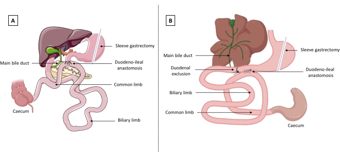
Figure 1: Representation of single anastomosis duodeno-ileal bypass with sleeve gastrectomy (SADI-S). (A) In humans, the duodenum is cut proximally from the main bile duct. A latero-terminal duodeno-ileal anastomosis is performed with the remnant duodenum, defining a biliary limb (before the anastomosis) and a common limb which measures one-third of the total length of the small bowel (after the anastomosis). (B) In mice, the duodenum is excluded by ligature proximally to the main bile duct, and a latero-lateral duodeno-ileal anastomosis is performed. The figure was created with BioRender.com and Servier Medical Art templates which are licensed under a Creative Commons Attribution 3.0 Unported License; https://smart.servier.com/. Please click here to view a larger version of this figure.
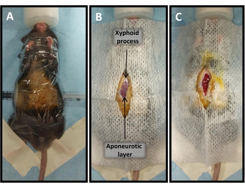
Figure 2: Mouse installation for SADI-S. (A) General installation. (B) Skin opening from the xyphoid process (sternal base) to the middle of the abdomen. (C) Musculo-aponeurotic layer and peritoneal opening. Please click here to view a larger version of this figure.
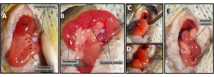
Figure 3: Duodenal exclusion. (A) Avascular window between duodenal arteries (blue dotted circle) on the posterior side of the duodenum, localized before the main bile duct (black arrows). (B) Avascular window between duodenal arteries (blue dotted circle) on the anterior side of the duodenum. (C,D) Duodenal exclusion using 6-0 non-absorbable suture. (E) Final view of excluded duodenum. Please click here to view a larger version of this figure.
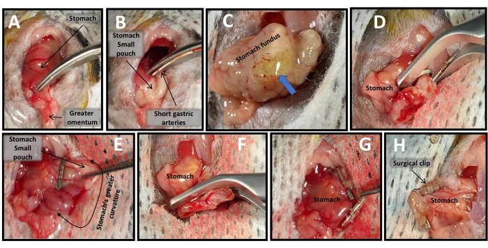
Figure 4: Sleeve gastrectomy. (A) Greater omentum removal. (B) Incision of short gastric arteries. (C) Initial gastrotomy (blue arrow). (D–G) Stomach cardiac region removal using two surgical clips. (H) Surgical clips anchoring using 6-0 non-absorbable suture. Please click here to view a larger version of this figure.
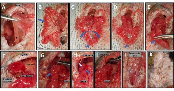
Figure 5: Duodeno-ileal anastomosis. (A) Identification of the last ileal loop (asterisk). (B) Count 10 cm (one-third of the total length of the small bowel) from the last ileal loop (asterisk) to the site of the future anastomosis (blue arrow). (C,D) Small bowel rotation around the site of the future anastomosis (blue arrow). (E) Ileal enterotomy. (F) Duodenotomy (white arrow). (G–I) Side-to-side anastomosis in two layers between the duodenotomy (white arrow) and the ileal enterotomy (blue arrow). (J) Musculo-aponeurotic layer closure. (K) Skin closure. Please click here to view a larger version of this figure.
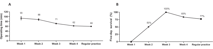
Figure 6: The SADI-S procedure learning curve. (A) The effect of training on the duration of the operation. Data are presented as the mean value ± SEM. (B) The effect of training on five-day survival. Data are presented as percentages. Please click here to view a larger version of this figure.
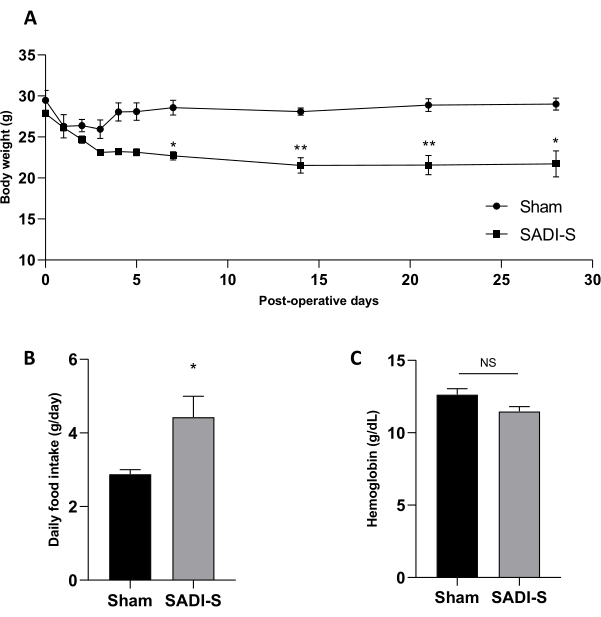
Figure 7: General parameters after SADI-S. (A) Postoperative body weight, (B) food intake measured for 24 h at day 14, and (C) blood hemoglobin concentrations were compared between SADI-S and sham control mice. Data are presented as the mean ± SEM. Statistical comparisons were made with two-way ANOVA (with Sidak's multiple comparisons test) or Mann-Whitney non-parametric tests. * p < 0.05; ** p < 0.01. Please click here to view a larger version of this figure.
Bariatric surgeries, whose techniques are constantly evolving, appear to be currently the most effective treatment for obesity and associated metabolic comorbidities3,19,20. The SADI-S procedure, firstly described in 20074, is a promising procedure associated with greater metabolic effects than other malabsorptive surgeries. Animal models, particularly mice that allow the rapid generation of genetically modified models, are strongly needed to fully understand the mechanisms underlying these improvements. Here we describe a reliable and reproductible model of SADI-S in mice.
The first critical step of the SADI-S procedure is the exclusion of the duodenum, allowing only the bile and pancreatic secretions to travel into the duodenum and the first two-thirds of the small intestine. In humans, the duodenum is cut, allowing end-to-side duodeno-ileal anastomosis4. In the rat SADI-S model described by Montana et al.15, exclusion of the duodenum by a non-absorbable suture or surgical clamp is imperfect in a few cases, resulting in duodenal repermeabilization (i.e., reintroduction of the bolus into the original digestive tract). However, a section of the duodenum followed by end-to-side anastomosis is difficult to transpose in mice, leading us to prefer duodenum ligation. Indeed, the short length of the duodenal vessels limits the duodenal mobilization if the duodenum is completely transected, making it difficult to perform termino-lateral anastomosis. Initial experiments (data not shown) showed high mortality, even with trained and skilled experimenters. Only one case of repermeabilization has been observed in this study. Special attention must be given to the duodenal arteries during this step. Circumferential devascularization of the duodenum leads to death in all cases, but mice can be expected to recover from a small devascularized area caused by distal vessel ligation. The anatomical variability of the duodenal vascularization in mice prevents us from describing a constant localization to perform this exclusion. However, 0.5 cm of the duodenum after the pylorus must be available to permit the end-to-side anastomosis.
Another critical step when performing the anastomosis is to display the bowel in a way that the biliary limb comes to the site of the duodeno-ileal anastomosis from the left side. Otherwise, the food will oppose the bile flow, causing the biliary limb to distend, the bile to diffuse to the abdominal cavity, and the mice to die from biliary peritonitis around postoperative day 2. This condition resembling an afferent loop syndrome21 can be prevented by performing a loop of the small intestine centered on the anastomosed zone of the ileum. This is necessary because, contrary to humans, the caecum is positioned on the left side of the abdomen in 80% of cases in mice22.
In humans, the common limb measures approximately 250 cm to limit malnutrition, which corresponds to approximately one-third of the total length of the small bowel23. Prior to surgeries, we measured the total length of the small intestine of the mouse model under similar feeding conditions (C57BL6/J under a chow diet) to determine the size of the common limb. As the small bowel length could vary between mice of different genetic backgrounds or following different feeding conditions, we strongly encourage future surgeons to perform a pilot study to measure the bowel size. The same size should be used for each mouse of the same background, as systematically exteriorizing the totality of the bowel for a complete measurement during surgery should be avoided (as there is increased risk of dehydration, hypothermia, and visceral injury).
The sleeve gastrectomy is part of the original SADI-S technique, allowing restriction in addition to malabsorption4. Several models of sleeve gastrectomy in mice are available in the literature12,24,25,26. The use of surgical clips instead of sutures alone allows a significant gain of time24 and reduces blood loss, two necessary conditions for surgical success. Anchoring the surgical clip using a 8-0 running suture prevented intragastric clip migration in all cases in our experiment. By removing the cardiac region, this technique allows the removal of about 80% of the stomach12. In this model, however, SADI-S was associated with overfeeding compared with the sham control mice, aiming (probably) to compensate for the malabsorption caused by bowel derivation. Other models suggested that sleeve gastrectomy in mice preferentially modified the food intake behavior instead of the absolute quantity of food ingested per day in the long term11,26. This limited restrictive effect is a limitation of this model.
This protocol has a 75% survival rate. It is worth noting that the 5-day survival was a strong predictor for long-term survival, as no late death occurred during our experiment. No anastomotic stenosis was observed. However, reaching this survival rate required at least 3 weeks of intensive microsurgical training by an experimenter specialized in animal surgery; the increased survival over time correlated with a decreased operating time. Perioperative care is one of the keys to the success of this technique. A strict analgesic protocol is needed in addition to systematic antibiotic-based therapy, and alimentation must be introduced progressively, using only a gel diet for 3 days. As previously described12, supplementation with vitamins B1, B9, B12, and liposoluble vitamins (A, D, E, K) is necessary after malabsorptive surgery, as well as iron supplementation, which prevented anemia in our experiment, but has not been described for the SADI-S model yet15.
In conclusion, SADI-S can be transposed successfully in mice, with a few modifications from its description in humans. This technique requires training and a strict perioperative protocol. Adapting this surgery to mice could allow for a better understanding of the mechanisms underlying the strong metabolic effect of this promising procedure in comparison with former models and could help to better define its surgical indications.
The authors have nothing to disclose.
We thank Ethicon (Johnson and Johnson surgical technologies) for kindly providing the suture cord and surgical clips. This work was supported by grants from the NExT Talent Project, Université de Nantes, CHU de Nantes.
Agagani needle 26 G | Terumo | 050101B | 26 G needle |
Betadine dermique | Pharma-gdd | 3300931499787 | Povidone solution |
Betadine scrub | Pharma-gdd | 3400931499787 | Povidone solution |
Binocular microscope | Optika Microscopes Italy | SZN-9 | Binocular stereomicroscope |
Buprecare | Animalcare | 3760087151244 | Buprenorphin |
Castroviejo, straight 9 cm | F.S.T | 12060-02 | Micro scissors |
Castroviejo, straight 9 cm | F.S.T | 12060-02 | Needle holder |
Chlorure de sodium Fresenius 0.9% | Fresenius Kabi | BE182743 | NaCl 0.9% |
Clamoxyl | Med'vet | 5414736007496 | Amoxicilline |
Cotton buds | Comed | 2510805 | Cotton swabs |
Element HT5 | Scilvet | Element HT5 | Automated hematology analyzer |
Emeprid | CEVA | 3411111914365 | Metoclopramid |
Extra Fine Graefe Forceps, curved (tip width: 0.5 mm) | F.S.T | 11152-10 | Surgical forceps |
Extra Fine Graefe Forceps, straight (tip width: 0.5 mm) | F.S.T | 11150-10 | Surgical forceps |
Fercobsang | Vetoprice | QB03AE04 | Iron, multivitamins and minerals |
Forane | Baxter | 1001936060 | Isoflurane |
Graefe forceps, straight (tip width: 0.8 mm) | F.S.T | 11050-10 | Forceps |
Graphpad Prism version 8.0 | GraphPad Software, Inc. | Version 8.0 | Software for statistical analysis |
Heat pad | Intellibio innovation | A-2101-00300 | Heat pad |
Incubator | Bioconcept Technologies | Manufactured on demand | Incubator |
Lighting | Optika Microscopes Italy | CL-30 | Lighting for microscopy |
Ocrygel | Med'vet | 3700454505621 | Carboptol 980 NF |
Pangen 2.5 cm x 3.5 cm | Urgovet | A02978 | Haemostatic collagen compress |
Prolene 6/0 | B.Braun | 3097915 | Optilene 6/0 (0.7 metric) 75 cm 2XDR13 CV2 RCP, suture cord |
Prolene 8/0 | Ethicon | 8732 | 2 x BV175-6 MP, 3/8 Circle, 8 mm, suture cord |
Scissors | F.S.T | 146168-09 | Surgical scissors |
Sterile compresses | Laboartoire Sylamed | 211S05-50 | Non-woven sterile compressed |
Terumo Syringe | Terumo | 50828 | 1 mL syringe |
Titanium hemostatic clip | Péters Surgical | B2180-1 | Surgical clip |
Vannas Wolff | F.S.T | 15009-08 | Micro scissors |
Vita Rongeur | Virbac | 3597133087611 | Vitamin supplementation |
Vitaltec stainless | Péters Surgical | PB 220-EB Medium | Surgical clip applier |