Here, we discuss clinical and radiological features of endolymphatic sac tumors and report the methodology and results of a surgical removal in a case in care at our otologic referral center.
Endolymphatic sac tumors (ELST) are low-grade papillary adenocarcinoma originating from the endolymphatic sac. Usually slow-growing, with local aggressiveness and a low risk of distant metastases, ELST can be sporadic but also frequently associated with von Hippel Lindau disease. The current treatment of ELST is primarily surgical resection. A 55-year-old woman accessed our otologic tertiary level referral center for a sudden worsening of hearing loss in her left ear and vertigo. A magnetic resonance (MRI) and computer tomography scan study subsequently showed a mass in the petrous bone; hence, the presence of an ELST was hypothesized. After embolization of the mass, the patient underwent surgical removal of the lesion. The resection of the mass was done through a translabirinthine approach, with an uneventful procedure. No residual disease remained after surgery. After 24 months of radiologic follow up with MRI, there are no signs of recurrence disease. This paper reports the management of this sporadic ELST, as well as the follow up results, providing clinicians this protocol for the handling of such a challenging otologic skull base surgery and rare disease.
Endolymphatic sac tumors (ELST) are neoplasms originating from the endolymphatic sac, a neuroectoderm-derived organ located in the posteromedial surface of the temporal bone. Histologically, ELST are characterizable as low-grade papillary adenocarcinoma1. Usually, ELST are slow-growing, with local aggressiveness and a low risk of distant metastases2,3,4. ELST can be sporadic, but they are also frequently associated with von Hippel Lindau disease (VHL)4.
Given the slow growing rate, the paucity of the clinical signs, and the late onset of the symptoms, they are difficult to diagnose in the early stages, especially when not associated with VHL5,6. The typical symptomatology at diagnosis of ELST includes hearing loss (79.8%), tinnitus (52.6%), and disequilibrium (45.5%), due to endolymphatic hydrops or intralabyrinthine hemorrhages4,7,8. Further and less frequent symptoms are facial palsy (25.6%), headache (13.8%), otalgia (5.1%), and trigeminal symptoms (3.2%)8.
The current treatment of ELST is primarily surgical resection. The tumor's residuals can be treated with stereotactic radiosurgery, which can be considered if the patient cannot tolerate the operation9,10. The surgical approach and the prognosis are mostly related to the tumor extension6. Even if there is no official staging system for ELST11,12, a recent grading system proposed by Li and colleagues13, based on imaging data and intraoperative findings, defined four main grades, according to the tumor extension. This and other previously proposed staging systems are reported in Table 1.
Article | ELST Staging / Grading | |||
Bambakidis et al.11 | Stage I: confiled to the temporal bone and middle ear | |||
Stage II: extended to the posterior fossa | ||||
Stage III: extended to the middle cranial fossa | ||||
Stage IV: extended to the clivus and/or sphenoid wing | ||||
Schipper et al.12 | Type A: limited to the dura of the posterior cranial fossa | |||
Type B: infiltration of lateral semicircular canal and/or cochlea | ||||
Type C: infiltration of sigmoid sinus and/or jugular bulb | ||||
Li et al.13 | Grade I: confined to the posterior petrous bone between posterior semicircular canal and posterior fossa, may involve facial nerve, no inner ear and jugular foramen invasion | |||
Grade II: extended anteriorly with invasion of semicircular canals, cochlea, IAC, middle ear, without cerebellopotine angle and jugular foramen involvement | ||||
Grade IIIa: involvement of jugular foramen or internal carotid artery or clivus | ||||
Grade IIIb: extensive intracranial invasion or involvement of cavernous sinus or jugular foramen | ||||
Wu et al.6 | Type I: confined to the endolymphatic sac region | |||
Type II: extended to at least another one anatomic structure |
Table 1: Previously published tumor staging systems. An overview of previously published grading/staging systems.
To date, only ~300 cases were reported, mostly as single clinical cases or small case series5,6. Recurrence rates and survival estimates have not been clearly defined yet8. In the present paper, we report the clinical and surgical management of a single case of sporadic ELST, as well as the follow-up results, providing clinicians our experience of the handling of such a challenging otologic skull base surgery and rare disease. Here, we describe ELST in a 55-year-old woman who accessed our otologic tertiary level referral center from a peripheric hospital. The patient had been hospitalized 3 days before for a sudden worsening of the left hearing loss, which began 3 months earlier, accompanied by an episode of acute vertigo with nausea and vomiting. The anamnesis was positive for hypertension and hypercholesterolemia, both under treatment, and smoking (3 pack-years). No personal or familiar history of VHL disease or compatible symptoms were reported by the patient.
The local ethical committee approved the publication of this clinical case. A written informed consent form was collected from the patient.
1. Clinical and audiological evaluation
2. Imaging study
3. Surgical management
4. Postsurgical course
From the audiologic assessment, it was possible to identify a moderately severe left sensorineural hearing loss (Figure 1), with a reduction of word discrimination. The tympanometry was bilaterally normal, and the stapedial reflexes were evocable with ipsilateral and contralateral stimulus. At the videonystagmography , it was possible to assess a grade II horizontal spontaneous nystagmus beating on the right side, which grew in intensity after head shaking. From the clinical and audiological assessment, the case seemed to be associated with a peripheric cochlear and labyrinthic deficit, without signs of central pathology.
Form the MRI study it was possible to identify an occupation of the left mastoid cells by high-intensity mucous material, conditioning a swollen appearance of the cortex bone in the posterior side of the petrous part with obliteration of the ipsilateral pontocerebellar cistern and minimal imprint on the left cerebellar gyri, which was compatible with the presence of an ELST (Figure 2).
Figure 2: Preoperative MRI. T1-weighted SPGR sequence after gadolinium contrast agent administration showed an isointense mass with foci of hyperintensity, with heterogeneous enhancement. Please click here to view a larger version of this figure.
The patient was then submitted to a high-resolution computer tomography (HRCT) of the petrous bone, with a 64-slice scan that confirmed the presence of an isodense mass on the posterior wall of the petrous bone with erosion and an infiltrative "moth-eaten" pattern (Figure 3).
Figure 3: Preoperative HRCT. Isodense mass on the posterior wall of the left petrous bone with erosion of petrous bone and an infiltrative "moth-eaten" pattern. Please click here to view a larger version of this figure.
According to the Li grading system13, in this case, it was possible to suspect the presence of a grade 2 ELST. The diagnostic digital subtraction angiogram showed a vascular tumor with tumor blush due to blood supply from the ascending pharyngeal, posterior auricular, and occipital branches of the left external carotid artery (Figure 4A).
Post embolization angiogram showed complete obliteration of the tumor blush (Figure 4B). There was no complication during and after the procedure.
Figure 4: Left external carotid artery angiogram lateral view. (A) Tumor blush, (B) complete obliteration of the tumor blush after embolization. Abbreviation: ECA = external carotid artery. Please click here to view a larger version of this figure.
After the complete exposure of the dura of the middle and posterior cranial fossa, in the endolymphatic sac area, an easily bleeding neoplasm was identified. The neoplasm was indivisible from the dura of the posterior cranial fossa, so the dura was cut and removed en-bloc with the tumor.
The neoplasm had reached and invaded the IAC, which had to be opened to completely remove the tumor. The intraoperative extemporaneous histological examination indicated the neoplasm as a papillary tumor. Inside the IAC, the facial nerve seemed to be intact, and it was possible to electrically stimulate it, with good muscular responses. There was no appearance of post-surgical cerebrospinal fluid (CSF) leak, and the blood loss due surgery was minimal.
Post surgery, the patient experienced self-limiting vertigo for a week, a total loss of the left residual hearing, but no other complications. In particular, the patient did not present CSF leakages or any grade of facial nerve palsy. One week after the surgery, the definitive histological examination confirmed the papillary nature of the tumor, confirming the diagnosis of ELST. The genetic study excluded the presence of a VHL disease. The patient is in good health. She undergoes radiological follow-up with MRI every 6 to 8 months. After 24 months, she does not present radiological or clinical signs of recurrence disease.
Figure 1: Tonal audiometry. Left sensorineural moderately severe hearing loss. Please click here to view a larger version of this figure.
ELST are rare and locally aggressive neoplasms that arise on the posteromedial surface of the temporal bone. Due to their slow growth rate, and the initial paucity of the symptoms, the diagnosis is often delayed, resulting in an advanced tumor stage. Since the surgery morbidity as well as the oncologic outcomes are strongly correlated with the tumor dimensions and extension6,8,13,15, an early diagnosis is necessary.
The first step for a prompt diagnosis is the clinical evaluation of patients. The medical and familiar history of a patient that may refer to the presence of a VHL has to be thoroughly investigated. Anyway, in the case reported herein, no history of known VHL was reported from the patient, as well as previous suggestive symptoms.
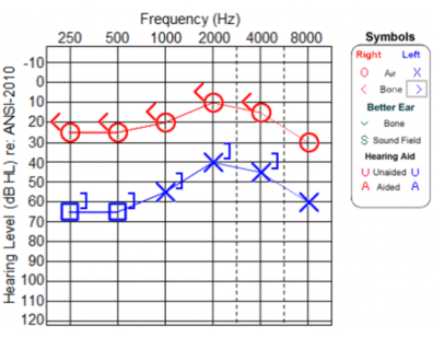
Further, since hearing loss and vertigo are a frequent association of symptoms underlying a number of peripheric clinical conditions of the ear, ranging from hydropic diseases of the inner ear to the acute unilateral vestibulopathy, but also central disturbances, is important to rule out neurological and neuroradiologic examinations in every patient reporting Meniere-like symptoms. In fact, even when the clinical picture seems to indicate a pure peripheric lesion, an imaging study of the brain, the brainstem, and the petrous bones should be always advised, even to exclude central diseases leading to peripheric deficit, but, mainly, for the study of IAC and the PCA. Those aspects are even more important when a patient presents a confirmed diagnosis of VHL or occurrences compatible with such a disease.
In the case herein reported, the diagnosis was appropriately suspected based on a rapid imaging study after the symptom’s onset and intraoperatively confirmed shortly afterward. As shown here, an early diagnosis based on the clinical signs and symptoms and prompt imaging studies can lead to a more sustainable surgery, with low risk of postoperative residuals or recurrence of disease.
Even if hearing loss and vertigo are a frequent association of symptoms underlying many peripheric clinical conditions of the ear, neuroradiologic examination have to be promptly ruled out to exclude misdiagnosed central disturbances, as well as IAC or PCA diseases. Furthermore, an accurate anamnesis should investigate for occurrences that are compatible with VHL, such as prior diagnosis of tumors such as pheochromocytoma, cerebellar hemangioblastoma, or retinal angioma; prior comparison of cysts at kidneys, liver, pancreas, or genital tract; and symptoms like headache, asthenia, hypertension, ataxia, or vision disturbances14.
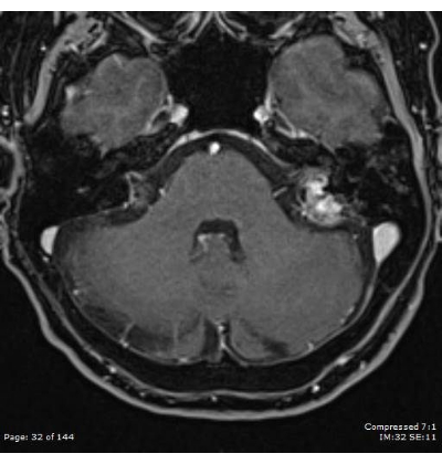
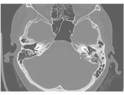
In our third level referral hospital, all the patients reporting cochlear or vestibular symptoms promptly undergo an imaging study, preferably a high-field MRI study of the brain and the brainstem and the petrous bone with contrast agent, for an accurate evaluation of those structures, the IAC and the PCA, and a HRCT scan study, for the evaluation of the bony structures of the petrous bone, if needed. The occurrence of a mass arising from the posterior face of the petrous bone, especially if associated with a suspected or known diagnosis of VHL, is highly suggestive of ELST.
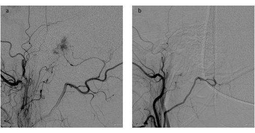
Before the surgical resection of such a mass, the principal decisions to be made from the clinicians are the execution of a preliminary embolization of the tumor and the surgical access to the lesion. There is no global consensus on the necessity of a preliminary embolization6,8,15, especially in low-staging masses, but this procedure may lead to a reduction in the bleeding of the mass during the resection, ensuring a cleaner surgical field and so an easier removal of the mass.
In this protocol for the managing of ELST, preoperative embolization is always proposed, even for limited and low-staging masses, since it leads to reduced surgery time, reduced blood loss, easier surgery and therefore, reduced morbidity.
The early diagnosis, based on an accurate and prompt clinical assessment and imaging study in patients reporting Meniere-like symptoms, can guarantee an early-stage surgery in case of an ELST. Further, thanks to a low staging of disease at the diagnosis, an accurate embolization and a precise surgical resection of the mass, it is usually possible to achieve good oncologic results, without disease recurrence and long disease-free survival period, avoiding or reducing the incidence of the main complications of major petrous bone surgery, such as the facial nerve palsy or the postoperative CSF leaks.
In the presence of an ELST, the imaging assessment, which should include an MRI and an HRCT study, make it possible to stage the tumor. According to the stage of the tumor and the audiologic assessment of the patient, it is possible to define a surgical approach and estimate a prognosis15.
To date, in fact, surgical resection is considered the treatment of choice in patients with an ELST. Another therapeutic option is the radiation therapy8,15. However, since the role of adjuvant radiation therapy is not well reported yet8,16, it should be considered for patients with residual disease after surgical resection, or in inoperable patients due to morbidity or the excessive tumor extension8,10,15.
As reported by Nelson and colleagues15, another therapy for inoperable patients could be represented by pazopanib, a multikinase inhibitor and antiangiogenic agent. Anyway, there is no evidence currently supporting the use of this drug as primary or adjuvant therapy for ELSTs.
Before the surgical resection, clinicians can propose a preliminary embolization of the mass. Even if recommended by some authors6,8, especially in situations where vascularity of the tumor could otherwise preclude a complete resection, ELST preoperative embolization is not constantly reported in literature13. Even for small masses, in accordance with other authors (e.g., Wu and colleagues6), an accurate preoperative embolization is always ruled out in our center, since it can reduce the difficulty and the morbidity of the surgery, in addition to the risk of severe intraoperative blood loss. In fact, since the main goal in the treatment of ELST is full surgical eradication8,15, small masses must be treated with reduced bleeding as one of the key points in managing this pathology.
As reported, a low-staging mass with controlled bleeding can be removed through an exclusive posterior labyrinthectomy approach, guaranteeing little postsurgical morbidity and good oncologic results. After the surgical removal of the mass, clinicians must initially deal with the postoperative symptoms and monitor the patient for complications. The eventual specific complications of such a major surgery on the petrous bone include CSF leakages and facial nerve palsy.
According to the literature, postsurgical complications, as far as residual disease after resection are, comprehensibly, more frequent when the tumors are extensive and their resection requires a highly invasive surgery6,8,15. In the case described herein, no major postsurgical complications, as well as no residual disease, were reported. Further, after the histologic confirmation of an ELST, patients with such a tumor should exclude the coexistence of a VHL through specific genetic studies. Genetic studies excluded VHL for the patient herein discussed.
In a recent systematic review by Tang and colleagues8, from the analysis of a total of 253 unique tumors in 247 patients, reported recurrence or progression after surgery was 21.8%, after a mean time of 46.7 months (from 3 to 180 months) and the disease-free survival was 47.6 ± 46.4 months in patients with sporadic disease and 49.8 ± 48.3 months in patients with VHL-associated disease.
Even if there is no consensus on the follow-up of the patients after the resection of an ELST, an MRI study every 6 to 8 months could effectively identify a recurrence or a progression of the disease after surgery. It is not clear how long the MRI follow-up should be. In their systematic review, Tang and colleagues reported a recurrence even after 15 years from surgery, while Poletti et al.10 and Sykopetrites15 reported cases with recurrence after more than 10 years. Thus, a very lasting and accurate protocol of follow up should be considered based on the possibility of a recurrence of the disease after such a long period.
In conclusion, the case reported herein demonstrates the importance of managing ELST through early diagnosis based on an accurate clinical and radiological assessment. Further, an eventual preventive embolization of the mass and an accurate surgery on a small, low-grade tumor, can lead to good oncologic results with limited postoperative surgical morbidity. Despite this, a long-lasting MRI follow up should be always advised, since recurrence of the disease is possible even after a long time after surgery.
The authors have nothing to disclose.
None.
AC40 | Interacoustic | Clinical audiometer | |
AT235 | Interacoustic | Clinical tympanometry | |
Innova 3131 | GE Healthcare | Biplane angiograph | |
LightSpeed VCT | GE Healthcare | 64 Slice CT system | |
S2 πDrive | Striker | Drilling system for Otosurgery | |
Signa HDX | GE Healthcare | 1.5 T MRI system | |
SYNAPSYS VNG | Inventis | Video nystagmograph |