Here, we present a feasibility study to assess a portable amplitude-integrated electroencephalogram (aEEG) recording system during the transport of infants with suspected hypoxic ischemic encephalopathy (HIE).
Infants at risk of HIE require early identification and initiation of therapeutic hypothermia (TH). Earlier treatment with TH is associated with better outcomes. aEEG is frequently used when making the decision whether to commence TH. As this is often limited to tertiary centers, TH may be delayed if the infant requires transport to a center that provides it. We aimed to provide a method for the application of amplitude-integrated electroencephalogram (aEEG) and to determine the feasibility of acquiring clinically meaningful information during transport. All infants ≥35 weeks, at risk of HIE at referral, were eligible for inclusion. Scalp electrodes were placed in the C3-C4; P3-P4 position on the infant's scalp and connected to the aEEG amplifier. The aEEG amplifier was, in turn, connected to a clinical tablet computer with EEG software to collect and analyze aEEG information. Recordings were reviewed by the chief principal investigator and two independent reviewers (blinded) for background trace and artifact. Predefined criteria for data quality were set to movement artifacts and software impedance notifications. Surveys were completed by healthcare staff and parents for acceptability and ease of use.
Impaired oxygen delivery or blood flow to the brain around the time of birth can cause brain injury (hypoxic ischemic encephalopathy; HIE). This is a leading cause of death and disability in full-term babies1. HIE is reported to have a worldwide incidence of 2 per 1000 deliveries, which is likely to be higher in low to middle-income countries. In Western Australia, approximately 50 babies are born each year who have a clinical diagnosis of HIE (32308 live births in 2022, an incidence of approximately 1.5 per 10002).
HIE is diagnosed with criteria based on the circumstances of birth, changes in the infant's blood acid balance (from umbilical cord or baby after birth), and changes in neurological status3. The treatment for moderate or severe HIE is whole-body hypothermia of 33-34 °C. The earlier this is commenced, the likely greater the benefit to the baby's neurological outcome3,4,5. Amplitude-integrated EEG (aEEG) is an essential part of assessment and monitoring, which in many regions is only available in tertiary centers. Electrodes (EEG safety DIN connector or DIN 42802 with 1.5 mm diameter pin) are inserted into the scalp to measure and record brain electrical activity. Electrodes are then connected to an amplifier, which is in turn connected to a clinical tablet computer to display and record brainwave activity. aEEG can provide information on brain wave activity and be used to assist with the diagnosis of HIE6. The combination of neurological examination and aEEG may enhance the ability to identify infants with moderate or severe HIE7,8,9.
The Newborn Emergency Transport Service of Western Australia (NETS WA) provides intensive care transport to approximately 1100 sick babies per year. Healthcare staff in local hospitals can contact NETS WA through a free phone number and are immediately connected via a conference call with a neonatal specialist doctor and nurse to request a transfer. The NETS team travels either by specialized road ambulance or fixed-wing aircraft with a purpose-built intensive care transport equipment cot. On arrival of the transport team to the referring hospital, the baby is assessed, and treatment is continued or escalated. The team then transfers the infant to a tertiary neonatal intensive care unit (NICU) in the state capital (Perth). In Western Australia, owing to the often-large distances between referring hospitals and centralized specialist services10 (a maximum distance of 1381 miles), aEEG may not applied for up to many hours after birth, leading to a delay in treatment.
Many monitoring systems have been designed for the intensive care unit but owing to their size and power requirement, they are impractical for the transport environment. Ward-based equipment is prone to movement artifacts, which may record insufficient quality data in the transport environment. Ambulatory EEG amplifier technology, designed primarily to be worn by patients at home, can connect to a computer tablet with EEG detection software and be applied to a newborn during transport11. This can be carried by the neonatal transport team to a baby with suspected HIE. The software is capable of being viewed remotely via a portable internet uplink and web-based viewer. Currently, there are no reports on the suitability or readability of aEEG in the transport environment.
Applying and monitoring aEEG in the transport of infants with HIE has not been previously reported, and there are no existing protocols for its use. There is no evidence to support feasibility or utility in the transport environment. We present a protocol for aEEG use in neonatal transport. It aims to assess whether aEEG in babies with HIE who require transport after birth is feasible and can provide readable clinical information.
The study was approved by the Child and Adolescent Health Service (CAHS) Human Research Ethics Committee (HREC, Approval Number RGS0000004988) and adhered to the tenets of the Declaration of Helsinki.
1. Identification of patients for inclusion in the study
2. Obtaining consent from the parents
3. Requesting consent from the transport doctor and nurse
4. aEEG application
5. Setting up EEG amplifier and tablet
6. aEEG monitoring during transport.
7. Data interpretation
8. Analysis
Between 1st September 2022 and 5th June 2023, 25 babies were transported in Western Australia with possible HIE and were eligible for inclusion. Of the total, 20 infants were consented, recruited, and successfully had aEEG applied during their transport. A total of 5 patients were not recruited and 3 were missed for consent. In one case, the device had started an automatic software update and was unusable; in one case, the baby was not at risk of HIE and was deemed ineligible for recruitment. Of the 20 infants, 12 (12/20; 60%) were male and 8 (8/20; 40%) were female. A total of 13 infants (13/20; 35%) were primarily transported by fixed-wing aircraft and 7 (7/20; 65%) by road. In total, 38 h of aEEG traces were recorded. The median postnatal age at which the recording commenced was 5.7 h (3.9-6.5 h) h. Infants primarily transported by fixed-wing aircraft compared to the road had aEEG commenced at 8.9 h (6.2-10.8 h) vs. 4.2 h (3.0-5.5 h); p = 0.0048. The median duration of EEG data recorded per patient was 1.9 h (1.6-3.1 h), with a range of 0.9-6.4 h. There were no impedance notifications in 16/20 recordings. Four recordings had a mean (range) of 4 (1-6) impedance notifications per recording. Despite this, all recordings had information that could be analyzed; examples are shown in Figure 9. The technology was acceptable to parents and staff.
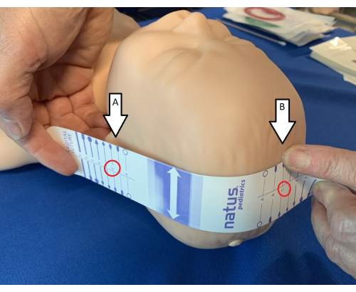
Figure 1: Identification of landmarks for measuring tape. Using the electrode measuring tape, identify the ear tragus (A) and sagittal suture (B) and ensure the tape is lined up so that the letters match (in the mannequin shown in the figure, it is "C"). Please click here to view a larger version of this figure.
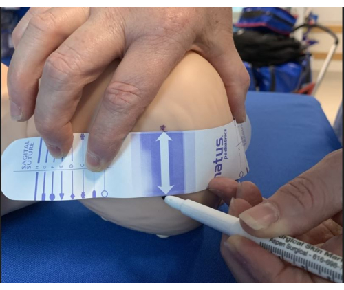
Figure 2: Identifying sites for electrode placement. Once the measuring tape is appropriately positioned to landmarks, mark electrode sites on both sides of the arrow using a marker pen. Please click here to view a larger version of this figure.
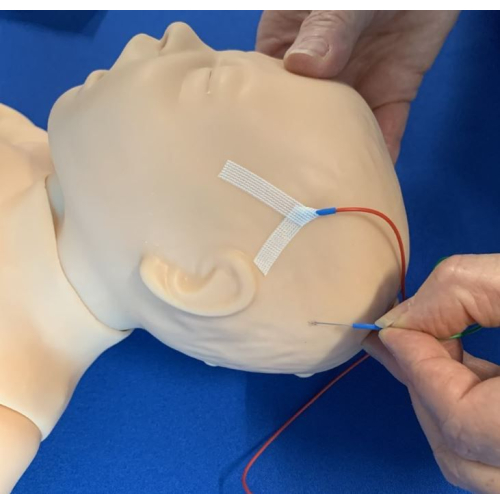
Figure 3: Insertion of electrodes. The electrodes were inserted subcutaneously and positioned caudally, taping with the steristrips Chevron technique. Please click here to view a larger version of this figure.
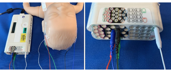
Figure 4: Electrodes connected to the corresponding site on the aEEG amplifier. Please click here to view a larger version of this figure.
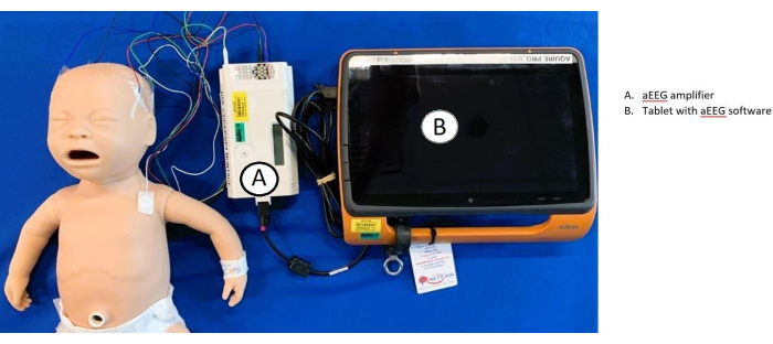
Figure 5: Overview of aEEG set-up and connectivity. Electrodes are connected to the baby and (A) amplifier. The amplifier is then connected to the (B) tablet, which uses aEEG software. Please click here to view a larger version of this figure.
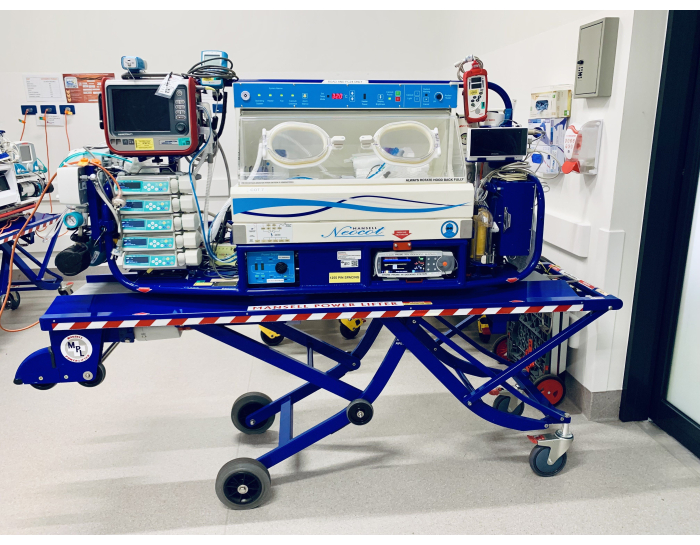
Figure 6: Mansell cot. Please click here to view a larger version of this figure.
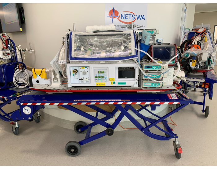
Figure 7: Voyager cot. Please click here to view a larger version of this figure.
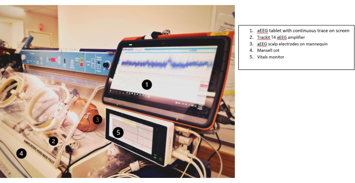
Figure 8: Tablet secured to the cot and visible to the transport team. (1) aEEG tablet with continuous trace on the screen, (2) Trackit T4 aEEG amplifier, (3) aEEG scalp electrodes on the mannequin, (4) Mansell cot, and (5) vitals monitor. Please click here to view a larger version of this figure.
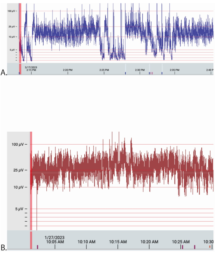
Figure 9: Examples of aEEG traces from 2 patients extracted from the online Stratus EEG Analysis software. The Y-axis is electrical amplitude (µV), and the X-axis is date and time in 5 min increments. The time base was 30 mm/s for each, and the sensitivity was 70 µV/cm for both traces. The traces are montages of the C4-C4, P3-P4 as interpreted by 'experts'. (A,B) Patient A was transported by road and patient B by air. Please click here to view a larger version of this figure.
This novel study describes the application and early data acquisition of aEEG in infants with suspected HIE who require transport soon after birth. aEEG application during neonatal transport has not been reported previously. aEEG has been used under novel circumstances, such as remote monitoring under telehealth neurocritical care consultation in Brazil and the Paediatric ED15,16.
The aEEG needle application is similar to that used in ‘static’ neonatal intensive care17 and is positioned on the scalp to achieve optimum signal for the aEEG. The positioning of this device in the neonatal transport environment may differ depending on the configuration of the transport cot. There were no issues with the needle positioning in the scalp during transport in the 20 cases; no additional trauma or bleeding was noted. It was noted on one occasion that a needle became displaced, but this is no more common than would be in a ‘static’ NICU environment in a normally moving baby. All aEEG recordings were made using an ethernet cable attached to the clinical tablet to the amplifier. This device has Bluetooth wireless capability, the next phase of method refinement. This needs to be tested in the aircraft environment.
The main limitation of aEEG remains the subjective and interpretative nature of the output, and the risk of misinterpretation cannot be underestimated18. In our methodology, we used a combination of expert consensus review, trace time > 100 µV, and the number and magnitude of impedance. Traces were separated into 15 min segments to simplify the interpretation between ‘experts’. Each section was assessed for background pattern, and the whole trace was assessed. No portion was not assessed. The aEEG interpretation of the baseline was considered as an artifact or not if 2/3 ‘experts agreed. This consensus approach was used owing to the lack of objective assessment in aEEG interpretation and to assure traces were robustly readable. The normal range of human brain activity on the aEEG scale is 10-25 µV; >100 µV was chosen as prolonged portions above this value are more likely to be related to non-biological activity (i.e., external movement). Impedance was measured as a standard to understand opposition to the electrical signal. We believe the combination of these measures provides a holistic overview of the aEEG trace.
There were some limitations of these methods. This study was initially designed to include video as well as an EEG recording. At the time of this study, the camera equipment was not available. We were unable to assess the functionality of video recordings in transport. To refine the method further, a video camera will be located on the cot to record baby movement alongside the EEG in the future. This may support seizure identification during transport. aEEG currently cannot be objectively reviewed. We believe the proxy measures we used to demonstrate feasibility are robust. As machine learning and artificial intelligence progress, more objective methods may be available and integrated into the software to provide definitive scoring in the future19.
The current tablet does not have mobile capabilities. The Newborn Emergency Transport Service of Western Australia (NETS WA) is upgrading to a 5G sim-operated device, which can transmit data via the software cloud to the receiving hospital. Specialists can then access and review aEEG traces while the transport is in progress, except during flights, where only the senior registrar can view these.
The method is unique and has not been reported previously; it has similarities to the technique in ‘static’ NICU with some important differences. The technique has important clinical and research applications as the study of brain function in transported babies is very limited. Early clinical decision-making and prognosis are important in perinatal asphyxia, and babies who are not born in tertiary institutions should have this opportunity also. Applying aEEG in transport may allow the study of babies with HIE (in early identification and prognosis), seizure presentation, and monitoring over distance. It may promote the optimal neuromonitoring of babies who require sedation during transport.
In summary, aEEG in neonatal transport is both feasible and produces sufficient data for clinical interpretation. Having the ability to make early clinical decisions in time-critical conditions (such as HIE) is important. Future studies should focus on how the new technology can improve patient outcomes utilizing real-time aEEG traces and include video monitoring for seizures during transport.
The authors have nothing to disclose.
We thank the Perth Children’s Hospital Foundation for their generous support in purchasing the EEG equipment used in this project. We thank Gardar Thorvardsson (Kvikna) and Ieesha Sparks (Temple Medical and Scientific) for their support in the technical aspects of the study project.
Stratus EEG Centrum, Acquire Pro Software for Microsoft Windows |
Stratus Software Solutions LLC Kvikna Medical Lyngháls 9 110 Reykjavik Iceland |
Version 4.2 | |
Trackit T4PCU24+8 | Lifelines Neuro 7 Clarendon Court Over Wallop StockBridge Hants, UK |
SN: T4-170046 Issue: 4 C169 |
|
Ultra Subdermal Needle Electrode | Natus 3150 Pleasant View Road Middleton WI 53562 USA |
019-476600 | 14 mm x 0.38 mm SST Needle DIN 42802 connector |
.