1. Preparation of nanoparticles
- Nano-herbal encapsulation
- Prepare 50 mL of 1% (v/v) acetic acid.
CAUTION: Glacial acetic acid is an irritant, which can cause severe skin burns and eye damage. Wear a full-length lab coat, nitrile gloves, and goggles, and work under a fume hood. - Prepare chitosan solution (1.2% w/v) by dissolving 0.6 g of chitosan flakes (medium molecular weight) in 50 mL of 1% acetic acid (prepared above). Agitate overnight (O/N) at room temperature (R/T) to get a homogeneous emulsion.
- Add 0.5 g of Tween 80 and stir (1,000 rpm) at 60 °C for 2 h to get a homogeneous solution. Bring the solution to R/T before adding bioactive compounds (carvacrol or thymol).
- Add 0.75 g of carvacrol (or thymol) dropwise or gradually while stirring (1,000 rpm) the mixture for 20 min at R/T. The weight ratio of chitosan to bioactive compound is 1:1.25.
- Add 50 mL (0.5% w/v) of TPP dropwise to the mixture while stirring at R/T. Continue stirring for 30 min to get a homogeneous emulsion.
- Prepare a negative control by following the same procedure but without the addition of bioactive compounds.
- Prepare 50 mL of 1% (v/v) acetic acid.
- Purification
- Centrifuge the emulsion at 10,000 × g for 30 min at 4 °C and collect the formed NPs (pellet) after decanting the supernatant. Reserve the supernatant to test the encapsulation efficacy.
- Wash the particles (from step 1.2.1) using aqueous Tween 80 (1% v/v) with double the volume of pellets formed to remove the unbound or free bioactive compounds. Make sure to disturb the pellet by vortexing until a homogeneous solution is formed in each wash step.
- Wash the particles (from step 1.2.2) with deionized water twice to get rid of impurities.
- Reconstitute the NPs by resuspending the pellet (from step 1.2.3) in 30 mL of deionized water. Store the NPs at 4 °C for up to 6 months.
NOTE: The experiment can be paused at this stage.
- Characterization
- Dilute the NPs in deionized water until the ultraviolet-visible (UV-Vis) readings between 250 to 400 nm fall within the range (absorbance >1).
- Record the UV-Vis absorption spectra of the NPs over the wavelengths ranging from 250 to 400 nm using a UV-Vis spectrophotometer.
- Store an aliquot (1 mL) of NPs at R/T for a range of durations (e.g., 1 to 6 months) and test the antimicrobial effect by the cylinder plate method along with a fresh sample.
- Coating on fabric
- Apply NPs on a woven cotton fabric using a pad-dry-cure method7,21.
- Immerse the fabric swatches in the solution containing NPs mixed with a fabric binder for 3 min until soaked. Then, pass the swatches through a two-roller laboratory padder to remove excess liquid.
- Oven-dry the swatches at 100 °C for 30 min and cure at 130 °C for 10 min.
- Finally, wash the swatches in an ultrasound bath for 15 min to remove unbound NPs.
NOTE: Alternatively, follow the simplified method described below.
- Cut the cotton fabric (or lab coat swatches) into 10 cm x 10 cm squares.
- Immerse the cut pieces in 2 mL of the synthesized NP (10%) in a plastic container (12 cm x 12 cm) for 2 min at R/T. Remove excess liquid by air drying.
- Heat press at 100 °C for 3 min to bind the NPs.
- Remove unbound NPs by rinsing in aqueous Tween 80 (2% w/v) and dry in the oven at 100 °C for 30 min.
NOTE: This antimicrobial fabric can be stored for 2 years at R/T.
- Apply NPs on a woven cotton fabric using a pad-dry-cure method7,21.
- Wash durability
- Cut the fabric into 50 mm x 50 mm square swatches.
- Place the swatches in 50 mL of warm tap water (40 °C) in a glass/plastic container and add two drops of regular detergent (non-antimicrobial).
- Rinse on a rocking platform shaker or magnetic stirrer for 30 min.
- Air dry or incubate at 60 °C for 2 h and test for antimicrobial efficacy.
2. Cylinder plate assay for screening of nanoparticles
- Prepare trypticase soy agar (TSA), trypticase soy broth (TSB), antibiotic base agar (ABA), and antibiotic seed agar (ASA), according to manufacturers' instructions, and sterilize the media by autoclaving at 121 °C at 15 psi for 15 min.
- Subculture the microbes (from stock) of interest, against which the efficacy tests are performed, on freshly prepared TSA plates (or any appropriate media) by the three-way streak plate method and incubate to produce purity plates. (Recommended species: S. aureus, E. coli, P. aeruginosa, and C. albicans.)
- Inoculate the cultures from fresh purity plates in TSB (10 mL tubes) at 35 °C O/N with moderate shaking (100-200 rpm).
- Aliquot 5.0 mL of the molten ASA in each of the test tubes. The number of tubes corresponds to the number of microorganisms tested.
- Pour 20 mL of presterilized molten ABA into each Petri plate (100 mm x 20 mm) aseptically to form the base layer. The number of agar plates corresponds to the number of microorganisms to be tested. Wait until the media get solidified.
- After solidification of the base layer, inoculate each molten ASA with an overnight culture from step 2.3 (1.0 mL, prewarmed to 35 °C) and immediately transfer the contents (mixture of ASA and culture) to the surface of an ABA plate. Swirl the plate to distribute the molten agar layer evenly. Ensure that the seed layer appears smooth and free from bumps or bubbles.
- Place up to six stainless steel cylinders (6 mm x 6 mm x 10 mm; preautoclaved), evenly spaced on a hexagonal pattern, per agar plate using a pair of sterile tweezers (Figure 1).
- Load the cylinders with the synthesized NPs of different volumes (30 µL, 50 µL, 75 µL, and 100 µL) to be screened for antimicrobial effects. The negative control is the one without any bioactive compounds.
- Incubate common bacterial pathogens (e.g., E. coli, S. aureus, P. aeruginosa) at 35 °C for 24 h and fungal species (e.g., C. albicans) at R/T for 3 days.
- Measure the diameter of the clear zone and compare the effectiveness of the synthesized NPs.
NOTE: This test can be used to prescreen the best NPs to be coated on the fabric.
3. Parallel streak method (modified from AATCC 147)
- Material preparation
- Cut the NP-treated fabric into 50 mm x 25 mm swatches.
- Prepare Mueller-Hinton agar (MHA), TSA, and TSB, according to manufacturers' instructions, and sterilize the media by autoclaving at 121 °C at 15 psi for 15 min.
- Subculture the microbes (from stock) of interest, against which the efficacy tests are performed, on freshly prepared TSA plates (or any appropriate media) by the three-way streak plate method and incubate at 35 °C for 2 days for bacterial stains and at R/T for 5 days for fungal strains to produce purity plates. (Recommended species: S. aureus, E. coli, P. aeruginosa, and C. albicans.)
- Spiking of microbial cultures
- Inoculate the cultures from fresh purity plates in TSB (10 mL tubes) at 35 °C O/N with moderate shaking (100-200 rpm).
- Dilute the O/N cultures to 1.5 × 10⁸ colony forming units (CFU)/mL or corresponding to the turbidity of 0.5 McFarland standard (usually a 1/10 to 1/20 dilution for most healthy cultures).
- Inoculate the diluted culture above using a 4 mm sterile loop as follows. Load a loopful of broth culture and transfer it to the surface of the MHA agar plate by making five parallel streaks, each 6 cm in length and 1 cm apart, according to Figure 2. Do not refill the loop.
- Gently press the fabric swatch using a sterile spatula across the five streaks so that the fabric is in the center and touches all five streak lines. Incubate at 35 °C for 24 h.
- Qualitative evaluation of antimicrobial efficacy
- Examine the incubated plate for the interruption of growth along the streaks beyond the edges of the fabric (clear zone indicates inhibition of growth).
- Calculate the average width of a zone of inhibition along the streak line (W) on either side of the fabric swatch using equation (1).
W = (T – D)/2 (1)
W = width of clear zone; T = total length of the clear zone, including the swatch width; D = width of the fabric swatch (25 mm).
4. Quantitative log reduction method (modified from AATCC 100)
- Experimental preparation
- Cut the fabric into 50 mm x 50 mm square swatches.
- Prepare TSA, TSB, Letheen broth, and phosphate-buffered saline (PBS), according to manufacturers' instructions, and sterilize the media by autoclaving.
- Prepare O/N cultures of microbes of interest, against which the efficacy tests are performed, by inoculating isolated colonies from purity plates in sterile TSB and incubating at 35 °C for 18-24 h. (Recommended species: S. aureus, E. coli, P. aeruginosa, and C. albicans.)
- Spiking of microbial cultures
- Dilute the O/N cultures to 1.5 × 10⁸ CFU/mL or corresponding to the turbidity of 0.5 McFarland standard (usually a 1/10 to 1/20 dilution for most healthy cultures).
- Determine the appropriate volume of cultures for spiking by measuring the liquid holding capacity of the fabric as follows.
- Add a series of volumes of the diluted broth culture (e.g., 100-500 µL) onto fabric swatches (in separate Petri plates) and choose the volume such that the fabric swatch absorbs the water fully and leaves no residual/free liquid. The liquid holding capacity differs from the type and thickness of the fabric. For regular cotton lab coats, it is roughly 200 µL.
- Spike the volume (liquid-holding capacity determined above [e.g., 200 µL]) of each culture onto the swatches placed in sterile Petri plates. The number of swatches corresponds to the number of tests (e.g., fabric tested immediately and after washing [and the laundering cycles tested]), and the antimicrobial effects tested over the contact time (after a set time/day [e.g., 30 min, 2 h, Day 1-3]).
NOTE: Use a micropipette with aerosol filter tips (to prevent contamination of the pipette in subsequent uses) to inoculate the cultures on the swatches with even distribution. - For the negative control, use the same type of untreated fabric swatches. Perform the negative control for each corresponding test-microbial species, contact period, different fabrics, and wash cycles.
- Recovery of microbes by viable plate counts
- Allow the inoculated swatches (both treated and untreated) to air-dry inside the Petri plates (lids ajar) at R/T for the required contact period/s to be tested (e.g., 0 min, 30 min, 60 min, etc., or even days for long-term effects). Always include a "0 min" to represent an immediate effect and neutralizing efficacy.
- Transfer the swatches aseptically to separate sterile centrifuge tubes (50 mL) and screw the caps tightly.
- Add Letheen broth (any relevant neutralizing buffers) to make a 1/100 dilution (e.g., 19.8 mL for an inoculum of 200 µL).
- Close the centrifuge tubes with screw caps tightly and vortex for 1 min at medium speed.
- Serially dilute the suspension with the sterile PBS in subsequent 1/10 dilutions, such that the colony counts of the "untreated" group becomes too low to count (TLTC).
- Plate the dilutions (0.1 mL) on appropriate media plates, which support the growth of the microorganism (e.g., TSA for bacteria or Sabouraud dextrose agar [SDA] for fungi) or any media that optimize the growth and provide good contrast to make the colony counting accurate.
- Incubate the bacterial plates at 35 °C for 2 days and the fungal plates at R/T for 5 days.
- Count the viable CFUs directly using a colony counter or using imaging software (e.g., CFU AI).
- Calculate the microbial log reduction (R) due to antimicrobial fabric using equation (2):
R = × 100 (2)
A = log value of the number of CFUs recovered from untreated fabric; B = log value of the number of CFUs recovered from treated fabric.
Initial screening of the synthesized NPs
Following the two-step oil-in-water emulsion technique16, the bioactive compounds (carvacrol and thymol) were successfully encapsulated in chitosan. This was confirmed by UV-Vis spectrophotometry for the peak absorption of the respective bioactive compounds compared to controls, which were the chitosan NPs without any bioactive compounds. The constituted NPs were homogeneous and stable over 12 months at 4 °C. The initial screening of the antimicrobial effectiveness was verified by the cylinder plate method (Figure 1). This is a qualitative method as the zone of clearance is influenced by multiple factors, such as agar thickness, the strength of the inoculum, and the concentration of the test samples. Many simpler methods, such as the Kirby-Bauer disk diffusion method and well diffusion method, can be employed for this purpose, but the cylinder plate method provides the opportunity to vary the concentrations (Figure 1A) by diluting the NPs, and each cylinder can hold the test sample volume up to 200 µL. In addition, the agar overlay with cultures forms a smooth inoculum, enabling the determination of the clear zones with better precision17. The results demonstrated that the clear zones are proportionate to the progressively increasing concentrations (Figure 1A). This adds validity to the data and differentiates it from any artifacts or abnormal zones. Based on the size of the clear zones (usually over 20 mm), the correct concentration of NPs can be selected for coating. Any previously characterized or old NPs, which were stored appropriately (4 °C), can also be verified by the large zones (Figure 1B,C) before coating onto the fabric.
Qualitative screening of the treated fabric samples
Despite the antimicrobial effectiveness of the encapsulated NPs confirmed by large clear zones, the NP-coated fabrics must be tested. This is because the antimicrobial agents may work differently when applied to the fabric compared to their original formulations. Many factors, such as the properties of the fabric (thickness, hydrophobicity), coating efficacy, and degradation of bioactive compounds while coating, influence the effectiveness20. As such, the parallel streak method was used to evaluate the antimicrobial effects of the treated fabrics qualitatively. The negative controls (untreated fabrics) demonstrated no antimicrobial effects by the uninterrupted microbial growth along all five streak lines (Figure 3 A,B). The treated fabric showed interrupted microbial growth along the streak lines (Figure 3 C,D), which was due to the diffused bioactive compounds from the fabric. However, the average width of the clear zones was low (<5 mm) as the test samples were subjected to 10 wash cycles before testing. As the inoculum concentrations decrease along the parallel steaks from the first to the fifth streak (Figure 2), the clear zones are prominent in the subsequent streaks. If the first streak (high inoculum) shows a clear zone, the antimicrobial potential of the fabric is usually high. Some irregularities, such as the fifth line in Figure 3D, may occur due to the qualitative nature of the test. The clear zone (interrupted growth) due to the bacteriostatic activity provides an indication of the antimicrobial potential, but does not give an adequately sensitive guideline18, which is calculated by the "log reduction test". However, the parallel streak method is useful for a relatively quick and easily executed procedure to screen a large number of swatches6,7,20, particularly when testing for a wash durability test over many cycles.
Quantitative analysis of the treated fabric samples
The log reduction test (also known as the percentage reduction test) demonstrated a significant (<0.001) reduction of microbial cultures upon contact with the treated fabric for 30 min (Figure 4 and Figure 5). The antimicrobial fabric swatches were significantly effective (>99%) against a Gram-positive bacterium (S. aureus), Gram-negative bacteria (E. coli and P. aeruginosa), and a skin fungal species (C. albicans). Since the untreated fabric (negative control) had no antimicrobial activity, the recovered CFUs were too numerous to count until diluted to extinction, up to 106 dilutions (Figure 4). The number of CFUs recovered from the treated fabrics at "0" contact time (plated immediately upon inoculation and neutralization) was very similar to that of the untreated fabric, and data were omitted for simplicity. The neutralizer used (Letheen broth) is effective in neutralizing the effects of phenolic derivatives such as carvacrol and thymol. Compared to carvacrol NPs, thymol NPs showed slightly higher antimicrobial effects against all four microbes (Figure 5). Both the thymol and carvacrol-coated fabric worked equally effectively against three microbes (S. aureus, E. coli, and C. albicans) with over a 4-log reduction (99.99%), except P. aeruginosa, which ranged from a 2.8- to a 3.2-log reduction (99.9). This was expected, as P. aeruginosa is intrinsically resistant to a range of antimicrobials22. The wash durability test demonstrated that the treated fabric was able to exhibit effective antimicrobial resistance (>99%) against three species (S. aureus, E. coli, and C. albicans) and moderate resistance against P. aeruginosa after 10 wash cycles.
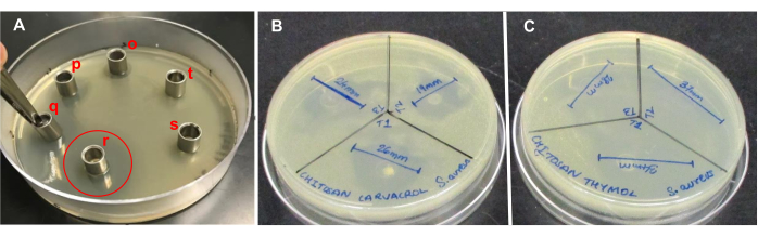
Figure 1: Cylinder plate assay of synthesized nanoparticles with a range of concentrations tested against bacteria. (A) Serially diluted thymol NPs for an initial screening against E. coli showing the placement of cylinders and clear zones after 18 h of incubation. Progressively increasing concentrations "o" to "r" resulted in proportionately higher zones. The clear zone produced by the highest concentration is indicated by a red circle. The negative control (chitosan NPs without the bioactive compounds) is represented by "t" and the supernatant extracted during purification is represented by "s". (B) Two out of three concentrations of carvacrol NPs (12 months old, stored at 4 °C) screened for the fabric treatment showing effective zones (>20 mm) against S. aureus. (C) Three concentrations of thymol NPs (12 months old, stored at 4 °C) screened for the fabric treatment showing effective zones against S. aureus. Abbreviation: NP = nanoparticle. Please click here to view a larger version of this figure.
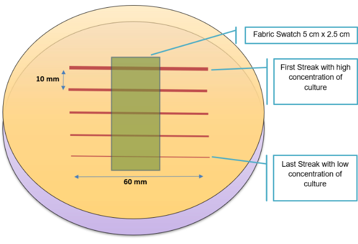
Figure 2: Parallel streak method layout. Placement of a fabric swatch on Mueller-Hinton agar inoculated with five subsequent parallel streaks. Please click here to view a larger version of this figure.
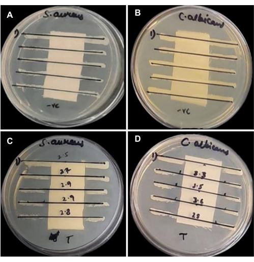
Figure 3: Parallel streak method results showing clear zones for the treated fabric swatches. The swatches were placed on top of the inoculum (bottom row) compared to untreated (top row). (A) Untreated fabric against S. aureus, (B) untreated fabric against C. albicans, (C) thymol NP-coated fabric (after 10 wash cycles) against S. aureus, and (D) thymol NP-coated fabric (after 10 wash cycles) against C. albicans. Please click here to view a larger version of this figure.
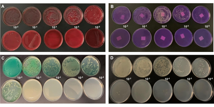
Figure 4: Log reduction results of antimicrobial fabric coated with thymol encapsulated chitosan nanoparticles tested agaisnt four pathogens. The lactose agar plates in panel B were donated by the Canadian Food Inspection Agency, which include their labels at the back side and look like swatches. Other agar plates were prepared in the lab, without labels. Fabric swatches were not placed on top of the agar plates, as in the experiment shown in Figure 3. Rather, the microbes were recovered from the swatches (after vortexing in PBS) and plated. (A) S. aureus on blood agar, (B) E. coli on purple lactose agar, (C) P. aeruginosa on cetrimide agar, (D) C. albicans on SDA. The pathogens were spiked on "untreated" (top row) and "treated" (bottom row) swatches for 30 min and recovered upon neutralization and dilution plating. The dilution ratios are shown between the treated and untreated series. Please click here to view a larger version of this figure.
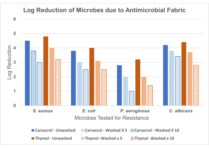
Figure 5: Log reduction of three bacteria (S. aureus, E. coli, and P. aeruginosa) and one fungus (C. albicans) due to the contact of antimicrobial fabrics impregnated with two bioactive compounds (carvacrol and thymol separately). The antimicrobial efficacy is weakened after washed cycles (five times and 10 times, respectively) for both carvacrol- and thymol-coated fabrics. Please click here to view a larger version of this figure.
