This video demonstrates the protocol for performing colonoscope-guided pinch biopsy in the mouse. This technique helps visually analyze the colon lumen and collect tissue samples for detailed studies.
Protocol
All procedures involving animal models have been reviewed by the local institutional animal care committee and the JoVE veterinary review board.
1. Colonoscopy and wound induction
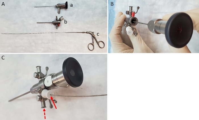
Pre-assemble the components of the endoscope by first inserting the 1.9 mm rigid bore endoscope into the sheath (Figure 1A-B). Attach the air pump (to provide colonic insufflation) using the tubing provided, to the gas valve on the left side of the sheath next to the working channel (Figure 1C). NOTE: Although the lens described here is 0°, a 30° lens can also be used for this purpose.
Ensure that the working channel is in the open position and insert 3 Fr biopsy forceps through the working channel and advance it until the end of the sheath (while ensuring that it does not protrude out of the sheath) (Figure 1C). Attach the assembled endoscope to the light source and video imaging device per manufacturer’s instructions.
Anesthetize the mouse with 5% isoflurane with oxygen in an induction chamber. Then move the mouse to an endoscopic staging platform containing a heating system (to prevent hypothermia) on its ventral side and maintain under anesthesia using a nose cone with 2% isoflurane with oxygen. Add vet ointment to the eyes to prevent dryness while under anesthesia. Ensure that the mouse is fully anesthetized by gently pinching the rear foot to test for a reflex. NOTE: Any strain or either sex of mice can be used with this technique; however, it is preferable that mice are at least 8 weeks of age, so they are large enough for the procedure.
Add vet ointment to the eyes to prevent dryness while under anesthesia.
Ensure that the mouse is fully anesthetized by gently pinching the rear foot to test for a reflex.
Fill a 3 mL syringe with an attached rat gavage needle with room temperature phosphate-buffered saline (PBS). Insert needle approximately 1 cm into the mouse’s anus and gently infuse PBS to clear fecal material. Several fecal pellets should exit the mouse along with the PBS that was infused. NOTE: Instillation of an excessive volume of PBS can result in foam formation in the lumen which could obscure viewing of the lumen.
Insert the assembled endoscope 0.5 cm into the anus of the mouse. Advance the biopsy forceps into the lumen of the rectum until the full ‘jaws’ (including hinge) of the forceps are beyond the end of the sheath (as observed on the video monitor). Turn the forceps 90° so that the jaws open in an east-west orientation.
To biopsy, open the forceps and advance approximately 1 cm, close the forceps and in one smooth motion quickly pull back on the forceps while leaving them closed.
Avoid fully insufflating the colon when performing the biopsy. Therefore, leave the right side of the gas valve open during this step; although it should be noted that upon opening the forceps there is a risk of damaging the mucosa when the colon is in this position.
Representative Results
Figure 1: Items needed to carry out biopsies. (A) Image of lens (a), sheath (b) and biopsy forceps (c). (B) Insertion of the lens into the sheath. (C) Insertion of forceps through the working channel of the sheath (solid arrow). Dashed arrow indicates correct location for attachment of air pump.
Colonoscopic Guided Pinch Biopsy in a Mouse Model: A Technique to Visualize and Extract Tissue Samples from Mouse Colon. J. Vis. Exp. (Pending Publication), e20810, doi: (2023).