A subscription to JoVE is required to view this content. Sign in or start your free trial.

Method Article

Measuring Microglial Uptake of Intravitreally-Delivered Fluorescent Particles Using Flow Cytometry

866 views

⸱

July 8th, 2025

In This Article

Abstract

Source: Murinello, S. et al., Assessing Retinal Microglial Phagocytic Function In Vivo Using a Flow Cytometry-based Assay. J. Vis. Exp. (2016)

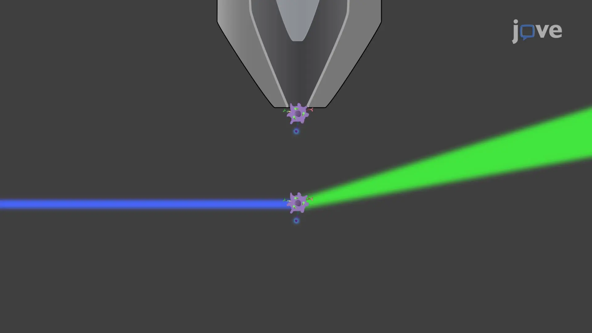
This video demonstrates flow cytometry for measuring microglial uptake of fluorescent bioparticles after intravitreal delivery in a mouse eye. Flow cytometry detects live cells expressing CD11b and distinguishes phagocytic microglia cells exhibiting green fluorescence from other myeloid cells, including Ly6C-expressing monocytes and Ly6G-expressing neutrophils.
Protocol

All procedures involving animal models have been reviewed by the local institutional animal care committee and the JoVE veterinary review board.

1. Preparation of Materials for Injection

  1. Sterilize a 33 G needle and syringe: disassemble and autoclave at 115 οC. Prepare needles for injection by rising in sterile phosphate-buffered saline (PBS).
  2. Thaw fluorescently labeled particles at room temperature for 5 - 10 min. Prepare particle solution for injection by reconstituting to a 50 mg/ml solution in sterile PBS with calcium ion and magnesium ion (Ca2+/Mg2+).

Access restricted. Please log in or start a trial to view this content.

Results

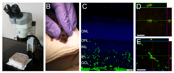
Microscope, animal handling, retinal cell layers, and confocal microscopy in biological research.
Figure 1: Intravitreal Injection Setup and Representative Retinal Section showing Fluorescently Labeled Particles in Retinal Layers. (A) A gel pack is positioned under the dissection microscope. (B) A rodent is held as sho.......

Access restricted. Please log in or start a trial to view this content.

Disclosures

No conflicts of interest declared.

Materials

List of materials used in this article
NameCompanyCatalog NumberComments
StereomicroscopeNikonDiscontinued
Hamilton syringe, 600 seriesSigma26702
33 gauge, Small Hub RN NDL, 0.5 in, point style 4 - 12oHamilton7803-05
Zymosan A (S. cerevisiae) BioParticles, Alexa Fluor 488 conjugateThermoFisher ScientificZ-23373Prepare immediately before injection
DPBSCorning21-030-CV
Dumont #5/45 ForcepsFine Science Tools11251-35Need two
Dumont #5SF ForcepsFine Science Tools11252-00
Vannas Spring Scissors - 3mm Cutting EdgeFine Science Tools15000-10Curved
Neural Tissue Dissociation Kit – Postnatal NeuronsMiltenyi Biotec130-094-802
5 mL Polystyrene Round-bottom TubeFalcon352054
96 well U-bottom plateFalcon353077
Stain Buffer (BSA)BD Biosciences554657
CD11b-BV650 AntibodyBioLegend101259
Ly6C-APC-Cy7BioLegend128025
Ly6G-PE-Cy7BioLegend127617
Propidium IodideBD Biosciences556463
Purified anti-mouse CD16/32 AntibodyBioLegend101301

Explore More Articles

Intravitreal DeliveryRetinal Myeloid CellsCD11b AntibodiesLy6C Ly6GPropidium IodideCell SuspensionFc Receptor Blocking