Summary

내측 측두엽 구조의 수동 분할을위한 포괄적 인 프로토콜

Published: July 02, 2014
doi:

Summary

The present work provides a comprehensive set of guidelines for manually tracing the medial temporal lobe (MTL) structures. This protocol can be applied to research involving structural and/or combined structural-functional magnetic resonance imaging (MRI) investigations of the MTL, in both healthy and clinical groups.

Abstract

편도체, 해마 및 관련 parahippocampal 영역 (entorhinal perirhinal, 적절한 parahippocampal) : 본 논문은 측두엽 (MTL)를 포함 뇌 영역의 설정을 수동으로 추적을위한 포괄적 인 프로토콜을 설명합니다. 사용할 수있는 대부분의 다른 추적 프로토콜과 달리, 일반적으로 특정 MTL 영역 (예를 들어, 편도 및 / 또는 해마)에 초점을 맞추고, 현재 추적 지침에 의해 채택 된 통합적인 관점에서 모든 MTL 하위 영역의 명확한 지역화 할 수 있습니다. 현존하는 추적 프로토콜이 다양 MTL 구조, 조직 학적 보고서 및 뇌 도해 타겟팅 및 예시 시각적 재료의 보수와 같은 다양한 소스로부터 정보를 통합함으로써, 본 프로토콜은 이해를위한 정확하고, 직관적이며 편리한 가이드를 제공한다 MTL 해부학. 이러한 추적 지침의 필요성또한 자동 및 수동 분할 프로토콜의 가능한 차이를 설명하기에 의해 강조된다. 이 지식은 구조 MRI 조사뿐만 아니라 관련 연구쪽으로 적용뿐만 아니라, 모두 건강하고 임상 군에서 해부학 적 정의의 ROI에서의 구조 – 기능 colocalization을과의 fMRI 신호를 추출 할 수 있습니다.

Introduction

측두엽 (MTL)은 감각 정보 1의 가장 높은 수준의 통합의 추정 지역은, 대상 분석의 빈번한 대상이되어왔다. 예를 들면, 해마와 연관된 parahippocampal 영역은 광범위 메모리 연구 2-5에서 연구되어왔다. 또한, 편도의 역할은 자주 감정 처리 및 감정 -인지 인터랙션 6-11 검사 연구에서 강조되었다. 최근, 다양한 MTL 지역은 12 형질 성격의 개인차에 이들과 다른 뇌 영역의 구조와 기능을 연결 성격 신경 과학의 신흥 분야에서 주목을 받았다. MTL 구조의 해부학과 기능을 평가하는 특정 구조 및 기능 이상이 다른 MTL 구조에서 발생할 수있는 퇴행성 질환의 진단을 촉진 중요 할 수있다. 예를 들어, 알츠하이머 질환 (AD), 중요한entorhinal 피질 및 해마의 트로피 13,14을 관찰 할 수 있고, 해마의 위축 AD (15)에 가벼운인지 장애의 전환을 예측할 수있다. 자동 분할 알고리즘은 최근 피질 및 피질 하 구조를 분할에 대한 인기를 끌고있다, 그러나 어떤 도구로,이 프로그램은 필연적으로 어떤 경우에 오류가 발생합니다. 이러한 경우에 연구원은 지식과 MTL 구조의 해부학 적 경계를 인식하는 가이드 라인을 모두 장착해야합니다. 현존하는 문학의 경향은 해마 16-19에 집중하는 경향이 많은 프로토콜로, 개별 MTL 하위 영역에게 16-21을 목표로하고있다.

MTL 추적에 사용할 수있는 게시 된 가이드 라인의 대부분과 달리,이 의정서는 모든 MTL 하위 영역의 명확한 현지화 수 있도록 가이드 라인의 포괄적 인 세트를 제공합니다. 다음 MTL 구조에 대한 추적 지침을 설명합니다 : 편도 (AMY), 해마 (HC), perirhinal 피질 (중화 인민 공화국), entorhinal 피질 (ERC), 그리고 parahippocampal 피질 (PHC). 에이미와 HC는 첫 번째 추적 된 후 parahippocampal 이랑 (PHG) 구조 다음에 있습니다. 일반적인 용어의 HC가 적절한 HC를 포함 HC 형성, subiculum의, 그리고 uncus 22 ~ 24의 후방 부분을 참조 여기에 사용합니다. 또한, PHG는 두 세그먼트, 전방 부와 후방 부로 구분 될 수 있음을주의한다. PHG의 전방 부분 내에서, 각각 더 그의 피질 영역 PRC 및 ERC 대응 측면 및 내측 전방 PHG,로 분할 될 수있다. PHC, PHG의 후방 부분의 피질 영역은 적절한 parahippocampal 피질에 해당합니다. 단순 이유로, 우리는 후방 PHG를 참조 PRC와 ERC는 측면과 중간 전방 PHG를 참조하는 용어를 사용하고, PHC됩니다. segme각 구조에 대한 ntation는 anterior-posterior/rostro-caudal에서 다음 관상면에서 각 슬라이스 실제 수행 다음에 추적 관련 랜드 마크와 함께 전방 및 후방 경계의 거친 현지화로 시작 방향. 모든 경우에, 시상 및 축 섹션은 밀접하게 해부학 적 경계와 랜드 마크의 국산화를 지원하기 위해 모니터링됩니다.

이러한 추적 지침에 대한 필요성 또한 자동 및 수동 분할 프로토콜 출력 가능한 차이를 표시하는 도면에 도시된다. 현재 비주얼 형식 MTL 구조의 모두를 설명 프로토콜의 장점은 테두리 정의에 영향을 미칠 수있는 해부학 (예 담보 고랑 [CS] 깊이)의 변화는 주변 해부학 (으로 문맥으로 기술 될 수 있다는 점이다 예 , 중화 인민 공화국과 ERC 내측 및 외측 경계는 CS (25)의 깊이에 따라 위치 변경 </s까지>). 이것은 단지 하나 또는 별도의 구조를 추적하고, 우리의 지식, 같은 시각 포괄적 인 가이드 라인이 존재하지 않는 경험이 추적이나 숙련 된 추적을 지우거나 이해하지 않을 수 있습니다.

본 프로토콜은 이전의 조사가 구조적 자기 공명 (MR) 영상에서 최근 개발이 허용 고해상도 뇌 영상에 적응, 감정 (26)의 메모리 향상 효과 MTL 하위 영역에서 차등 기여를 식별 MTL 추적에 사용되는 가이드 라인을 명시 적으로 프리젠 테이션입니다 . 추적은 3T MR 스캐너를 사용하여, (24 세, 여성) 건강한 지원자에서 얻은 검사에 설명되어 있습니다. AC-PC에 인수 각도 평행 해부학 적 이미지는 3D MPRAGE (복셀 크기 = 1 × 0.5 × 0.5 mm, TE = 2.26 밀리,, FOV = 256 X 256mm TR = 1800 밀리 초)로 취득 하였다. 화상 데이터 등 경사 방향과 같은 다른 수집 각도로 취득하는 경우, 데이터는 등록해야AC-PC에 평행 또는 수직 방향으로 ridded, 해부학 적 랜드 마크의 설명은 적절하게 번역하도록. 이미지는 다음 수동 추적을위한 분할 소프트웨어 (27)에 NIFTI 형식 및 입력에 번역되었다. 현재 프로토콜에 사용되는 스캔 데이터는 임상 시험 심사위원회에 의해 승인 된 연구의 일부가 서면 동의를 제공하는 자원 봉사자로 수집 하였다.

이러한 구조 18-22,28-31,뿐만 아니라 해부학 적 분석과지도 책 23,32,33에서에 대한 다양한 별도의 추적 프로토콜 정보를 그림으로,이 의정서는 현존하는 문헌에 불일치를 해결 가이드 라인의 포괄적 인 세트를 제공합니다. 첨부 된 영상 자료에 의해 보완,이 작품은 MTL 구조의 명확한 이해를 증진하고, MTL 추적의 기본 방법 또는 supplementa으로 하나, 수동 분할을 채택하고 향후 연구의 관심을 자극 할 것으로 예상된다자동 분할에 공예의 방법. MTL 해부학을 이해하는, 정확하고 직관적이며 편리한 가이드를 제공함으로써,이 프로토콜은 연구원은 일부 MTL 구조를 구체적으로 분석 대상이되는 경우에도, 자신의 주변 구조를 기준으로 모든 MTL 하위 영역의 위치를​​ 식별하는 데 도움이됩니다. 이것은 현지화의 정확성을 증가뿐만 아니라, 추적기는 MTL의 가능성이 높다 형태 학적 변화의 경우에 결정을 내릴 도움이 될 것입니다. 이 지침은 건강한 그룹의 부피 분석 및 이상 뇌 검출뿐만 아니라, 기능적, 해부학에 대한 지역화 절차 및 tractographic 분석을 포함하여 MTL의 구조 및 / 또는 기능적 MRI의 조사, 관련 연구에 적용 할 수 있습니다. 이 의정서는 또한 주요 해부학 적 랜드 마크가 상대적으로 보존하는 경우, 환자 (위축 예를 들면, 환자)에 대한 MTL 구조의 분할을 알릴 수 있었다. 임상 피사체를 추적s '의 데이터가 위축 및 / 또는 해부학 적 변화의 정도에 따라, 추가 시간과 노력이 걸릴 수 있습니다.

그것은 투자 수익 (ROI)을 정의 할 때 뇌회와 외피 사이의 차이를 고려하는 것이 중요합니다. 피질 문제 만 회색으로 의미하면서 해부학, 여기 이랑 흰색 물질과 회색 물질 모두를 의미한다. 투자 수익 (ROI)의 사용 목적에 따라 세분화 흰색 물질을 포함하거나 제외 할 수 있습니다.

우리는 한 번에 순차적으로 수행 할 수있는 추적, 하부 구조에 의해 하부 구조, 하나의 반구를 추천합니다. 특정 소프트웨어 패키지 (34)는 이후의 조각, 작업 속도 향상 기능에 붙여 넣을 수 한 조각에 설명 국경을 추적하는 수 있습니다. 또한 필요에 따라 (해부학 적 검출의 예) 양측에 걸쳐 일관성을 확인하기 위해서, 대향하는 반구를 참조하는 것이 바람직하다. 두 개의 반구 내에서 동일한 구조 또는 병렬 추적들도 수행 할 수 있습니다. 에 관계없이 프로세스가 완료되면 추적이 순차적 또는 병렬 여부, 추적기는 최종 결과를 다시 확인하고 필요에 따라 조정을, 두 반구 여러면보기를 참조해야합니다. 트레이서의 경험 및 촬상 데이터의 해상도에 따라, 건강한 대상 데이터 MTL 수동 세분화에서 3-4 시간에, 트레이서 초보자의 경우, 80-10 시간 이상으로부터 취할 수 경험 한 경우.

그림 1
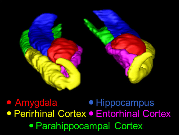
본 프로토콜을 사용하여 추적 MTL의 그림 1. 3D 개요. 여기에 표시된 구조는 AMY (적색), HC (파란색), 중화 인민 공화국 (노란색), ERC (핑크), 및 PHC (녹색)입니다 .

Protocol

1. 하멕 AMY의 전방 조각 전두엽과 측두엽의 백질 연결이 연속 30 보이는 곳 limen의 섬엽이 처음 나타나는 AMY의 첫 번째 조각을 식별합니다. 관상보기에서 AMY의 inferolateral 국경으로 각 번들을 사용합니다. AMY의 모양의 랜드 마크로서 시신경 시신경 교차를 찾습니다. 주변 uncus에서 초기 조각에서 AMY를 구분하는 축 방향 및 시상 뷰를 사용합니다. entorhinal 영역 (32)을</…

Representative Results

수동 및 자동 분할 가능한 차이의 그림 AMY, HC, PRC, ERC, 및 PHC에 대한 설명서 세그멘테이션의 3D 모델은도 1에 도시되고, 세그먼트의 시상 단면도는도 2에 도시된다. 수동 및 자동간에 극단적 가능한 차이를 예시의 목적 트레이싱은 잘못된 자동 분할과 대표 주제에서 AMY의 조각 (아래 그림 3 참조) 수동 추적과 함께 나란히했다. …

Discussion

전통적으로, 수동 분할은 많은 연구자들에 의해 황금 표준으로 간주하고있다. 그럼에도 불구하고, 각각의 구조의 정확한 묘사는 MTL 구조의 높은 변수 형태로 복잡하고, 주변 신경 조직 및 비 – 신경 영역에 대하여 이러한 구조의 약함 일반적 MRI 대조 순서되었습니다. 역사적으로, 일부 MTL 구조에 대한 문헌에서 충돌에 대한 설명이 있었다. 40,41 중단으로 PRC를 분할의 경우에서, 예를 들어, ?…

Offenlegungen

The authors have nothing to disclose.

Acknowledgements

This research was supported by funds to FD. MM was supported by an IGERT Fellowship under National Science Foundation Grant No. 0903622. The authors wish to thank the Dolcos Lab members for assistance with data collection and preparation.

Materials

ITK-SNAP ITK-SNAP Team at University of Pennsylvania and University of Utah ITK-SNAP v2.2
FSL Functional Magnetic Resonance Imaging of the Brain (FMRIB) Analysis Group FSL v4.1
3T Siemens Trio MR Scanner Siemens 3T Trio

Referenzen

  1. Amaral, D. G. Introduction: what is where in the medial temporal lobe. Hippocampus. 9, 1-6 (1999).
  2. Squire, L. R., Zola-Morgan, S. The medial temporal lobe memory system. Science. 253 (5026), 1380-1386 (1991).
  3. Eichenbaum, H., Otto, T., Cohen, N. J. The hippocampus: what does it do. Behavioral & Neural Biology. 57 (1), 2-36 (1992).
  4. Henke, K., Buck, A., Weber, B., Wieser, H. G. Human hippocampus establishes associations in memory. Hippocampus. 7 (3), 249-256 (1997).
  5. Tulving, E., Markowitsch, H. J. Episodic and declarative memory: role of the hippocampus. Hippocampus. 8 (3), 198-204 (1998).
  6. Dolcos, F., Iordan, A. D., Dolcos, S. Neural correlates of emotion–cognition interactions: a review of evidence from brain imaging investigations. Journal of Cognitive Psychology. 23 (6), 669-694 (2011).
  7. Davidson, R. J., Irwin, W. The functional neuroanatomy of emotion and affective style. Trends in Cognitive Sciences. 3 (1), 11-21 (1999).
  8. Lindquist, K. A., Wager, T. D., Kober, H., Bliss-Moreau, E., Barrett, L. F. The brain basis of emotion: a meta-analytic review. The Behavioral and Brain Sciences. 35 (3), 121-143 (2012).
  9. Phan, K. L., Wager, T., Taylor, S. F., Liberzon, I. Functional neuroanatomy of emotion: a meta-analysis of emotion activation studies in PET and fMRI. Neuroimage. 16 (2), 331-348 (2002).
  10. Wager, T. D., Phan, K. L., Liberzon, I., Taylor, S. F. Valence, gender, and lateralization of functional brain anatomy in emotion: a meta-analysis of findings from neuroimaging. Neuroimage. 19, 513-531 (2003).
  11. Zald, D. H. The human amygdala and the emotional evaluation of sensory stimuli. Brain Research Reviews. 41, 88-123 (2003).
  12. DeYoung, C. G., Hirsh, J. B., Shane, M. S., Papademetris, X., Rajeevan, N., Gray, J. R. Testing predictions from personality neuroscience: brain structure and the big five. Psychological Science. 21 (6), 820-828 (2010).
  13. Visser, P. J., Verhey, F. R., Hofman, P. A., Scheltens, P., Jolles, J. Medial temporal lobe atrophy predicts Alzheimer’s disease in patients with minor cognitive impairment. Journal of Neurology, Neurosurgery, & Psychiatry. 72 (4), 491-497 (2002).
  14. Ezekiel, F., et al. Comparisons between global and focal brain atrophy rates in normal aging and Alzheimer disease. Alzheimer Disease & Associated Disorders. 18 (4), 196-201 (2004).
  15. de Leon, M. J., et al. Imaging and CSF studies in the preclinical diagnosis of Alzheimer’s disease. Annals of the New York Academy of Sciences. 1097, 114-145 (2007).
  16. Boccardi, M., et al. Survey of protocols for the manual segmentation of the hippocampus: preparatory steps towards a joint EADC-ADNI harmonized protocol. Journal of Alzheimer’s Disease. 26, 61-75 (2011).
  17. Konrad, C., Ukas, T., Nebel, C., Arolt, V., Toga, A. W., Narr, K. L. Defining the human hippocampus in cerebral magnetic resonance images-an overview of current segmentation protocols. Neuroimage. 47 (4), 1185-1195 (2009).
  18. Hasboun, D., et al. MR determination of hippocampal volume: comparison of three methods. American Journal of Neuroradiology. 17 (6), 1091-1098 (1996).
  19. Pantel, J., et al. A new method for the in vivo volumetric measurement of the human hippocampus with high neuroanatomical accuracy. Hippocampus. 10, 752-758 (2000).
  20. Entis, J. J., Doerga, P., Barrett, L. F., Dickerson, B. C. A reliable protocol for the manual segmentation of the human amygdala and its subregions using ultra-high resolution MRI. Neuroimage. 60 (2), 1226-1235 (2012).
  21. Goncharova, I. I., Dickerson, B. C., Stoub, T. R., deToledo-Morrell, L. MRI of human entorhinal cortex: a reliable protocol for volumetric measurement. Neurobiology of Aging. 22, 737-745 (2001).
  22. Watson, C., et al. Anatomic basis of amygdaloid and hippocampal volume measurement by magnetic resonance imaging. Neurology. 42 (9), 1743-1750 (1992).
  23. Duvernoy, H. The human hippocampus: functional anatomy, vascularization, and serial sections with MRI. Third Edition. , (2005).
  24. Amaral, D. G., Witter, M. P. The three-dimensional organization of the hippocampal formation: a review of anatomical data. Neurowissenschaften. 31 (3), 571-591 (1989).
  25. Insausti, R., et al. MR volumetric analysis of the human entorhinal, perirhinal, and temporopolar cortices. American Journal of Neuroradiology. 19 (4), 659-671 (1998).
  26. Dolcos, F., LaBar, K. S., Cabeza, R. Interaction between the amygdala and the medial temporal lobe memory system predicts better memory for emotional events. Neuron. 42 (5), 855-863 (2004).
  27. Yushkevich, P. A., et al. User-guided 3D active contour segmentation of anatomical structures: significantly improved efficiency and reliability. Neuroimage. 31 (3), 1116-1128 (2006).
  28. Bonilha, L., Kobayashi, E., Cendes, F., Li, M. L. Protocol for volumetric segmentation of medial temporal structures using high-resolution 3-D magnetic resonance imaging. Human Brain Mapping. 22 (2), 145-154 (2004).
  29. Bronen, R. A., Cheung, G. Relationship of hippocampus and amygdala to coronal MRI landmarks. Magnetic Resonance Imaging. 9 (3), 449-457 (1991).
  30. Pruessner, J. C., et al. Volumetry of temporopolar, perirhinal, entorhinal and parahippocampal cortex from high-resolution MR images: considering the variability of the collateral sulcus. Cerebral Cortex. 12 (12), 1342-1353 (2002).
  31. Pruessner, J. C., et al. Volumetry of hippocampus and amygdala with high-resolution MRI and three-dimensional analysis software: minimizing the discrepancies between laboratories. Cerebral Cortex. 10 (4), 433-442 (2000).
  32. Duvernoy, H. . The human brain: surface, three-dimensional sectional anatomy with MRI, and blood supply. Second Edition. , (1999).
  33. Amaral, D. G., Lavenex, P., et al. . in The hippocampus book. Hippocampal neuroanatomy. , (2006).
  34. Blaizot, X., et al. The human parahippocampal region: I. temporal pole cytoarchitectonic and MRI correlation. Cerebral Cortex. 20 (9), 2198-2212 (2010).
  35. Ding, S. -. L., Van Hoesen, G. W. Borders, extent, and topography of human perirhinal cortex as revealed using multiple modern neuroanatomical and pathological markers. Human Brain Mapping. 31 (9), 1359-1379 (2010).
  36. Ding, S. -. L., Van Hoesen, G. W., Cassell, M. D., Poremba, A. Parcellation of human temporal polar cortex: a combined analysis of multiple cytoarchitectonic, chemoarchitectonic, and pathological markers. The Journal of Comparative Neurology. 514 (6), 595-623 (2009).
  37. Frankó, E., Insausti, A. M., Artacho-Pérula, E., Insausti, R., Chavoix, C. Identification of the human medial temporal lobe regions on magnetic resonance images. Human Brain Mapping. 35 (1), 248-256 (2014).
  38. Lehmann, M., et al. Atrophy patterns in Alzheimer’s disease and semantic dementia: a comparison of FreeSurfer and manual volumetric measurements. Neuroimage. 49 (3), 2264-2274 (2010).
  39. Winterburn, J. L., et al. A novel in vivo atlas of human hippocampal subfields using high-resolution 3T magnetic resonance imaging. Neuroimage. 74, 254-265 (2013).
  40. Malykhin, N. V., Bouchard, T. P., Ogilvie, C. J., Coupland, N. J., Seres, P., Camicioli, R. Three-dimensional volumetric analysis and reconstruction of amygdala and hippocampal head, body and tail. Psychiatry research. Neuroimaging. 155 (2), 155-165 (2007).
  41. Patenaude, B., Smith, S. M., Kennedy, D. N., Jenkinson, M. A Bayesian model of shape and appearance for subcortical brain segmentation. Neuroimage. 56 (3), 907-922 (2011).
  42. Smith, S. M., et al. Advances in functional and structural MR image analysis and implementation as FSL. Neuroimage. 23, (2004).
  43. Woolrich, M. W., et al. Bayesian analysis of neuroimaging data in FSL. Neuroimage. 45, (2009).
  44. Smith, S. M. Fast robust automated brain extraction. Human Brain Mapping. 17 (3), 143-155 (2002).
  45. Morey, R. A., et al. A comparison of automated segmentation and manual tracing for quantifying hippocampal and amygdala volumes. Neuroimage. 45 (3), 855-866 (2009).
  46. Baldassano, C., Beck, D. M., Fei-Fei, L. Differential connectivity within the parahippocampal place area. Neuroimage. 75, 228-237 (2013).
  47. Shrout, P. E., Fleiss, J. L. Intraclass correlations: uses in assessing rater reliability. Psychological Bulletin. 86 (2), 420-428 (1979).
  48. Bland, J. M., Altman, D. G. A note on the use of the intraclass correlation coefficient in the evaluation of agreement between two methods of measurement. Computers in Biology and Medicine. 20, 337-340 (1990).
  49. Yushkevich, P. A., et al. A high-resolution computational atlas of the human hippocampus from postmortem magnetic resonance imaging at 9.4 T. Neuroimage. 44 (2), 385-398 (2009).

Play Video

Diesen Artikel zitieren
Moore, M., Hu, Y., Woo, S., O’Hearn, D., Iordan, A. D., Dolcos, S., Dolcos, F. A Comprehensive Protocol for Manual Segmentation of the Medial Temporal Lobe Structures. J. Vis. Exp. (89), e50991, doi:10.3791/50991 (2014).

View Video