This study presents a highly reproducible large animal model of renal ischemia-reperfusion injury in swine using temporary percutaneous bilateral balloon-catheter occlusion of the renal arteries for 60 min and reperfusion for 24 h.
Acute kidney injury (AKI) is associated with higher risk for morbidity and mortality post-operatively. Ischemia-reperfusion injury (IRI) is the most common cause of AKI. To mimic this clinical scenario, this study presents a highly reproducible large animal model of renal IRI in swine using temporary percutaneous bilateral balloon-catheter occlusion of the renal arteries. The renal arteries are occluded for 60 min by introducing the balloon-catheters through the femoral and carotid artery and advancing them into the proximal portion of the arteries. Iodinated contrast is injected in the aorta to assess any opacification of the kidney vessels and confirm the success of the artery occlusion. This is furtherly confirmed by the flattening of the pulse waveform at the tip of the balloon catheters. The balloons are deflated and removed after 60 min of bilateral renal artery occlusion, and the animals are allowed to recover for 24 h. At the end of the study, plasma creatinine and blood urea nitrogen significantly increase, while eGFR and urine output significantly decrease. The need for iodinated contrast is minimal and does not affect renal function. Bilateral renal artery occlusion better mimics the clinical scenario of perioperative renal hypoperfusion, and the percutaneous approach minimizes the impact of the inflammatory response and the risk of infection seen with an open approach, such as a laparotomy. The ability to create and reproduce this clinically relevant swine model eases the clinical translation to humans.
Acute kidney injury (AKI) is a commonly diagnosed condition among surgical patients associated with significant morbidity and mortality1,2. Available data show that AKI can affect even half of all hospitalized patients worldwide and leads to 50% mortality rate in patients in the intensive care unit1,3. Despite its high prevalence, current AKI therapy remains limited to preventive strategies, such as fluid management and dialysis. Therefore, there is an ongoing interest in exploring alternative therapies for AKI4,5,6.
AKI is typically classified into pre-renal, intrinsic, and post-renal based on its etiology4,5,6. The majority of surgical patients with AKI are associated with pre-renal causes due to hypovolemia, resulting in ischemia-reperfusion injury (IRI) of the kidneys2. Clinically, urine output decreases, and creatinine levels increase due to decreased renal function. The kidney is a high-metabolic-rate organ and susceptible to ischemia. A highly reproducible large animal model of renal IRI is necessary to obtain a better insight into the pathophysiology of AKI and its potential therapeutic approaches5.
To mimic the clinical scenario of kidney hypoperfusion peri-operatively, a model of bilateral renal artery occlusion is deemed suitable. Previously described models entailing unilateral renal artery occlusion with or without resection of the contralateral kidney do not provide sufficient clinical applicability7,8. Although these models are sufficient for causing AKI, they do not resemble real-life clinical scenarios neither in terms of type nor duration of injury.
The aim of this paper is to present a porcine model of percutaneous bilateral temporary occlusion of the renal arteries by balloon-catheter occlusion under angiography. Bilateral renal artery occlusion mimics the clinical scenario of renal hypoperfusion, followed by the subsequent removal of the balloon for reperfusion9,10. The technical steps are described, including catheterization, catheter guidance, angiography, and hemodynamic monitoring. This method not only allows for a highly controlled and replicable occlusion of the renal arteries, but the percutaneous approach minimizes the impact of the inflammatory response by limiting the amount of insult to the body compared to an open approach.
All in vivo studies were conducted in accordance with the National Institutes of Health's guidelines on animal care and use and were approved by the Boston Children's Hospital's Animal Care and Use Committee (Protocol 18-06-3715). All animals received humane care in compliance with the Guide for the Care and Use of Laboratory Animals. Figure 1 shows the timeline including anesthesia, surgical preparation, and timepoints for primary outcome measurements of this study.
1. Induction, anesthesia and intubation
2. Surgical preparation and vascular access
3. Induction of renal ischemia-reperfusion injury
4. Animal recovery
5. Functional assessment
6. Euthanasia
Functional analysis
The representative results of this study arise from 6 animals and the data shown are mean ± standard error of the mean.Renal function is assessed by determining the urine output, estimated glomerular filtration rate (eGFR), plasma creatine, and blood urea nitrogen (BUN). The biomarkers of renal function are assessed using a portable chemistry analyzer. eGFR is calculated according to the following formula: eGFR =1.879 × BW1.092/PCr0.6 (BW: body weight in kg; PCr: plasma creatinine in mg/dL)10,11.
Following 60 min of bilateral renal artery occlusion, urine output was significantly decreased from 3.6 mL/kg/h ± 0.5 mL/kg/h to 0.2 mL/kg/h ± 0.1 mL/kg/h (p < 0.01). This decrease remained significant at 6 h (1.2 mL/kg/h ± 0.1 mL/kg/h; p = 0.02 vs. baseline) and 24 h (1.3 mL/kg/h ± 0.4 mL/kg/h; p = 0.02 vs. baseline) following reperfusion. Similarly, a significant decrease was observed in eGFR, which dropped from 2.5 mL/kg/h ± 0.1 mL/kg/h at baseline to 1.7 mL/kg/h ± 0.1 mL/kg/h (p < 0.001) at the end of ischemia and to 1.5 mL/kg/h ± 0.1 mL/kg/h (p < 0.001), 1.2 mL/kg/h ± 0.1 mL/kg/h (p < 0.001) and 0.9 mL/kg/h ± 0.1 mL/kg/h (p < 0.001) at 2 h, 6 h and 24 h reperfusion, respectively (Figure 2A-B).
Plasma creatinine was significantly increased at 2 h (2.7 mg/dL ± 0.2 mg/dL; p < 0.01), 6 h (3.7 mg/dL ± 0.3 mg/dL; p < 0.001) and 24 h (5.6 mg/dL ± 0.7 mg/dL; p < 0.001) of reperfusion compared to baseline (1.1 mg/dL ± 0.1 mg/dL). BUN was 6.5 mg/dL ± 0.8 mg/dL at baseline and increased to 17.8 mg/dL ± 3.3 mg/dL (p < 0.001) and 36.2 mg/dL ± 2.9 mg/dL (p < 0.001) at 6 h and 24 h of reperfusion, respectively (Figure 2C-D).
Gross anatomy and histology
There were evident necrotic and hemorrhagic areas which were unevenly distributed in both kidneys at the end of the 60 min of bilateral renal ischemia and the 24 h of reperfusion (Figure 3A). Masson's Trichrome staining revealed confluent coagulative necrosis which was located at the proximal tubules of the renal cortex. (Figure 3B). Plastic embedded sections (1 µm) were also assessed since they provide significant details of the histology (Figure 3C). All Masson's Trichrome slides were evaluated for cell necrosis, loss of brush border, cast formation, and tubule dilatation. Then, a semi-quantitative scoring system for acute tubular necrosis (ATN) was implemented as follows: 0 if none; 1 if less than 10%; 2 if between 11%-25%; 3 if between 26%- 45%; 4 if between 46%-75%; and 5 if greater than 76%. ATN scoring showed significant injury in the renal cortex (score of 4.5 ± 0.3) and considerable injury in the medulla (score of 2.7 ± 0.4).
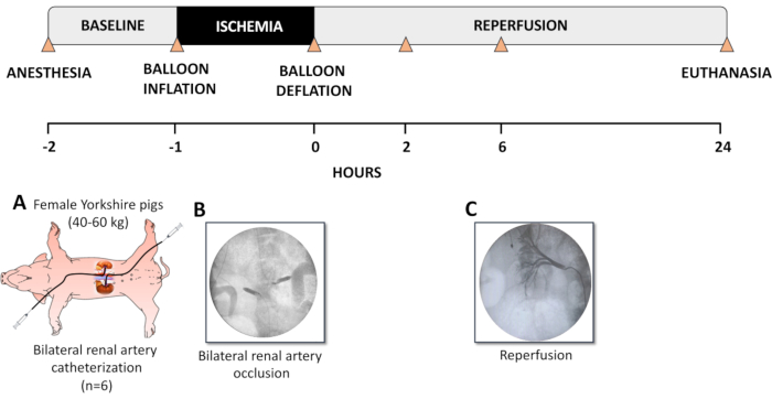
Figure 1. Description of the experimental model. (A) Female Yorkshire pigs (40-60 kg) were sedated and intubated. The left femoral artery and the right carotid artery were catheterized with a 5F angiography sheath. Right jugular venous lines and a Foley urinary catheter were also placed. Selective catheterization of the renal arteries was performed using a 5F multipurpose guide-catheter. (B) Occlusion of the renal arteries was performed using a 5F percutaneous transluminal angioplasty (PTA) dilatation catheter inflated in the proximal portion of the renal artery, totally occluding the blood flow to the kidneys for 60 min. Confirmation of the occlusion was acquired by injection of iodinated contrast medium in the aorta and by checking for any opacification of the vessels of the kidneys. (C) Following 60 min of occlusion, the balloons were deflated and carefully removed. Angiography was performed to confirm renal artery patency and the establishment of renal reperfusion. The animals were then allowed to reperfuse the kidneys under physiological conditions for the next 24 h and were subsequently euthanized. Blood and urine samples were collected right before and after bilateral renal ischemia, at 2, 6, and 24 h after occlusion (timepoints indicated with triangles). This figure has been modified from Doulamis et al11. Please click here to view a larger version of this figure.
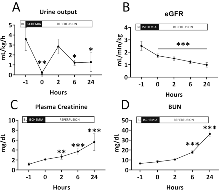
Figure 2. Renal function before and after renal ischemia-reperfusion injury. (A) Urine output; (B) Estimated glomerular filtration rate (eGFR); (C) Plasma creatinine and (D) Blood urea nitrogen (BUN). All results are shown as mean and standard deviation for each timepoint. A significant decrease can be seen in the urine output and the eGFR following ischemia-reperfusion injury. Accordingly, a significant increase is noted in plasma creatinine and BUN. Data were analyzed by two-way repeated measures ANOVA with the Benjamini and Hochberg's false discovery rate (n=6). *p < 0.05 vs Baseline; **p < 0.01 vs Baseline; ***p < 0.001 vs Baseline. Please click here to view a larger version of this figure.
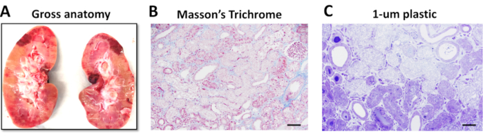
Figure 3. Gross kidney anatomy and renal tissue injury at 24 hours of reperfusion following renal ischemia-reperfusion injury. (A) Gross anatomy of the left kidney showing pale areas indicative of infarction and red hemorrhagic areas following 60 min of bilateral renal artery occlusion and 24 h of reperfusion. (B) Renal Cortex of vehicle shows extensive coagulative necrosis of primarily proximal tubules, following 60 min of ischemia and 24 h of reperfusion (Masson's Trichrome, original magnification 20x). (C) These 1 µm plastic (araldite-epon) embedded sections demonstrate in greater detail the confluent tubular necrosis consisting primarily of matrix with swelling and degenerative changes of organelles (Toluidine blue, original magnification 40x). Scale bar = 200 µm. This figure has been modified from Doulamis et al11. Please click here to view a larger version of this figure.
AKI is a common clinical disorder affecting up to 50% of hospitalized adult patients worldwide6,12. A clinically relevant animal model is needed to further investigate the pathophysiology of the disease and potential therapeutic targets. Although there are several murine models replicating AKI, these do not completely mimic their respective clinical scenarios and the anatomy of the human kidney. This study proposes a clinically relevant swine model to allow for translation to humans13.
Here, the protocol describes a percutaneous approach which is not only clinically relevant but also minimizes the inflammatory response and the risk for infection that accompanies an open approach. It should also be highlighted that a consistent hydration protocol should be used for all animals in order to achieve optimal hemodynamic control and avoid renal hypoperfusion11. This can be easily done when the animal is anesthetized but cannot always be accurately performed during the recovery period when water is provided ad libitum.
Iodinated contrast medium should be used cautiously in order to avoid contrast-induced nephrotoxicity. This can be achieved by 1:1 or 1:2 dilution with normal saline. In this study, we used a dose which is 10 times lower than the estimated safety threshold for humans (3.33 mL/kg)9,14.
Among others, the study uses eGFR for the assessment of renal function based on a formula accounting for the body weight and plasma creatinine levels10,11. It should be noted that although the use of inulin for the determination of GFR has been previously documented, its use was deferred in the current protocol due to severe hypotensive vasospastic reaction after inulin infusion. This can be avoided by using steroids or epinephrine prior to inulin administration. However, the use of these drugs may not be appropriate according to each study design. For this reason, a validated formula to estimate the eGFR based on plasma creatinine and body weight was used11.An alternative way for determining GFR would be using the formula: (urine creatinine x urine flow rate) / (plasma creatinine x kidney weight).
For the assessment of the ATN score, the use of Masson’s Trichrome staining is preferable to conventional hematoxylin and eosin staining as it can better trace tissue injury. Another alternative may be the use of plastic embedded sections, which provide greater details as they allow for thinner slicing of the sample11. This preclinical model of AKI can be used to mimic several clinical scenarios such as kidney transplantation, renal hypoperfusion following cardiogenic shock (e.g., myocardial infarction, aneurysm rupture, aortic dissection), transcatheter procedures at high risk of renal ischemia and cardiovascular procedures with prolonged cardiocirculatory arrest times.
This study has some limitations. The study used only female animals. This was done to reduce any possible effects related to urinary catheterization, which is less traumatic in females than males. In addition to this limitation, the study used young, otherwise healthy animals, thus eliminating confounding variables that may be related to coexisting diseases. In conclusion, the current study describes a highly reproducible large animal model of renal IRI, which can be used to decrease the burden of AKI.
The authors have nothing to disclose.
We would like to thank Dr. Arthur Nedder for his help and guidance. This work was supported by the Richard A. and Susan F. Smith President's Innovation Award, Michael B. Klein and Family, The Sidman Family Foundation, The Michael B. Rukin Charitable Foundation, The Kenneth C. Griffin Charitable Research Fund, and The Boston Investment Council.
0.9% sodium chloride injection, usp, 100 ml viaflex plastic container | Baxter | 2B1302 | For animal hydration |
Agent contrast 100.0ml injection media btl ioversal 74% | CARDINAL HEALTH | 133311 | For visualizing the vasculature |
Bard Bardia Closed System Urinary Drainage Bag | BARD Inc | 802001 | For urine collection |
BD Vacutainer K2 EDTA | BD | 367841 | For blood sample storage |
BD Vacutainer Lithium Heparin | BD | 366667 | For blood sample storage |
Betadine | Henry Schein | 6906950 | For skin disinfection |
Bookwalker retractor | Codman | For skin retraction | |
Bupivacaine 0.25% | Hospira | Administer at incision site for analgesia | |
Buprenorphine SR | Zoo Pharm | 10mg/ml bottle, Dose: 0.2mg/kg SC | |
Cath angio 5.0 Fr x100.0 cm 0.038 in JR4 | MERIT MEDICAL SYSTEM INC | 7523-21 | For identification of the renal arteries |
Cuffed endotracheal tube | Emdamed | To establish a secure airway for the duration of the operation | |
EKG Medtronics- Physiocontrol LifePak 20 Oxygen saturation monitor | GE Healthcare Madison WI | For oxygen saturation monitoring | |
Encore 26 inflator | BOSTON SCIENTIFIC | 710113 | For inflating the balloon catheters |
Ethanol 95% (Ethyl alcohol) | Henry Schein | For skin disinfection | |
Fentanyl patch | Mylan | Dose: 25-50mcg/hr, TD | |
Gold silicone coated Foley | TELEFLEX MEDICAL INC | 180730160 | For urine collection |
Heparin sodium | LEO Pharma A/S | Dose: 200 IU/kg IV | |
i33 ultrasound machine | Phillips | Use ultrasonographic guidance for femoral catherization if necessary | |
Inqwire diagnostic guide wire – 0.035" (0.89 mm) – 260 cm (102") – 1.5 mm j-tip | MERIT MEDICAL SYSTEM INC | 6609-33 | For guiding the balloon catheters to the renal arteries |
Intravenous catheter, size 20 gauge | Santa Cruz Biotechnology | Inc SC-360097 | For fluid administration |
Isoflurane | Patterson Veterinary Supply, Inc. | 21283620 | Dose: 3%, INH |
Metzenbaum blunt curved 14.5 cm – 5(3/4)" | Rudolf Medical | RU-1311-14M | For tissue dissection and cutting |
Neonatal disposable transducer kit with 30ml/hr flush device and double 4-way stopcocks for continuous monitoring | Argon Medical | 041588505A | For pressure measurement |
Powerflex pro PTA dilatation catheter 6 x 20 mm – shaft length (135cm) | CARDINAL HEALTH | 4400602X | For occlusion of the renal arteries |
Pressure monitoring lines mll/mll – 12" clear, mll/mll | Smiths Medical | B1571/MX571 | For pressure measurement |
Procedure pack | Molnlycke Health Care | 97027809 | Surgical drape, gauze pads, syringes, beaker etc |
Protamine | Henry Schein | 1044148 | For heparin reversal |
Scalpel blade – size #10 | Cardinal Health (Allegiance) | 32295-010 | For the skin incisions |
Stopcock iv 4 way lrg bore rotg male ll adptr strl | Peoplesoft | 1550 | For connecting tubings |
Straight lateral retractor | Codman | For skin retraction | |
Suture perma hnd 18in 2-0 braid silk blk | CARDINAL HEALTH 1 | A185H | For suturing incision site and securing catheters |
Syringe contrast injection 10ml fixed male luer red | MERIT MEDICAL SYSTEM INC | MSS111-R | To administer the contrast agent |
Syringe medical 60ml ll plst strl ltx free disp | CARDINAL HEALTH 1 | BF309653 | For urine collection and flushing of the angiocath |
Tilzolan (tiletamine/zolazepam) | Patterson Veterinary Supply, Inc. | 07-893-1467 | Dose: 4-6 mg/kg, IM |
Xylazine | Putney, INC | Dose: 1.1-2.2 mg/kg, IM |