Pretargeted Radioimmunotherapy: A Method for Targeted Delivery of Therapeutic Radionuclides to Tumors
Abstract
Source: Membreno, R., et al. Pretargeted Radioimmunotherapy Based on the Inverse Electron Demand Diels-Alder Reaction. J. Vis. Exp. (2019).
This video describes the technique of pretargeted radioimmunotherapy or PRIT, in which immunoconjugates followed by radioligands are administered for treating tumors. This radioimmunotherapy reduces radiation doses to healthy tissues and utilizes radionuclides with very low half-lives incompatible with antibody-based vectors.
Protocol
All procedures involving animal models have been reviewed by the local institutional animal care committee and the JoVE veterinary review board.
1. The Preparation of huA33-TCO
NOTE: The synthesis of huA33-TCO has been previously reported. However, for the ease of the reader, it is replicated here with adjustments for optimal conditions.
- In a 1.7 mL microcentrifuge tube, prepare a 125 μL solution of (E)-cyclooct-4-enyl 2,5-dioxo-1-pyrrolidinyl carbonate (TCO-NHS) in dry dimethylformamide (DMF) at a concentration of 40 mg/mL (0.15 M). This solution can be aliquoted and frozen at -80 °C for use in future experiments.
- In a separate 1.7 mL microcentrifuge tube, prepare a 5 mg/mL solution of huA33 in 1 mL of phosphate buffered saline (PBS; 2.7 mM potassium chloride, 137 mM sodium chloride, and 11.9 mM potassium phosphate, pH 7.4).
- Using small aliquots (<5 μL) of 0.1 M Na2CO3, adjust the pH of the antibody solution from Step 1.2 to 8.8-9.0. Use either pH paper or a pH meter with a microelectrode to monitor the pH and exercise care that the pH does not exceed 9.0.
- To the antibody solution described in Step 1.3, slowly add a volume corresponding to 40 molar equivalents of TCO-NHS relative to the amount of antibody. For example, if 5 mg (30 nmol) of huA33 is in the solution, add 9.0 μL (1.20 μmol) of the 40 mg/mL (0.15 M) solution of TCO-NHS.
NOTE: TCO-NHS is hydrophobic. When adding it to the antibody solution, add it slowly and with agitation to prevent precipitation. Do not exceed 10% DMF by volume in the final reaction solution. - Allow the reaction to incubate at 25 °C on a thermomixer for 1 h with mild agitation (500 rpm).
- After 1 h, purify the huA33-TCO immunoconjugate using a pre-packed disposable size-exclusion desalting column.
- Equilibrate the size exclusion column as described by the supplier to remove any preservatives present in the column during storage. A typical procedure involves washing the column 5x with a volume of PBS that corresponds to the volume of the column: 5 x 2.5 mL of PBS.
- Add the reaction mixture to the size exclusion column noting the volume of the reaction mixture.
- After the reaction mixture has entered the column, add an appropriate amount of PBS to bring the total volume of solution added to the column up to 2.5 mL. For example, if the conjugation reaction resulted in a total volume of 1.3 mL, add 1.2 mL of additional PBS to the column.
- Collect the product using 2 mL of PBS as the eluent.
NOTE: This step will yield the final construct huA33-TCO in 2 mL of PBS, pH 7.4.
- Measure the concentration of the huA33-TCO using a UV-Vis spectrophotometer monitoring the 280 nm wavelength. The molar extinction coefficient for most IgGs (ε280) is 210,000 M-1cm-1.
- If a solution with a higher concentration of immunoconjugate is desired, concentrate the huA33-TCO solution using a centrifugal filter unit with a 50,000 molecular weight cut-off following manufacturer instructions.
NOTE: Many antibodies have been known to aggregate or precipitate during concentration. When attempting this procedure with a new antibody, researchers should defer to the literature or their own experience handling the antibody in question. - Store the completed huA33-TCO immunoconjugate at 4 °C in the dark if it is needed immediately. If it is to be used more than 4 days in the future, store it at -80 °C in the dark.
NOTE: This is an acceptable stopping point in the procedure. The completed huA33-TCO immunoconjugate should be stable for at least 6 months storage at -80 °C in the dark.
2. The Synthesis of Tz-PEG7-NHBoc
- In a 1.7 mL microcentrifuge tube, dissolve 5 mg of tetrazine N-hydroxysuccinimidyl ester (Tz-NHS; 12.6 μmol) in 0.15 mL of anhydrous dimethyl sulfoxide (DMSO).
- In a separate 1.7 mL microcentrifuge tube, dissolve 8 mg of the Boc-protected amino PEG polymer, O-(2-Aminoethyl)-O'-[2-(Boc-amino)ethyl]hexaethylene glycol (NHBoc-PEG7-NH2; 17.1 μmol) in 0.15 mL of anhydrous DMSO.
- To the solution of Tz-NHS, add 3 μL (21.3 μmol, 1.7 molar equivalents) of triethylamine (TEA) and mix thoroughly.
- Add the pink tetrazine solution to the NHBoc-PEG7-NH2 solution from Step 2.2 and incubate the reaction mixture for 30 min at room temperature (RT) with mild agitation.
- After 30 minutes, dilute the reaction mixture 1:1 in H2O and purify it using reversed-phase C18 high-performance liquid chromatography (HPLC) to separate any unreacted Tz-NHS from the Tz-PEG7-NHBoc. Use solvents without acid to prevent the premature removal of the Boc protecting group. Monitor both Tz-PEG7-NHBoc and Tz-NHS at a wavelength of 525 nm.
NOTE: Retention times are highly dependent on the HPLC equipment of each laboratory (pumps, columns, tubing, etc.), and appropriate controls should be run before purification. However, to present an example, if a gradient of 5:95 MeCN/H2O (both without any additive) to 95:5 MeCN/H2O over 30 min, a flow rate of 1 mL/min, and an analytical 4.6 mm x 250 mm C18 column are used, the retention times of Tz-NHS and Tz-PEG7-NHBoc are around 16 min and 18 min, respectively. - Freeze the collected HPLC eluent using liquid nitrogen and wrap the now-frozen collection tube in aluminum foil. Place the frozen collection tube on a lyophilizer overnight to remove the HPLC mobile phase. The product will be a characteristic bright pink solid.
NOTE: If liquid nitrogen is unavailable, freeze the collected HPLC eluent in dry ice or overnight in a -80 °C freezer.
3. The Synthesis of Tz-PEG7-NH2
- To the product from Step 2.6, Tz-PEG7-NHBoc, add 1.5 mL of dichloromethane (DCM) and transfer this solution to a small round-bottom flask.
- Add 0.25 mL of trifluoroacetic acid (TFA) dropwise to the pink solution from Step 3.1.
- Allow the reaction to incubate for 30 min at RT with mild agitation.
- After 30 min, evaporate the solvent via rotary evaporation. Do not exceed the water bath temperature of 37 °C.
- Reconstitute the pink viscous product in 0.5 mL of water.
- Purify the product using reversed-phase C18 HPLC to separate Tz-PEG7-NH2 from the Boc-protected precursor. Monitor both Tz-PEG7-NHBoc and Tz-PEG7-NH2 at a wavelength of 525 nm.
NOTE: Retention times are highly dependent on the HPLC equipment of each laboratory (pumps, columns, tubing, etc.), and appropriate controls should be run before purification. However, to present an example, if a gradient of 5:95 MeCN/H2O (both with 0.1% TFA) to 95:5 MeCN/H2O over 30 min, a flow rate of 1 mL/min, and an analytical 4.6 mm x 250 mm C18 column are used, the retention times of Tz-PEG7-NHBoc and Tz-PEG7-NH2 are around 18 min and 13 min, respectively. - Freeze the collected HPLC eluent using liquid nitrogen and wrap the now-frozen collection tube in aluminium foil. Place the frozen collection tube on a lyophilizer overnight to remove the HPLC mobile phase. The product will be a bright pink solid.
- Reconstitute the product from Step 3.7, Tz-PEG7-NH2, with 150 μL of DMSO and transfer to a 1.7 mL microcentrifuge tube.
- Measure the concentration of Tz-PEG7-NH2 using a UV-Vis spectrophotometer monitoring the 525 nm wavelength. The molar extinction coefficient for Tz-PEG7-NH2 (ε525) is 535 M-1cm-1.
4. The Synthesis of Tz-PEG7-DOTA
- Dissolve Tz-PEG7-NH2 (4.5 mg, 6.9 μmol) in 0.15 mL of DMSO (or proceed with the solution created in Step 3.8).
- Add 22 molar equivalents of TEA (21 μL, 0.15 mmol) to the tetrazine-containing solution from Step 4.1.
- Add 10 mg (14.2 μmol) of p-SCN-Bn-DOTA as a solid and vortex the solution for about 2 min or until the material is fully dissolved.
- Allow the reaction to incubate for 30 min at RT with mild agitation.
- After 30 min, dilute the reaction 1:1 in H2O and purify the product using reversed-phase C18 HPLC to remove unreacted p-SCN-Bn-DOTA. The p-SCN-Bn-DOTA can be monitored at a wavelength of 254 nm, while the Tz-PEG7-DOTA is best monitored at a wavelength of 525 nm.
NOTE: Retention times are obviously highly dependent on the HPLC equipment of each laboratory (pumps, columns, tubing, etc.), and appropriate controls should be run before purification. However, to present an example, if a gradient of 5:95 MeCN/H2O (both with 0.1% TFA) to 95:5 MeCN/H2O over 30 min and an analytical 4.6 x 250 mm C18 column are used, Tz-PEG7-DOTA and p-SCN-Bn-DOTA have retention times of around 15.6 min and 16.1 min, respectively. - Freeze the collected HPLC eluent using liquid nitrogen and wrap the now-frozen collection tube in aluminum foil. Place the frozen collection tube on a lyophilizer overnight to remove the HPLC mobile phase. The product will be a bright pink powder.
- Reconstitute the product in 0.15 mL of DMSO and measure the concentration using a UV-Vis spectrophotometer monitoring the 525 nm wavelength. The molar extinction coefficient for Tz-PEG7-DOTA (ε525) is 535 M-1cm-1.
- Analyze the final compound by nuclear magnetic resonance (NMR) and high-resolution mass spectrometry (HRMS) to verify that synthesis was successful. See Table 1 for the experimentally determined chemical shifts and molecular weights of all the compounds discussed in this work.
- Store the purified Tz-PEG7-DOTA solution in the dark at -80 °C.
NOTE: This is an acceptable stopping point in the procedure. The completed Tz-PEG7-DOTA precursor is stable for at least 1 year under these conditions.
5. 177Lu Radiolabeling of Tz-PEG7-DOTA
CAUTION: This step of the protocol involves the handling and manipulation of radioactivity. Before performing these steps — or performing any other work with radioactivity — researchers should consult with their home institution's Radiation Safety Department. Take all possible steps to minimize exposure to ionizing radiation.
NOTE: When working with small amounts of radiometals, it is recommended that all buffers be free from trace metals to prevent interference in coordination site binding.
- In a 1.7 mL centrifuge tube, add 200 μL of 0.25 M ammonium acetate buffer adjusted with aliquots of 1 M HCl to pH 5.5.
NOTE: If using less than 370 MBq of activity for the labeling, the volume of buffer used should be reduced to 100 µL. - Add the desired amount of [177Lu]LuCl3 to the buffer solution. The amount added will be dependent on the number of subjects in the experiment and the radioactive doses being administered. It is recommended that 1-2 extra doses worth of radioactivity be added as a precaution to compensate for the potential loss of radioactivity during purification steps.
- Add Tz-PEG7-DOTA in DMSO to the radioactive mixture in Step 5.2. The amount of Tz-PEG7-DOTA is dependent on the number of subjects being tested. More detail on this topic can be found in Step 6.2.2.2.
- Allow the solution to incubate at 37 °C for 20 min.
- Verify the radiolabeling is complete using radio instant thin layer chromatography (radio-iTLC) with 50 mM EDTA, pH 5.5 as the mobile phase. The labeled [177Lu]Lu-DOTA-PEG7-Tz will remain at the baseline — Rf = 0 — while free [177Lu]Lu3+ will be coordinated by the EDTA and will travel with the solvent front, Rf = 1.0 (Figure 1B).
- If quantitative labeling is not achieved, add ligands to coordinate the free radiometal. Alternatively, purify the labeled [177Lu]Lu-DOTA-PEG7-Tz using a C18 cartridge. Follow manufacturer instructions for use. A sample procedure is given below.
- Prime the cartridge by slowly passing 5 mL of ethanol through the cartridge with a large syringe. Then, pass through 5 mL of acetonitrile and then 5 mL of deionized (DI) H2O.
- Draw up the radioligand solution from Step 5.3 in a smaller syringe and inject it slowly onto the C18 cartridge. Then, wash the cartridge with 10 mL of DI H2O to remove any unbound [177Lu]LuCl3.
- Elute with 500 µL of ethanol. Remove any ethanol from the final product by passing over the vessel with a low flow rate of dry nitrogen or argon for 10-15 min. Subsequently, resuspend the radioligand in saline in a volume determined in Step 6.2.2.2.
6. In vivo Studies
CAUTION: As in Section 5, this step of the protocol involves the handling and manipulation of radioactivity. Before performing these steps, researchers should consult with their home institution's Radiation Safety Department. Take all possible steps to minimize exposure to ionizing radiation.
- Preparation of animals
- In female athymic nude mice, subcutaneously implant 5 x 10⁶ SW1222 colorectal cancer cells suspended in 150 μL of a 1:1 mixture of cell media and matrix (e.g., Matrigel) and allow these to grow into a 100-150 mm3 xenograft (14-18 days after inoculation).
- Sorting of animals for a biodistribution experiment
- Once the tumors are of sufficient size as determined by caliper measurement, sort the animals to ensure each cohort has approximately the same average tumor volume. The animals can be distinguished in each cage by markings on the tail with indelible ink (one band, two bands, etc.).
- Sorting of animals for a longitudinal therapy study
- Once the tumors are of sufficient size as determined by caliper measurement, attach ear tags to each of the animals to ensure correct tracking throughout the experiment.
NOTE: Numerical ear tags may fall off throughout the experiment. As a result, it is recommended to accompany these physical tags with ear notches in a systematic manner (i.e., right, left, bilateral, right x 2, left x 2).
- Once the tumors are of sufficient size as determined by caliper measurement, attach ear tags to each of the animals to ensure correct tracking throughout the experiment.
- Sort the animals so that the average tumor volume in each cohort is roughly equal. The following method for animal sorting can be performed using a spreadsheet program.
- List the animal identification numbers, ear notch pattern, and tumor volume in three separate columns.
- Sort the data from smallest to largest tumor volume.
- In a fourth column, assign each animal a cage number and cycle through the cages in a snakelike pattern. For example, if there are 5 cages, this column would be filled "5, 4, 3, 2, 1, 1, 2, 3, 4, 5…" until all the animals are assigned a cage.
- Once the cages are assigned, sort the data by cage number. If ear notches are being used, ensure that each animal in a given cage has a unique ear notch pattern. If duplicates (two or more of the same pattern) in a given cage, swap one mouse with one from another cage with the missing pattern until every cage has animals with unique ear notch patterns.
- Formulations and Injections
NOTE: The sequence of injection for both biodistribution and therapy study proceeds as follows: huA33-TCO is injected first, followed by [177Lu]Lu-DOTA-PEG7-Tz after a pre-determined interval.- Immunoconjugate
- Dilute an aliquot of the huA33-TCO solution from Step 1.9 to a concentration of 0.8 mg/mL in 0.9% sterile saline.
- Draw doses of 150 µL (100 µg) of huA33-TCO solution in syringes pretreated with 1% bovine serum albumin (BSA) in PBS and store these syringes on ice.
NOTE: BSA treatment reduces the non-specific binding of the antibody to the walls of the syringe. - Warm the animal under a heat lamp for 3 minutes to dilate the tail vein.
- Inject the huA33-TCO solution into the tail vein of the xenograft-bearing mouse. Allow 24 h (or a different pre-determined time interval) for the huA33-TCO to accumulate in the tumor of the mouse.
- Radioligand
- Radiolabel Tz-PEG7-DOTA as outlined in Section 5.
- Draw doses in 150 μL of 0.9% sterile saline containing 1.1 molar equivalents of Tz-PEG7-DOTA relative to the amount of huA33-TCO administered. As an example, if 100 μg (0.67 nmol) of huA33-TCO was injected into the animal and the [177Lu]Lu-DOTA-PEG7-Tz reaction mixture was made with a specific activity of 12.4 GBq/μmol, then doses will be drawn containing 9.14 MBq of activity each. This corresponds to 0.74 nmol of Tz (1.1 molar eq. relative to huA33-TCO).
- Warm the animals under a heat lamp for 3 min to dilate the tail vein.
- Inject the dose of radioligand into the tail vein of the xenograft-bearing mouse. The amount of activity to be injected is determined by the researcher. Published data have shown dose-dependent therapeutic effects on tumor size within a range of 7.4 – 55.5 MBq.
- Immunoconjugate
Representative Results
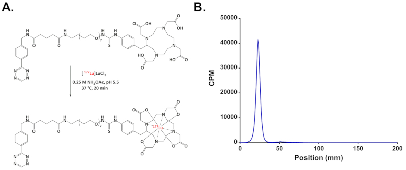
FIGURE 1: (A) Schematic of the radiolabeling of [ 177Lu]Lu-DOTA-PEG7-Tz; (B) Representative radio-iTLC chromatogram demonstrating the >98% radiochemical purity of [177Lu]Lu-DOTA-PEG7-Tz.
Divulgaciones
The authors have nothing to disclose.
Materials
| (E)-Cyclooct-4-enyl 2,5-dioxo-1- pyrrolidinyl carbonate (TCO-NHS) | Sigma-Aldrich | 764523 | Store at -80 °C |
| 2,5-Dioxo-1-pyrrolidinyl 5-[4-(1,2,4,5-tetrazin-3- yl)benzylamino]-5-oxopentanoate (Tz-NHS) | Sigma-Aldrich | 764701 | Store at -80 °C |
| Acetonitrile (MeCN) | Fisher Scientific | A998-4 | |
| Ammonium Acetate (NH4OAc) | Fisher Scientific | A639-500 | |
| Boc-PEG7-amine (O-(2- Aminoethyl)-O′-[2-(Bocamino)ethyl]hexaethylene glycol) | Sigma-Aldrich | 70023 | Store at -20 °C |
| Dichloromethane (DCM) | Fisher Scientific | D143-1 | |
| Dimethyl sulfoxide (DMSO), anhydrous | Fisher Scientific | D12345 | |
| EMD Millipore Amicon Ultra-2 Centrifugal Filter Unit | Fisher Scientific | UFC205024 | |
| GE Healthcare Disposable PD-10 Desalting Columns | Fisher Scientific | 45-000-148 | |
| N,N-Dimethylformamide (DMF), anhydrous | Fisher Scientific | AC610941000 | |
| Phosphate Buffered Saline (PBS) | Fisher Scientific | 70-011-044 | 10x Concentrated |
| p-SCN-Bn-DOTA | Macrocyclics | B-205 | Store at -20 °C |
| Triethylamine (TEA) | Fisher Scientific | AC157911000 | |
| Trifluoroacetic Acid (TFA) | Fisher Scientific | A116-50 | |
| Tumor measuring device | Peira | Peira TM900 |
