Colonoscopic Guided Murine Wound Model: A Method to Isolate Biopsy Induced Wound Bed from Colon to Understand the Healing Process

Published: April 30, 2023

Abstract

Source: Montrose, D. C., et al. Performing Colonoscopic-Guided Pinch Biopsies in Mice and Evaluating Subsequent Tissue Changes. J. Vis. Exp. (2021).

In this video, we describe a procedure to isolate the colonoscopic-induced wound bed at an appropriate time point for detailed molecular analysis. This analysis helps to understand the wound healing process in inflammatory conditions of the GI tract.

Protocol

All procedures involving animal models have been reviewed by the local institutional animal care committee and the JoVE veterinary review board.

1. Collection of the wound bed for molecular analysis

  1. Euthanize mouse using CO2 asphyxiation followed by cervical dislocation (or equivalent technique) on the selected day following biopsy.
  2. Harvest the distal region of the colon by opening the skin and abdominal muscle to expose the body cavity. Place closed scissors under the colon and gently lift to release it from the underlying mesentery and then cut the colon at its midpoint and at the anus to remove it from the mouse.
  3. Flush fecal content using a rat gavage needle attached to a 20 mL syringe filled with ice-cold 1x PBS then lay down the colon onto filter paper.
  4. Cut open the colon longitudinally on filter paper ensuring that the mesenteric side is face down against the filter paper. Apply 0.2% methylene blue to the mucosa using a squeeze pipet while tissue is still on the filter paper and then drain off excess methylene blue. View the colon under a dissecting microscope and locate the wound bed (Figure 1A).
  5. Using 4 inch micro iris scissors, cut around the edge of the wound bed (Figure 1A, dashed circle) being careful not to cut into the muscle layer (unless the muscle is desired) and transfer the dissected wound bed to a tube using fine point tweezers for snap-freezing or desired storage method.       
    NOTE: The amount of tissue collected in this manner is enough for extracting RNA for RNA-seq or equivalent analysis.

Representative Results

Figure 1
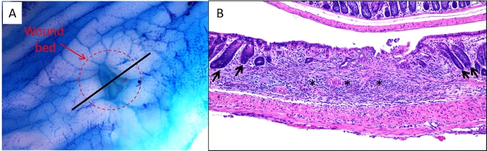
Figure 1: Ex vivo and sectioned images of wound bed. (A) A colon was harvested from a mouse 2 days following biopsy, stained with 0.2% methylene blue and imaged under a dissecting microscope. The dashed circle indicates the edges of the wound bed. The black line indicates the proper location to bisect the wound bed/colon prior to embedding for sectioning. (B) Representative image of an H&E-stained section of a wound bed. Asterisks indicate wound bed and arrows indicate intact crypts adjacent to wound bed indicating the borders of the injured region.

Divulgaciones

The authors have nothing to disclose.

Materials

Biopsy forceps, 3 Fr Karl Storz 61071ZJ
isofluorane Covetrus 2905
methylene blue Sigma-Aldrich M9140
micro iris scissors Integra 18-1619
scalpal with #10 blade Hill-Rom 372610

Tags

Play Video

Citar este artículo
Colonoscopic Guided Murine Wound Model: A Method to Isolate Biopsy Induced Wound Bed from Colon to Understand the Healing Process. J. Vis. Exp. (Pending Publication), e20811, doi: (2023).

View Video