Summary

In vivo Visualization of Synaptic Vesicles Within Drosophila Larval Segmental Axons

Published: October 15, 2010
doi:

Summary

This protocol discusses the live dissection of Drosophila larvae for the purpose of imaging the movement of GFP tagged axonal vesicles on microtubule tracks.

Abstract

Elucidating the mechanisms of axonal transport has shown to be very important in determining how defects in long distance transport affect different neurological diseases. Defects in this essential process can have detrimental effects on neuronal functioning and development. We have developed a dissection protocol that is designed to expose the Drosophila larval segmental nerves to view axonal transport in real time. We have adapted this protocol for live imaging from the one published by Hurd and Saxton (1996) used for immunolocalizatin of larval segmental nerves. Careful dissection and proper buffer conditions are critical for maximizing the lifespan of the dissected larvae. When properly done, dissected larvae have shown robust vesicle transport for 2-3 hours under physiological conditions. We use the UAS-GAL4 method 1 to express GFP-tagged APP or synaptotagmin vesicles within a single axon or many axons in larval segmental nerves by using different neuronal GAL4 drivers. Other fluorescently tagged markers, for example mitochrondria (MitoTracker) or lysosomes (LysoTracker), can be also applied to the larvae before viewing. GFP-vesicle movement and particle movement can be viewed simultaneously using separate wavelengths.

Protocol

1. Preparation of Reagents

  1. Prepare the dissection buffer preparation by adding the following compounds: 128 mM NaCl, 1mM EGTA, 4 mM MgCl2, 2mM KCl, 5mM HEPES, and 36 mM Sucrose in 1000 mL, PH to 7.2 and filter sterilize.

2. Preparation for the Dissection

  1. Siligard cube: The gel is cut to approximately one inch wide, 1 inch long and about 1cm high. The gel cube is placed on a glass slide during dissection. Cubes may be used more than once.
  2. Pins: Pins used for dissection need to be extra short. The sharp bottom portion of minutien pins works best in dissection.
  3. Need one sharp microdissection spring scissors.
  4. Need two fine tipped forceps.

3. Collection of Larvae

  1. Wandering 3rd instar larvae that are expressing a GFP tagged vesicle protein are collected onto a petri dish.
  2. Larvae are washed in deionized water to get rid of all the food.

4. Live dissection of larvae

  1. A larva is transferred to the dissection gel cube on a glass slide. Larva is immersed in dissection buffer.
  2. Larva is pined at the anterior and posterior using two fine pins with the dorsal side up.
  3. Using microdissection scissors, a small cut is made at the midline near the posterior end. From this incision a cut is made through to the anterior. Using two pins, pin the cut cuticle on the lateral edges.
  4. Add a drop of dissecting buffer. Carefully remove the gut, intestines and fat bodies with forceps. Most of these tissues normally ooze out during the dorsal cutting.
  5. Replace the buffer with fresh dissecting buffer. The larva should never be dry and should constantly be in dissecting buffer.
  6. Incubations using in vivo markers MitoTracker or LysoTracker are done at this time, by diluting the in vivo marker in dissection buffer. After the incubation of the in vivo marker wash the dissected larva several times (5 quick washes) using fresh dissecting buffer.
  7. After replacing the last wash push the pins into the syligard and immediately coverslip. The larvae is now ready for observation.

5. Live imaging of dissected larvae

  1. Place the coverslip and gel onto the microscope stage and image using an inverted microscope.
  2. We use a 100x lens to image tagged vesicles within larval segmental nerves. GFP expression in the cell bodies of the ventral ganglion is first evaluated before segmental nerves are imaged. If robust GFP staining is observed in the cell bodies in the ventral ganglion, the nerves are then imaged and used for evaluation.
  3. Using a dual view system with filters for CFP/YFP or RFP/YFP and split view software we can simultaneously image two tagged protein in vivo.

6. Representative Results

Figure 1
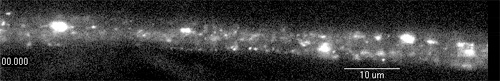
Figure 1: A representative image showing many axonal vesicles on many axonal tracks within a larval segmental nerve. Note the blobs of GFP, representing axonal accumulations.

Figure 2
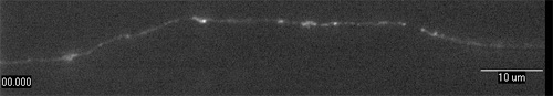
Figure 2: A representative image showing a single axonal track with many axonal vesicles within a larval segmental nerve.

Discussion

In vivo imaging of synaptic vesicle transport within Drosophila larval segmental nerves is a powerful tool for studying the mechanisms in axonal transport. We have previously used this protocol to evaluate the transport dynamics within larval nerves expressing expanded polyQ repeats to elucidate how expansion of repeats affect the dynamics of vesicle transport 3. Using this protocol the velocity of vesicle movement can be determined by converting the video streams to a kymograph. Using genetics, GFP vesicle transport can be visualized within a single axon or within several axons by using a specific GAL4 driver to drive the expression of GFP. Additionally the dynamics of transport can also be observed in larvae carrying mutations for specific genes. Further two vesicles tagged with different GFP variants (CFP or RFP) can be simultaneously visualized.

Divulgaciones

The authors have nothing to disclose.

Acknowledgements

SG is supported by funds from the State University of New York at Buffalo and from John R. Oishei Foundation.

MK was supported by a Student Research Award by CURCA at UB.

Materials

Material Name Tipo Company Catalogue Number Comment
1 Pair Micro Dissection Scissors   Fine Scientific   Spring scissors – 6mm blade. 0.125 mm tip diameter
2 Pair Forceps   Fischer Scientific   Fisherbrand Dissecting Micro-Adson serrated tip forceps
Box of Dissection Pins   Fine Scientific   Minutien Pins 0.1mm diameter

Referencias

  1. Brand, A. H., Perrimon, N. Targeted gene expression as a means of altering cell fates and generating dominant phenotypes. Development. 118, 401-415 (1993).
  2. Hurd, D. D., Saxton, W. M. Kinesin mutations cause motor neuron disease phenotypes by disrupting fast axonal transport in Drosophila. Genética. 144, 1075-1085 (1996).
  3. Gunawardena, S., Her, L. S., Brusch, R. G., Laymon, R. A., Niesman, I. R., Gordesky-Gold, B., Sintasath, L., Bonini, N. M., Goldstein, L. S. Disruption of axonal transport by loss of huntingtin or expression of pathogenic polyQ proteins in Drosophila. Neuron. 40, 25-40 (2003).
check_url/es/2151?article_type=t

Play Video

Citar este artículo
Kuznicki, M. L., Gunawardena, S. In vivo Visualization of Synaptic Vesicles Within Drosophila Larval Segmental Axons. J. Vis. Exp. (44), e2151, doi:10.3791/2151 (2010).

View Video