1. HL60 cell rolling
1.1. Fabrication of Patterned Substrates.
- Using microcontact printing (μCP)4-7 to make alternating self-assembled monolayers (SAMs) of PEG molecules on the gold-coated glass slides: Fabricate microcontact printing polydimethylsiloxane (PDMS) stamps that defined the receptor patterns with inclination angle of α = 10° by an SU-8 molding process. Clean the gold surface with piranha solution (3:1 mixture of sulfuric acid to 30% hydrogen peroxide) for 20 minutes and then rinse the surface with copious DI water at 24.5 °C prior to use. Ink the PDMS stamp with 5mM PEG solution in ethanol. Dry the stamp in air for 20 minutes. Gently put the stamp on the gold surface for 40 sec and make sure there is a good contact between the gold surface and the stamp. No excess pressure is required. Rinse the surface with ethanol and dry it under a stream of N2.
- Incubate the substrate within P-selectin solution (15 μg/mL in DPBS) using a perfusion chamber (Electron Microscopy Sciences) for 3 hours at 24.5°C to pattern the remaining areas with P-selectin. Rinse the surface with copious DPBS.
- Backfill the surface with BSA (1 mg/mL in DPBS) for 1 h to block non-specific interactions. Rinse the surface with copious DPBS.
1.2. Cell Rolling Experiments in a Flow Chamber.
- Flow a suspension of HL60 cells (~105 cells/mL) over the patterned surfaces in a rectangular flow chamber (Glycotech, Inc; width w = 1.0 cm; length = 6 cm; height h = 0.0127 cm) at 24.5°C. Use a syringe pump (World Precision Instruments, SP230IW) to generate flow rate of 75 μL/min, with corresponding shear stress around 0.5 dyn/cm2 (~0.05 Pa). Calculate shear stress τ by using the plane Poiseuille flow equation τ = 6μQ/wh2, where μ is the kinematic viscosity (0.001002 Pa s), Q is volumetric flow rate, w is width of the flow chamber, and h is height of the flow chamber.
- Use an inverted microscope (Nikon TE2000-U) with a mounted camera (Andor iXon 885) to record HL60 cells rolling interactions with adhesive P-selectin substrates using a 4× objective, typically at a rate of 1 frame per second for durations of 300 s. Perform three independent experiments, for each shear stress magnitude and pattern inclination angle. Present data as mean and standard deviation of the average values obtained from each experiment.
- Data Analysis.
Analyze the image sequences by a customized Matlab (Mathworks, Inc.) program that utilized a particle tracking freeware8 to generate tracks along the patterned line edges. Tracks extending till the end of a P-selectin band are selected and fitted with two straight line segments – one aligned with the flow, the other aligned with the pattern edge. These two segments are then used to calculate the edge tracking length, rolling velocity on the edge, and rolling velocity on the plain region.
2. Representative Results:
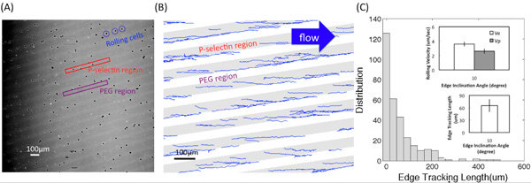
Figure (A) shows one of microscope images converted from the video of HL60 rolling interactions with adhesive P-selectin substrates using a 4× objective. Bright and dark regions correspond to P-selectin receptor and PEG regions, respectively. Figure (B) shows the tracks obtained using a customized Matlab program. The edge inclination angle was 10° and the shear stress was 0.5 dyn/cm2. The edge tracking length, le, displacement, d, and the rolling velocities on the edge and inside the bands, Ve and Vp, respectively, are described in Figure (C-1). Figure (C-2) shows the distribution (the number of recorded cells) of edge tracking length. Insets show the average value of le and the rolling velocity on the edge (Ve) and inside the bands (Vp) at the inclination angle α =10° and the fluid shear stress magnitude around 0.5 dyn/cm2. Error bars represent one standard deviation, where n = 3 replicate experiments (3 replicate surfaces) for each condition.
