1. Hexanethiolate Monolayer Protected Gold Clusters Synthesis
Hexanethiolate functionalized monolayer protected gold clusters (MPCs) are synthesized following a 2:1 1-hexanethiol (C6) to gold mole ratio to produce an average structure of Au225(C6)75. 4-9 Specific modifications to the Brust reaction, like ligand type, specific thiol-to-gold ratios, temperature, and reaction delivery rate, or post-synthesis treatments, 9-11 can yield a diverse range of MPCs with varying core sizes and functional protective groups, respectively.4 The MPC approximate (average) compositions of MPCs functionalized with various alkanethiol groups can be determined by proton (1H) nuclear magnetic resonance (NMR) analysis of iodine-decomposed samples.
- Dissolve 1.1 g tetraoctylammonium bromide (TOABr) in 30 mL of toluene with appropriate fume hood ventilation.
- Dissolve 0.38 g sodium borohydride (NaBH4) in ˜20 mL of 18 MΩ ultrapurified water (UP H2O) and allow to chill over ice for at least 30 min.
- Dissolve 0.31 g hydrogen tetrachloroaurate (HAuCl4) in ˜20 mL of UP H2O and quantitatively transfer the solution mixture to the TOABr-toluene solution using an additional ˜5 mL of UP H2O in order to phase-transfer the aqueous gold solution to the nonaqueous solution. Stir rigorously while lightly capped for 30 min so the burnt orange aqueous and clear nonaqueous phases are mixing well.
- Transfer both the clear aqueous and burnt orange nonaqueous phases to a separatory funnel. Discard the aqueous (bottom) layer and decant the nonaqueous (top) layer into a clean flask.
- Add C6 in a ratio of 2:1 with HAuCl4 to the nonaqueous solution. Stir for 30 min to form a Au(I) polymer, as detected by a color change from reddish orange to a pale yellow, nearly colorless solution.
- Transfer the reaction mixture to an insulated ice bath and chill to 0°C for at least 30 min with stirring.
- Quantitatively and quickly add the chilled NaBH4 solution to the reaction mixture in order to reduce Au(I) to a metallic gold in the presence of thiols, instantaneously forming a thick black solution of MPCs upon addition. Stir reaction overnight at 0°C.
- Transfer the reaction mixture to a separatory funnel, discard the aqueous (bottom) layer into a waste beaker, and rotary evaporate the nonaqueous (top) toluene layer to near complete dryness leaving a heavy black sludge in the flask.
- Precipitate the MPCs by adding acetonitrile and allowing to sit overnight.
- Collect MPCs by vacuum filtration using a glass frit of medium porosity with rubber fittings and side-armed flask with aspirator and rinse with a copious amount of acetonitrile.
- Allow MPCs to air dry, weigh product, characterize by transmission electron microscopy (TEM) and NMR analysis, and store capped for future use. Obtain TEM images by drop-casting MPCs dissolved in toluene onto formvar/carbon support film on copper grid (400 mesh) and operating the TEM instrument at 80-100 kV. The average core size can be estimated using image analysis software such as Image J (freeware).
2. Film Assembly: Dithiol-linked MPC Film Assembly for Protein Monolayer Electrochemistry
The gold substrate is first electrochemically cleaned and modified with a C6 SAM before immersing in alternating solutions of dithiol linking molecules and C6 modified MPCs to make up a “dip cycle,” which is repeated several times to ultimately form a dithiol-linked MPC film assembly. As described in prior studies,2 the original plasmid for the Pseudomonas aeruginosa azurin (AZ) protein was graciously given by Dr. Corey Wilson of Rice University and AZ was provided as a purified and lyophilized powder by the University of Richmond professor, Dr. Jonathan Dattelbaum, that was subsequently rehydrated with 4.4 mM potassium phosphate buffer (KPB, pH = 7.0, μ = 10 mM) to create a 5-10 μM solution as verified by ultraviolet visible (UV-Vis) analysis.
- Assemble the electrochemical (echem) sandwich cell in the following order from bottom to top: first Lucite retainer plate, gold substrate as a working electrode, brass electrical contact to gold working electrode, first rubber gasket, Viton o-ring that defines the electrode area (0.32 cm2), glass cell body, second rubber gasket, and second Lucite retainer plate. The entire cell is held together by threaded rods and carefully tightened wing nuts. The cell is fitted with a commercially purchased reference electrode that houses a glass barrel with 1 M saturated KCl, Ag/AgCl reference wire, and a Pt auxiliary electrode wire.
- Electrochemically clean the gold substrate by performing cyclic voltammetry (CV) in the potential windows from 0.2 to 0.9 V, 0.2 to 1.2 V, and 0.2 to 1.35 V (versus Ag/AgCl, KCl) at 100 mV/s in a solution of 0.1 M H2SO4 and 0.01 M KCl.
- Measure the charging current of the cleaned bare gold substrate by performing CV at “standard conditions,” including a potential window from 0.1 to 0.4 V (versus Ag/AgCl, KCl) scanned at 100 mV/s in KPB.1 Discard KPB and rinse successively with UP H2O, ethanol (EtOH), UP H2O, and EtOH.
- Expose the cleaned gold substrate to ˜300 μl of 5 mM C6 solution in EtOH and allow to sit overnight to form an ordered C6 SAM. Discard C6 solution from the cell and rinse successively with EtOH, UP H2O, EtOH, and UP H2O.
- Measure the charging current of the SAM at standard conditions. Discard KPB and rinse successively with UP H2O, EtOH, UP H2O, and EtOH. The charging current should be markedly decreased from that of the bare gold measurement (step 2.3).1
- Expose the SAM modified gold substrate to ˜300 μl of 5 mM 1,9-nonanedithiol (NDT) solution in EtOH and allow to sit for 1 hr to interdisperse NDT linking molecules within the C6 SAM. Discard NDT solution and rinse successively and thoroughly with EtOH, UP H2O, EtOH, UP H2O, and methylene chloride (CH2Cl2).
- Expose the gold substrate to a MPC solution of CH2Cl2 (˜1 mg/mL) with agitation by slowly bubbling with N2 gas for 1 hr. If necessary, replace evaporated MPC solution with more CH2Cl2. This is the anchoring MPC layer of the film assembly. Discard MPC solution and again rinse successively with CH2Cl2, UP H2O, and KPB.
- Measure the charging current of the MPC layer at standard conditions. Discard KPB and rinse successively with UP H2O and CH2Cl2.
- Expose the gold substrate to ˜300 μl of 5 mM NDT solution of CH2Cl2 with agitation by slowly bubbling with N2 gas for 20 min.
- Discard NDT and rinse thoroughly with CH2Cl2. Repeat steps 2.7 and 2.8 to deposit the second MPC layer of the film assembly.
- To deposit additional MPC layers, steps 2.9 and 2.10 are repeated. With each additional MPC layer a corresponding increase in the charging current is observed.
- After the networked MPC film is complete, rinse the film modified substrate with KPB. AZ protein is adsorbed on the MPC film assembly by injecting ˜150 μL of ˜5-10 μM AZ solution of KPB into the echem sandwich cell and allowing to sit capped and refrigerated for at least 1 hr.
- Remove the echem cell from the refrigerator and allow it to return to near room temperature. Thoroughly rinse with KPB, refill echem cell with KPB, and bubble KPB with N2 gas for 10 min.
- Protein monolayer electrochemical studies are performed as CV in the potential window from -0.25 V to 0.25 V (versus Ag/AgCl, KCl) scanned at 100 mV/sec in KPB.
3. Film Assembly: Dithiol-linked MPC Film Assembly for Optical Tracking
Prior to growing MPC films for optical evaluation, glass slide sections are pre-cleaned with Piranha solution (CAUTION! 2:1 concentrated H2SO4 and H2O2) and treated with (3-mercaptopropyl)-trimethoxysilane (3-MPTMS).1-2 MPC films are then assembled on these modified glass slides using the “dip cycle” technique as previously described above.
- Rinse a 3-MPTMS modified glass slide with CH2Cl2 and place it in a MPC solution of CH2Cl2 (˜1 mg/mL) for 1 hr while agitating on a shaker at low speed. This completes the first MPC layer of the film assembly by anchoring MPCs to the mercaptans endgroups of the silane. Rinse the slide thoroughly with CH2Cl2, and dry with N2 gas. Take a UV-Vis spectrum (from 400 to 1000 nm) of the slide, and rinse again with CH2Cl2.
- Place the slide in a 5 mM NDT solution of CH2Cl2 for 1 hr while agitating on a shaker at low speed. Rinse the slide with CH2Cl2.
- Place the slide in a MPC solution for 1 hr while agitating on a shaker at low speed. This completes the second MPC layer of the film assembly. Rinse the slide thoroughly with CH2Cl2, dry with N2 gas, and take a UV-Vis spectrum (from 400 to 1000 nm) of the slide. The absorbance across the spectrum should be increasing as additional MPC layers are adsorbed to the film assembly.
- To deposit additional MPC layers, steps 3.2-3.3 are repeated.
4. Characterization of Monolayer Protected Gold Cluster Film Assemblies by Cross Sectional Transmission Electron Microscopy
TEM cross sections are prepared by re-embedding en face embedded films.2, 12 This is done by first attaching a MPC film assembled on a 3-MPTMS modified glass slide onto a clean, standard microscope slides using Embed 812 epoxy resin to allow for improved handling during the procedure below. Use caution with the applied heat as higher temperatures will decompose the MPCs within the film.
- Mix Embed 812 epoxy resin and allow to thicken for at least 12 hr.
- Fill a “00” BEEM capsule with epoxy resin and invert on top of the MPC film sample (prepared in section 3). Place pressure on the capsule so that a bubble rises to the top of capsule, creating a seal between the epoxy resin and MPC film sample. Allow to polymerize for at least 18 hr at 60°C and then cool the mounted slides to room temperature.
- Heat mounted slides for 20 sec on a cast aluminum hot plate at 200°C in order to facilitate the removal of the block with attached MPC en face film.
- Cut the sample containing the film off of the BEEM capsule block using a jeweler’s saw.
- Re-embed the removed portion in a silicon flat mold with the MPC film side up facing the interior of the silicon well. Fill the silicon well with epoxy resin at room temperature and allow to polymerize for at least 18 hr at 60°C. Cool the sample to room temperature.
- Obtain thin sample sections of 60-80 nm on a Leica UCT ultramicrotome by using a diamond knife to cut sections perpendicular to the knife’s edge.
- Place sliced sections on formvar/carbon support film on copper grid (400 mesh) and take TEM images of prepared cross sections of MPC film assemblies.
5. Representative Results:
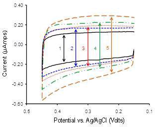
Figure 1 Double layer charging current monitoring of MPC film growth for a total of 5 dipping cycles (alternating exposure to MPC and NDT solutions). Charging current increased systematically with each dipping cycle, adding “layers” of MPC to the film (Fig. 2). The cyclic voltammograms were collected using a potential window from 0.1 to 0.4 V (versus Ag/AgCl, KCl) scanned at 100 mV/s in 4.4 mM potassium phosphate buffer (pH = 7.0, μ = 10 mM). Reprinted with permission from M.L. Vargo, C.P. Gulka, J.K. Gerig, C.M. Manieri, J.D. Dattelbaum, C.B. Marks, N.T. Lawrence, M.L. Trawick, and M.C. Leopold, Langmuir 26(1), 560-569. Copyright 2010 American Chemical Society.
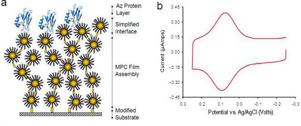
Figure 2 (a) Schematic representation of AZ protein adsorbed to a dithiol-linked MPC film assembly. (b) Typical cyclic voltammogram for AZ adsorbed to MPC film assembly collected using a potential window from -0.25 to +0.25 V (versus Ag/AgCl, KCl) scanned at 100 mV/s in 4.4 mM potassium phosphate buffer (pH = 7.0, μ = 10 mM).
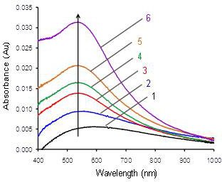
Figure 3 Representative UV-Vis spectral monitoring of a dithiol-linked MPC film growth on a 3-MPTMS modified glass slide. A dip cycle consists of an exposure of the glass slide to NDT linker solution followed an exposure to MPC solution. Each subsequent dip results in growth in film thickness and a concurrent absorbance increase. As the number of dip cycles increases, the surface plasmon band is gradually defined at ˜520 nm. Reprinted with permission from M.L. Vargo, C.P. Gulka, J.K. Gerig, C.M. Manieri, J.D. Dattelbaum, C.B. Marks, N.T. Lawrence, M.L. Trawick, and M.C. Leopold, Langmuir 26(1), 560-569. Copyright 2010 American Chemical Society.
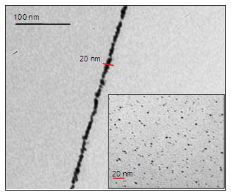
Figure 4 Transmission electron microscopy (TEM) cross sectional image analysis of a dithiol-linked MPC film assembly. Inset: Typical TEM image of hexanethiolate functionalized MPCs used in the film assembly. TEM analysis determined an average gold core diameter of the MPCs to be ˜2 nm using Image J analysis. Reprinted with permission from M.L. Vargo, C.P. Gulka, J.K. Gerig, C.M. Manieri, J.D. Dattelbaum, C.B. Marks, N.T. Lawrence, M.L. Trawick, and M.C. Leopold, Langmuir 26(1), 560-569. Copyright 2010 American Chemical Society.
