Introduction of small molecules to the developing Drosophila embryo offers great potential for characterizing biological activity of novel compounds, drugs, and toxins as well as for probing fundamental developmental pathways. Methods described herein outline steps that overcome natural barriers to this approach, expanding the utility of the Drosophila embryo model.
The Drosophila embryo has long been a powerful laboratory model for elucidating molecular and genetic mechanisms that control development. The ease of genetic manipulations with this model has supplanted pharmacological approaches that are commonplace in other animal models and cell-based assays. Here we describe recent advances in a protocol that enables application of small molecules to the developing fruit fly embryo. The method details steps to overcome the impermeability of the eggshell while maintaining embryo viability. Eggshell permeabilization across a broad range of developmental stages is achieved by application of a previously described d-limonene embryo permeabilization solvent (EPS1) and by aging embryos at reduced temperature (18 °C) prior to treatments. In addition, use of a far-red dye (CY5) as a permeabilization indicator is described, which is compatible with downstream applications involving standard red and green fluorescent dyes in live and fixed preparations. This protocol is applicable to studies using bioactive compounds to probe developmental mechanisms as well as for studies aimed at evaluating teratogenic or pharmacologic activity of uncharacterized small molecules.
The Drosophila embryo continues to be a premier model for investigation of fundamental mechanisms of development2. This powerful model is supported by a wide array of molecular genetic tools that permit manipulations of essentially any gene at any time point and within any developing organ. The small size, rapid development, and extensive characterization of morphogenesis of the Drosophila embryo make it a model of choice for genetic screens, many of which have uncovered fundamental developmental pathways3,4. Numerous phenotypes in the Drosophila embryo have been characterized and are easily interpretable, often providing a means to identify underlying molecular genetic mechanisms responsible for an abnormal trait.
Historically, a shortcoming of the fly embryo model has been the difficulty of introducing small molecules to embryonic tissues. This obstacle has posed limitations on: 1) using known bioactive small molecules as probes to interrogate developmental mechanisms and 2) using this established model to evaluate teratogenic or pharmacologic activity of uncharacterized small molecules. As a consequence, the screening potential of the fly embryo has been underutilized in characterization of small molecule activity.
Delivery of small molecules to the fly embryo can be achieved with two methods: 1) permeabilization of the eggshell and 2) microinjection. This article presents advances to the method of permeabilization that are easy to execute in the setting of a conventional Drosophila laboratory. It should be noted that recent advances in microinjection methods with microfluidics technology is also contributing to methods of introducing compounds to the embryo5,6. Introducing molecules to the embryo is prevented by a waxy layer of the eggshell7. The Drosophila eggshell consists of five layers. From the inside out they are: the vitelline membrane, the waxy layer, the inner chorionic layer, the endochorion and the exochorion8. The three outer chorionic layers can be removed by brief emersion of the embryo in dilute bleach, a step referred to as dechorionation. The exposed waxy layer can then be compromised by exposure to organic solvents, such as heptane and octane7,9, rendering the dechorionated embryo permeable, while it remains encased in the underlying vitelline membrane. However, use of these solvents introduces complications due to their toxicity and the difficulty in regulating their strong permeabilizing action, both of which have stark negative effects on embryo viability9,10.
A method of permeabilization using a composition termed embryo permeabilization solvent (EPS) has been previously described1. This solvent consists of d-limonene and plant-derived surfactants that enable the solvent to be miscible with aqueous buffers. The low toxicity of d-limonene and the ability to dilute the solvent to desired concentrations has yielded an effective method to generate permeable embryos with high viability1. However, two endogenous factors have continued to bring limitations to the application. First, embryos demonstrate heterogeneity in permeability after EPS treatment, even when care is taken to maintain close developmental staging. Second, embryos older than approximately eight hours have proven difficult to permeabilize, consistent with a hardening of the eggshell that occurs after egg laying11.
Described here are advances in the EPS method that: 1) assist in identifying and analyzing near-identically permeabilized embryos, even after fixation and immunostaining steps have been executed and 2) enable permeabilization of embryos at late developmental time points (>8 hr, stage 12 and older). Specifically, application of a far-red dye, CY5 carboxylic acid, is described that serves as a permeability indicator, which persists in the embryo during development and after formaldehyde fixation. In addition, it is shown that rearing embryos at 18 °C maintains the eggshell in an EPS sensitive state, enabling permeabilization of late stage embryos (stages 12-16).
These advances overcome the previously mentioned limitations to the EPS methodology. This application will therefore provide investigators with a means to introduce small molecules of interest to the embryo at distinct developmental time points while maintaining viability.
1. Preparation of Fly Cultures, Solutions, and Embryo Handling Devices
2. Staging, Dechorionation, and EPS Treatment of Embryos
3. Dye and Drug Treatment of Permeabilized Embryos
4. Identification of Permeabilized Viable Embryos
Embryo handling devices are pictured in Figure 1 to assist in visualizing the “home-made” devices for manipulation in the above Protocols. Results seen in Figure 2 illustrate the robust effect of rearing embryos at 18 °C on their ability to be permeabilized by EPS at late stages of development. This condition is applied in the protocol step 2.1. Efficacy of the CY5 carboxylic acid dye to reveal the various levels of permeability typically seen in EPS treated embryos is seen in Figure 3. The developmental dynamics of the dye distribution in the yolk is also seen in Figure 3, revealing a criterion used to assess viability, as described in Protocol step 4.2. The utility of the CY5 dye in determining embryo permeabilization subsequent to toxin treatment, formaldehyde fixation and immunostaining is illustrated by the result in Figure 4.
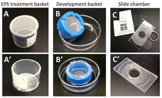
Figure 1. Embryo handling devices for the EPS method. The flat-bottomed basket is used in dechorination and EPS exposure steps (A,A'). The development basket is used for longer developmental exposures of permeabilized embryo (B,B'). The slide chamber is used for shorter developmental exposures and higher resolution imaging of live embryos (C,C'. See text for further description).
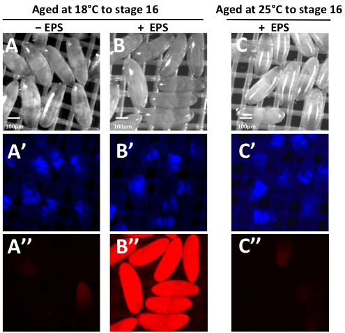
Figure 2. Effect of aging at 18 °C on EPS efficacy in late stage embryos. Embryos were collected for two hours followed by aging at 18 °C for 20 hr (Panels A-A”, B-B”) or at 25 °C for 10 hr (Panel C-C”). Embryos at 18 °C were then dechorionated and divided into two samples. The first sample was treated directly with 1 mM Rhodamine B dye in MBIM-T for 5 min, washed and visualized under brightfield and blue and red fluorescence channels (Panel A-A”). The second sample was treated with EPS (1:10 in MBIM for 1 min), washed and then treated with 1 mM Rhodamine B for 5 min and washed before visualization (Panel B-B”). Embryos raised at 25 °C were dechorionated and treated directly with EPS (1:10 in MBIM for 1 min), washed and then treated with 1 mM Rhodamine B for 5 min before visualization (Panel C-C”). Embryos were determined to be at stage 14 by the folds in the gut revealed by yolk autofluorescence in the blue channel (Panel A’, B’, C’). Embryos raised at 18 °C are impermeable prior to EPS treatment as seen by absence of Rhodamine B uptake (Panel A”). EPS treatment of 18 °C embryos yields a high degree of permeability as seen by Rhodamine B uptake (Panel B”). Embryos raised at 25 °C remain impermeable even with EPS treatment as seen by exclusion of Rhodamine B (Panel C”).
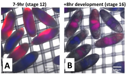
Figure 3. Incorporation of CY5 in permeable and viable embryos. Embryos were collected at 25 °C for 2 hr and aged for 14 hr at 18 °C (equivalent to 7-9 hr embryos at 25 °C, stage 12). After dechorination, EPS treatment was done (1:40 in MBIM for 1 min) followed by incubation in CY5 dye (50 µM in MBIM-T for 15 min). Embryos were washed three times in MBIM-T and transferred to development basket with MBIM in the reservoir. Development was allowed to proceed for 8 hr at room temperature. Uptake of CY5 (Red) is imaged in the far-red channel immediately after dye treatment and washing (Panel A) and after 8 hr development (Panel B). Distribution of the yolk is seen by autofluorescence in the blue channel. Dye uptake, hence permeability, is seen to vary from embryo to embryo. CY5 dye (red) is seen to localize to the yolk (blue), which becomes concentrated to the lumen of the gut at stage 16 (purple, Panel B).
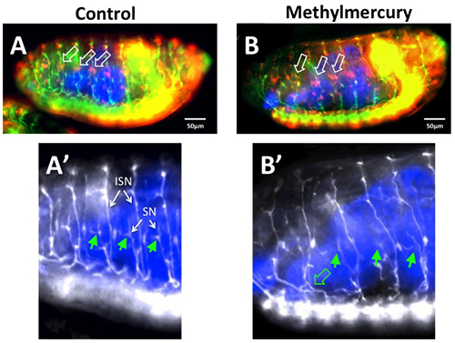
Figure 4. Determination of permeabilization and methylmercury effects in fixed and immunostained embryos. Embryos were collected at 25 °C for 2 hr and aged for 14 hr at 18 °C (equivalent to 7-9 hr embryos at 25 °C, stage 12). After dechorination, EPS treatment was done (1:40 in MBIM for 1 min) followed by incubation in CY5 dye (50 µM in MBIM-T for 15 min) together with methylmercury (50 µM MeHg, Panel B) or DMSO solvent control (0.1% final concentration, Panel A). Embryos were washed with MBIM-T and placed in a development basket with MBIM:M3 medium in the reservoir and aged for an additional 8 hr at room temperature. Embryos were then fixed in a two-phase 4% paraformaldehyde-heptane preparation by a standard protocol14. Staining was performed with anti-Fasciclin II (green in A,B and white in A’,B’) to label motor neurons and anti-elav antibodies (red in A, B) to label all neuron cell bodies. CY5 dye is revealed by direct fluorescence, which requires extended exposure due to diminished fluorescence intensity due to fixation (CY5 is pseudo-colored blue in all panels). The effects of MeHg are seen in the irregular patterning and clustering of the lateral chordotonal neuron cell bodies (elav-positive, labeled in red and denoted with white arrows in B versus A). In addition, a characteristic branching of the segmental (SN) (solid green arrows in A’) is seen to be highly variable with MeHg exposure (solid green arrows in B”) consistent with previously reported effects of MeHg on the embryo15. Projection of the intersegmental and segmental nerves at their roots are seen to be displaced posteriorly with MeHg exposure (open green arrow in B’). Note: Methylmercury is a potent neurotoxin. Care should be taken to wear gloves and eye protection when handling. Disposal should be done through an institutional environmental safety facility and service.
The above method outlines a means to obtaining viable Drosophila embryos that are accessible to small molecule treatments across a wide developmental range. This method introduces the novel and simple finding that aging embryos at 18 °C enables permeabilization of late stage embryos with the same efficacy as previously seen only in early stage embryos. In addition, use of the far-red dye CY5 carboxylic acid as a permeability indicator has proven effective in post-fix applications and does not interfere with conventional red and green fluorescent markers that can be used to reveal developmental phenotypes. These findings significantly advance the efficacy and utility of the EPS method.
This method is amenable to analyses of both living and fixed embryo preparations. Using brightfield microscopy and the slide chamber set up, typical features to score in the first half of embryo development are morphogenetic movements such cellularization of the blastoderm, cephalic furrow formation, germband elongation and germband retraction1. With GFP or RFP reporters more specific endpoints can be discerned, e.g. early segmentation patterns and formation of neural structures in later development1. GFP and RFP reports also enable observation of developmental events in living permeabilized embryos in both the slide chamber and developmental basket preparations. A simple method to determine gross toxicity of an applied drug or chemical is to monitor the pattern of yolk protein auto-fluorescence in the blue channel to determine a delay or cessation of development1.
The EPS methodology also has great potential for broadening the investigative tools for non-model insects, in particular the mosquito, which shares a similar architecture of the eggshell. Application of small molecules to embryos of other insect species would open up an avenue of investigation where standard genetic approaches to functional studies are currently lacking. Embryos developed in baskets can be processed for formaldehyde fixation, therefore opening up analyses to the wide array of immunostaining reagents available for Drosophila studies. However, this step requires that a post-fix determination of permeabilization be feasible to correlate phenotypes with embryos that had been made accessible to the drug. Application of CY5 carboxylic acid dye has proven highly effective for this approach. CY5 carboxylic acid is efficiently taken up in permeabilized embryos (Figure 3A). During development CY5 is concentrated in the yolk, which is ultimately sequestered in the lumen of the forming gut at stage 14 and later (Figure 3B). After fixation, CY5 fluorescence is markedly decreased, yet reliably detectable in the gut and serves as a marker of those embryos that were effectively permeabilized at the outset (see representative result Figure 4). It should be noted that CY5 detection at this stage requires longer camera exposures (e.g., 1-4 seconds) for detection. Thus, scoring of phenotypes with immunostaining patterns can proceed using the CY5 signal to confirm similarly permeabilized embryos together with tissue specific markers (e.g., neural specific antibodies seen in Figure 4).
The biggest challenge in this method is the sensitivity of embryo viability to the permeabilization process, something that has long troubled prior attempts to develop this method9,10,16. Viability subsequent to permeabilization is starkly age-dependent, and increases dramatically the older the embryo is upon permeabilization9. Yet, as we have shown previously, permeability becomes increasingly difficult with age1. A recent report now demonstrates the ability to permeabilize late stage embryos (stage 14) by re-invoking heptane as a solvent in conjunction with d-limonene17. The broad utility of this latter method is not clear as only one drug effect was characterized (nocodazole) and application to earlier stage embryos was not described17. Furthermore, application of the 18 °C development step outlined above yields late stage permeability and further avoids use of toxic organic solvents. The investigator who is new to the EPS protocol will experience variability in permeabilization and viability outcomes upon initial attempts. The steps outlined here give the investigator the tools to systematically vary conditions of permeabilization treatments and subsequent incubation steps to optimize conditions for specific strains of Drosophila they are working in their own laboratory setting.
An additional challenge with the method is the variability in chemical uptake seen from embryo to embryo. This variability is reflected in the heterogeneity of CY5 dye uptake seen in embryos immediately after dye treatment (Figure 3A). In contrast, Rhodamine B dye is more rapidly and evenly dispersed across the embryonic tissues than CY5 dye (Figure 2B”). Thus, some embryo-to-embryo variability may be harbored in the distribution properties of the chemical, drug or toxin of interest and is inherent to the method. Where quantification of dose is critical, it is recommended that uptake of the drug or toxin of interest is characterized through an alternative analytical method. Nonetheless, the ease of the above protocol, and the ability to screen hundreds of embryos, allows the investigator to evaluate dose responses and score characteristic phenotypes with little investment of resources, making for a powerful first approach to characterizing drugs or toxins in this highly developed model system.
The authors have nothing to disclose.
This work was supported by NIH/NIEHS R03ES021581 (awarded to M.D.R.) and by the University of Rochester Environmental Health Center (NIH/NIEHS P30 ES001247).
Fly Cage | Flystuff.com | 59-101 | http://flystuff.com/general.php |
Cocamide DEA [Ninol 11-CM] | Stepan Chemical | call for special order | http://www.stepan.com/ |
Ethoxylated alcohol [Bio-soft 1-7] | Stepan Chemical | call for special order | http://www.stepan.com/ |
d-limonene (Ultra high purity grade) | Florida Chemical Co. | call for special order | http://www.floridachemical.com/ |
Sodium hypochlorite | Fisher | SS290-4 | http://www.fishersci.com/ |
Tween-20 | Fisher | BP337 | http://www.fishersci.com/ |
PBS powder | Sigma | 56064C | http://www.sigmaaldrich.com/ |
Rhodamine B | Sigma | R6626 | http://www.sigmaaldrich.com/ |
CY5 carboxylic acid | Lumiprobe | #23090 | http://www.lumiprobe.com/p/cy5-carboxylic-acid |
DMSO | Sigma | 472310-100 | http://www.sigmaaldrich.com/ |
Shields and Sang M3 medium | Sigma | S8398 | http://www.sigmaaldrich.com/ |
Nitex Nylon mesh | Flystuff.com | 57-102 | http://flystuff.com/misc.php |
Dissolved oxygen (DO) membrane | YSI | #5793 | http://www.ysireagents.com/search.php |
25mm circular no.1 cover slip | VWR | 48380-080 | https://us.vwr.com/ |
Grape-agar plate mix | Flystuff.com | 47-102 | http://flystuff.com/media.php |
Nutator | VWR | 82007-202 | https://us.vwr.com/ |